. 2019 Dec;62(12):2365-2374.
doi: 10.1007/s00125-019-04998-4. Epub 2019 Oct 14.

Neutrophil elastase contributes to the pathological vascular permeability characteristic of diabetic retinopathy

Affiliations

Neutrophil elastase contributes to the pathological vascular permeability characteristic of diabetic retinopathy

Haitao Liu et al. Diabetologia. 2019 Dec.

Abstract

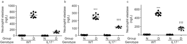
Aims/hypothesis: Levels of neutrophil elastase, a serine protease secreted by neutrophils, are elevated in diabetes. The purpose of this study was to determine whether neutrophil elastase (NE) contributes to the diabetes-induced increase in retinal vascular permeability in mice with streptozotocin-induced diabetes, and, if so, to investigate the potential role of IL-17 in this process.

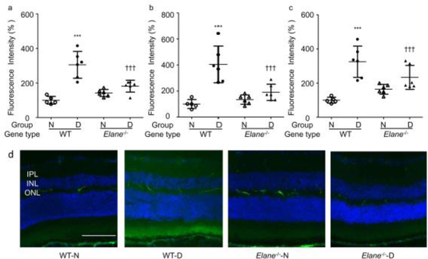
Methods: In vivo, diabetes was induced in neutrophil elastase-deficient (Elane-/-), Il-17a-/- and wild-type mice. After 8 months of diabetes, Elane-/- mice and wild-type age-matched control mice were injected with FITC-BSA. Fluorescence microscopy was used to assess leakage of FITC-BSA from the retinal vasculature into the neural retina. The level of NE in Il-17a-/- diabetic retina and sera were determined by ELISA. In vitro, the effect of NE on the permeability and viability of human retinal endothelial cells and the expression of junction proteins and adhesion molecules were studied.

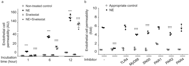
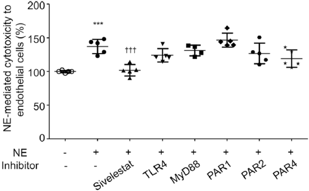
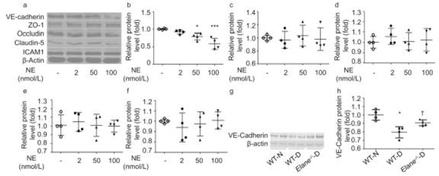
Results: Eight months of diabetes resulted in increased retinal vascular permeability and levels of NE in retina and plasma of wild-type animals. All of these abnormalities were significantly inhibited in mice lacking the elastase. The diabetes-induced increase in NE was inhibited in mice lacking IL-17. In vitro, NE increased retinal endothelial cell permeability, which was partially inhibited by a myeloid differentiation primary response 88 (MyD88) inhibitor, NF-κB inhibitor, and protease-activated receptor (PAR)2 inhibitor. NE degraded vascular endothelial-cadherin (VE-cadherin) in a concentration-dependent manner.

Conclusions/interpretation: IL-17 regulates NE expression in diabetes. NE contributes to vascular leakage in diabetic retinopathy, partially through activation of MyD88, NF-κB and PAR2 and degradation of VE-cadherin.

Keywords: Diabetic retinopathy; Elane; IL-17; Neutrophil elastase; Vascular permeability.


Conflict of interest statement

Duality of interest

The authors declare that there is no duality of interest associated with this manuscript.

Figures

Fig. 1
Fig. 1
Effects of Elane deletion on vascular permeability in diabetic mice. WT mice with diabetes (WT-D) of 8 months duration significantly increased accumulation of FITC-BSA in the IPL (a), INL (b) and OPL (c) layers of the retina compared with that in WT non-diabetic (WT-N) mice. (d) Representative images from each study group. Genetic deletion of Elane significantly inhibited the extravascular fluorescence in the IPL, INL and OPL of the retina in diabetic Elane−/−-D mice. FITC-BSA was injected intravenously, allowed to circulate for 30 min, and average fluorescence was measured from cross-sections from each layer, excluding microvessels. Data are shown as % relative to the fluorescence in WT-N mice. Scale bar, 100 μm. n=5 for WT-N, n=6 per group for the others. Data are expressed as mean ± SD. ***p<0.001 vs WT-N, p<0.05, †††p<0.001 vs WT-D
Fig. 2
Fig. 2
Effects of Elane deletion and diabetes on retinal thickness. (a) Optical coherence microscopy images of retinas, (b) graph of retinal thickness in experimental groups. Neither diabetes nor Elane−/− resulted in any significant change in retinal thickness or loss of photoreceptors at 8 months of diabetes (10 months of age). Scale bar, 100 μm, n=4 per group. Data are expressed as mean ± SD. Elane−/−-D, diabetic Elane-knockout mice; Elane−/−-N, non-diabetic Elane-knockout mice; WT-D, diabetic WT mice; WT-N, non-diabetic WT mice
Fig. 3
Fig. 3
Effect of human NE on hREC permeability, and possible mediators of this effect. (a) Human NE (50 nmol/l) directly increased hREC permeability, and this permeability could be inhibited by the NE inhibitor sivelestat. (b) The NE-induced increase in hREC permeability was significantly inhibited by 1 h incubation with sivelestat; it was significantly inhibited by MyD88 inhibitor peptide, NF-κB inhibitor and PAR2 inhibitor, but was not inhibited by TLR4 inhibitor, PAR1 inhibitor or PAR4 inhibitor. The complete names and concentrations of inhibitors used are listed in the Methods section. In the figure, replicates incubated with inhibitors but without NE are indicated by white circles, whereas replicates incubated with both the NE and the listed inhibitor are indicated by black circles. n=4 replicates for (a), n=3 replicates for (b). All samples were measured in duplicate on 2 days (a) or 1 day (b). Data are expressed as mean ± SD. ***p<0.001 vs non-treated control, †††p<0.001 vs human NE-treated control
Fig. 4
Fig. 4
NE-mediated cytotoxicity to hRECs. Human NE-mediated cytotoxicity towards hRECs was significantly increased after 12 h incubation compared with non-treated control. Sivelestat significantly inhibited the NE-induced cell death, but inhibition of TLR4, MyD88, PAR1, PAR2 and PAR4 did not inhibit cell death. n=5 in duplicate for each group; data are normalised to non-treated control and are expressed as mean ± SD. ***p<0.001 vs non-treated control, †††p<0.001 vs human NE-treated control
Fig 5
Fig 5
Effects of NE on the expression of VE-cadherin, the junction proteins ZO-1, occludin, and claudin-5, and ICAM-1. (a) Representative blots of effects of 12 h exposure of hRECs to various concentrations (2 nmol/l, 20 nmol/l and 100 nmol/l) of human NE on proteins. (b) VE-cadherin was degraded by human NE in a concentration-dependent manner. The expression of ZO-1 (c), occludin (d), claudin-5 (e) and ICAM-1 (f) did not seem to be affected by human NE. (g, h) Immunoblot from mouse retina showing decreased expression of VE-cadherin in diabetic animals vs non-diabetic control animals. Elane−/−-D, diabetic Elane-knockout mice; WT-D, diabetic WT mice; WT-N, non-diabetic WT mice Decreased expression of VE-cadherin was partially corrected in diabetic mice lacking NE. *p<0.05, ***p<0.001 vs non-treated control or WT-N, p<0.05 vs WT-D. n=4 replicates for (af). n=4 mice for each group in (g) and (h). Data are expressed as mean ± SD
Fig. 6
Fig. 6
Expression of NE in Il-17a−/− mouse retina and sera. Diabetes significantly increased the expression of NE in the non-perfused retina (a), perfused retina (b), and sera (c). They are all significantly decreased in Il-17a−/− mice. The protective effects on retinal vascular permeability from Il-17a−/− mice [44] may be followed by a reduction of NE. For the perfused retina, each sample was 7 retinas pooled. 3 separate pooled samples were run in triplicate. For the non-perfused retina, each sample was 5 retinas pooled; 3 separate pooled samples were run in triplicate. Each serum sample was pooled from lancet cheek draw from ten mice, drawn 2 months after they became diabetic; 3 separate pooled samples were run in triplicate. Data are expressed as mean ± SD. ***p<0.001 vs WT-N, †††p<0.001 vs WT-D

References

    1. Lee R, Wong TY, Sabanayagam C (2015) Epidemiology of diabetic retinopathy, diabetic macular edema and related vision loss. Eye and vision 2: 17. - PMC - PubMed
    1. Ogurtsova K, da Rocha Fernandes JD, Huang Y, et al. (2017) IDF Diabetes Atlas: Global estimates for the prevalence of diabetes for 2015 and 2040. Diabetes Res Clin Pract 128: 40–50 - PubMed
    1. Solomon SD, Chew E, Duh EJ, et al. (2017) Diabetic Retinopathy: A Position Statement by the American Diabetes Association. Diabetes care 40: 412–418 - PMC - PubMed
    1. Engelgau MM, Geiss LS, Saaddine JB, et al. (2004) The evolving diabetes burden in the United States. Annals of internal medicine 140: 945–950 - PubMed
    1. Klaassen I, Van Noorden CJ, Schlingemann RO (2013) Molecular basis of the inner blood-retinal barrier and its breakdown in diabetic macular edema and other pathological conditions. Progress in retinal and eye research 34: 19–48 - PubMed

Publication types

MeSH terms