. 2019 Oct 15;14(10):e0223790.
doi: 10.1371/journal.pone.0223790. eCollection 2019.

Exosomes and STUB1/CHIP cooperate to maintain intracellular proteostasis

Affiliations

Exosomes and STUB1/CHIP cooperate to maintain intracellular proteostasis

Joao Vasco Ferreira et al. PLoS One. .

Abstract

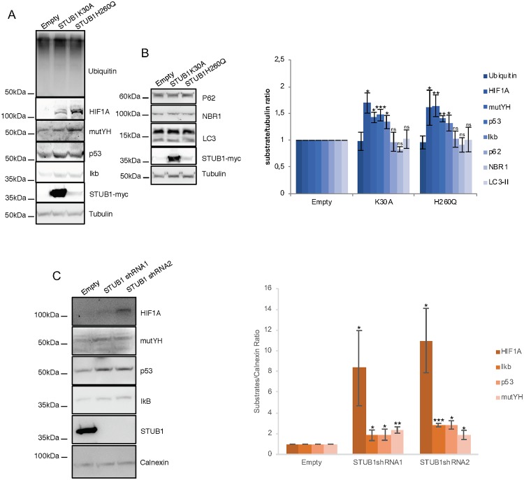
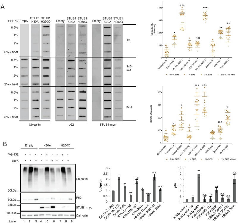
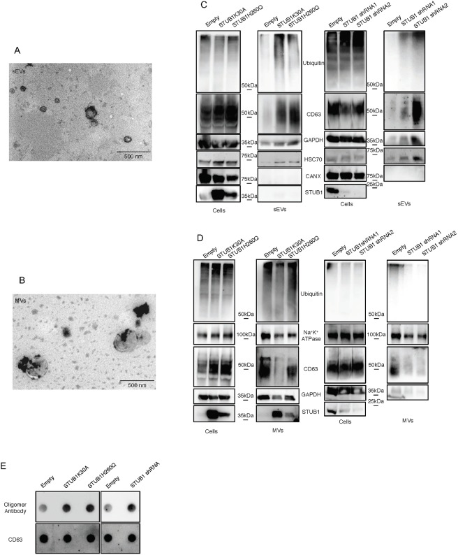
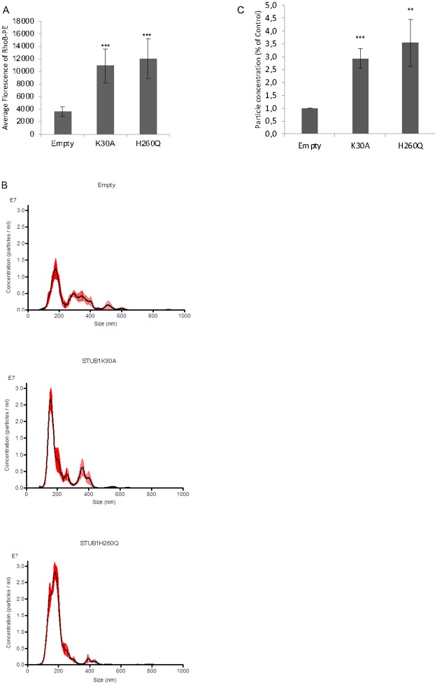
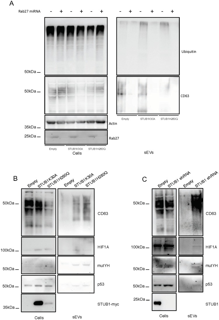
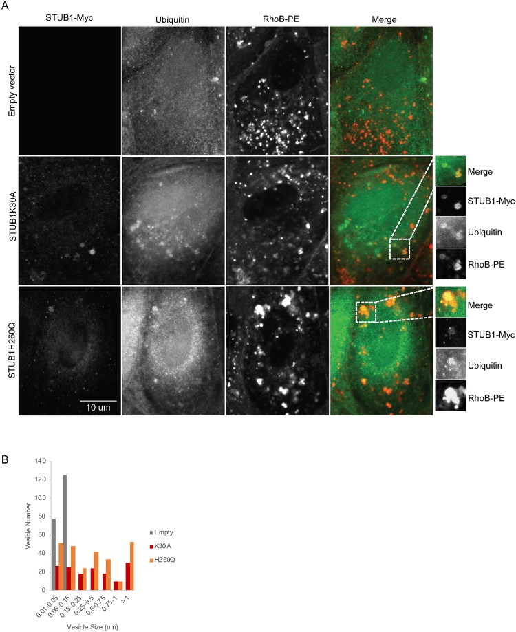
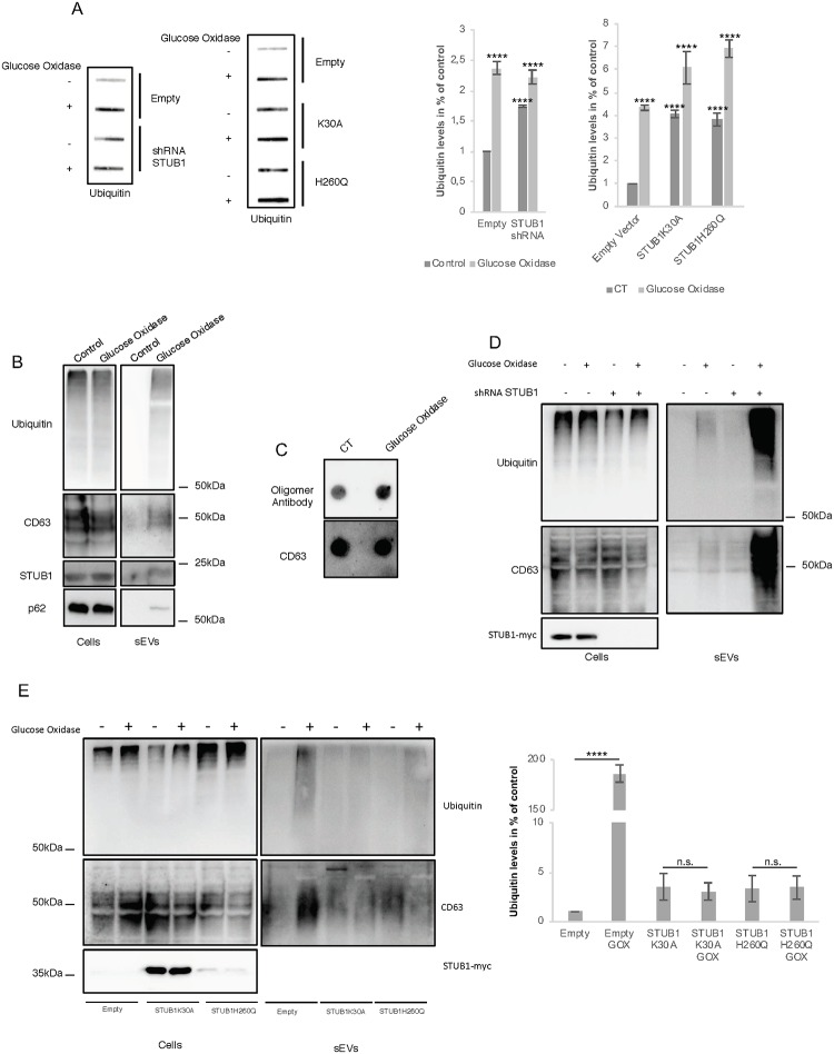
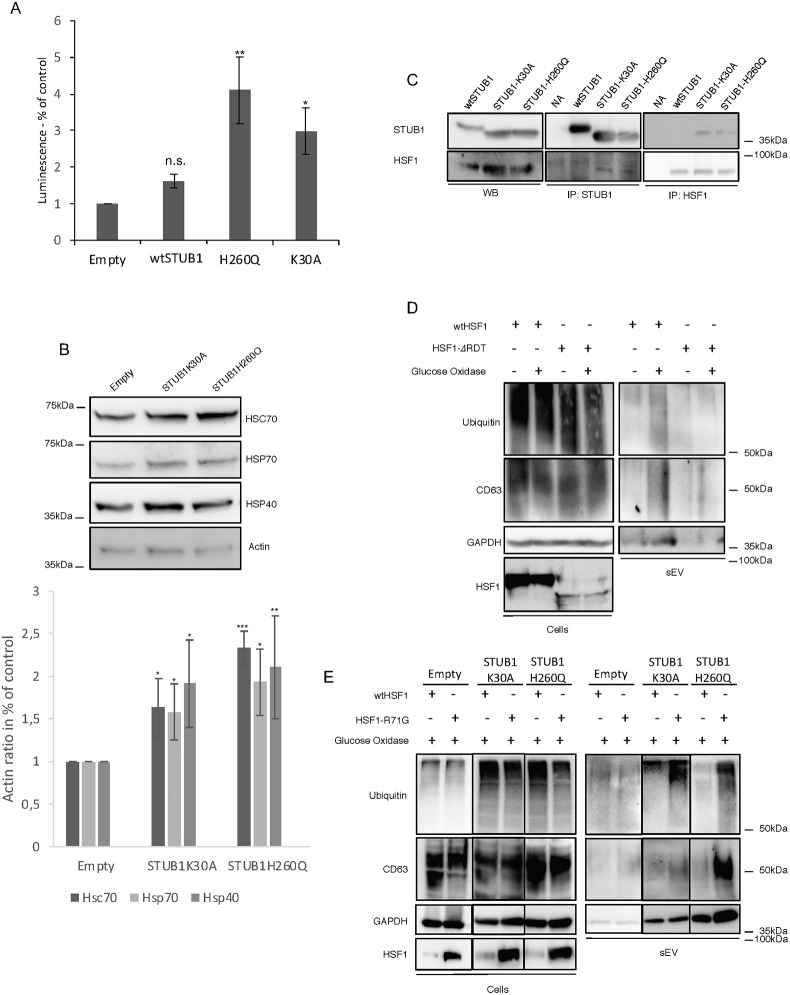
Deregulation of proteostasis is a main feature of many age-related diseases, often leading to the accumulation of toxic oligomers and insoluble protein aggregates that accumulate intracellularly or in the extracellular space. To understand the mechanisms whereby toxic or otherwise unwanted proteins are secreted to the extracellular space, we inactivated the quality-control and proteostasis regulator ubiquitin ligase STUB1/CHIP. Data indicated that STUB1 deficiency leads both to the intracellular accumulation of protein aggregates and to an increase in the secretion of small extracellular vesicles (sEVs), including exosomes. Secreted sEVs are enriched in ubiquitinated and/or undegraded proteins and protein oligomers. Data also indicates that oxidative stress induces an increase in the release of sEVs in cells depleted from STUB1. Overall, the results presented here suggest that cells use exosomes to dispose of damaged and/or undegraded proteins as a means to reduce intracellular accumulation of proteotoxic material.


Conflict of interest statement

The authors have declared that no competing interests exist.

Figures

Fig 1
Fig 1. Expression of STUB1-DN mutants induces stabilization of proteasomal substrates.
ARPE-19 cells were transduced using lentiviral particles containing vectors for the expression of either STUB1K30A or STUB1H260Q or with adenoviral particles containing shRNA against STUB1. Control cells were transduced with an empty vector. (A,B) Western blot of whole cell lysates with antibodies against: A) the proteasomal substrates HIF1A, mutYH, p53, IkB and B) the autophagic substrates LC3, p62, NBR1; ubiquitin, tubulin and myc tag. Expression of the STUB1 mutants increases the protein levels of proteasomal but not lysosomal substrates. C) Western blot of whole cell lysates with antibodies against STUB1 and the proteasomal substrates HIF1A, mutYH, p53, IkB. Depletion of STUB1 increases the protein levels of proteasomal substrates. All samples were analyzed under the same experimental conditions. The results represent the mean ±SD of N = 3 independent experiments (n.s. nonsignificant; *p < 0.05; **p < 0.01; ***p < 0.001).
Fig 2
Fig 2. Expression of STUB1DN mutants leads to the formation of protein aggregates.
ARPE-19 cells were transduced using lentiviral particles containing vectors for the expression of either STUB1K30A or STUB1H260Q. Control cells were transduced with empty vector. Cells were further incubated in the presence or absence of 10uM of MG-132 and 50nM of BafA for 6h. A) Cell extracts of proteins insoluble in increasing concentrations of SDS where filtered through a nitrocellulose membrane and trapped proteins were blotted with antibodies directed against ubiquitin, p62 and myc tag. Expression of STUB1-DN mutants increases the amount of protein aggregates trapped in the membrane both in the presence or absence of MG-132 or BafA. B) Western blotting for the proteins present in the supernatant (soluble proteins) shows a decrease in the levels of ubiquitin conjugates in the STUB1-DN mutant expressing cells (compare lane 1 with lane 4 and 7). All samples were analyzed under the same experimental conditions. The results represent the mean ±SD of N = 3 independent experiments (n.s. nonsignificant; *p < 0.05; **p < 0.01; ***p < 0.001).
Fig 3
Fig 3. STUB1 inactivation stimulates the secretion of extracellular vesicles positive for sEVs markers.
ARPE-19 cells were transduced using lentiviral particles containing vectors either for the expression of the STUB1K30A and STUB1H260Q mutants or with adenoviral particles containing shRNA against STUB1. Control cells were transduced with an empty vector. A,B) TEM images of sEVs (A) and MVs (B) isolated by sequential centrifugation. Size analysis of the vesicles shows that the 120,000g fraction contains particles with sizes smaller than 200nm (sEVs) and that the 16,500g fraction contains particles larger than 200nm (MVs). C,D) SDS-PAGE of cells extracts or vesicles isolated by sequential centrifugation from cell culture media were blotted with antibodies against ubiquitin, CD63, GAPDH, Hsc70, Calnexin (exosome negative marker [40, 41]), Na+K+ ATPase and myc tag or STUB1. STUB1-DN mutants expression and STUB1 depletion increases the release of sEVs, but not of other MVs, loaded with ubiquitinated proteins. E) Dot-Blot of 10ug of isolated sEVs incubated with an antibody that recognizes protein oligomers (A11) and the sEVs marker CD63. Both the expression of STUB1-DN mutants and the depletion STUB1 increases the levels of protein oligomers in sEVs. All samples were analyzed under the same experimental conditions.
Fig 4
Fig 4. STUB1 DN-mutants expression stimulates sEVs secretion.
ARPE-19 cells were transduced using lentiviral particles containing vectors either for the expression of the STUB1K30A and STUB1H260Q mutants. A) Cells were incubated with the MVE/exosome marker dye RhoB-PE prior to exosome isolation. Fluorescence quantification of the isolated exosomes shows an increase in cells expressing STUB1-DN mutants. B) Particle counting using nanoparticle tracking system (NanoSight). C) STUB1-DN mutants expression increases the number of exosomes released by ARPE-19 cells. All samples were analyzed under the same experimental conditions. The results represent the mean ±SD of N = 3 independent experiments (n.s. nonsignificant; *p < 0.05; **p < 0.01; ***p < 0.001).
Fig 5
Fig 5. Rab27 depletion prevents the secretion of ubiquitinated proteins by sEVs and STUB1 inactivation leads to an increase of proteasomal substrates in secreted sEVs.
ARPE-19 cells were transduced using lentiviral particles containing vectors for the expression of either STUB1K30A or STUB1H260Q, with adenoviral particles containing shRNA against STUB1 or with adenoviral particles containing miRNA against Rab27. Control cells were transduced with an empty vector. A) Western blot of whole cell lysates and sEVs samples with antibodies against ubiquitin, CD63 and GAPDH shows that depletion of Rab27 prevents the secretion of vesicles loaded with ubiquitinated proteins. B,C) Western blot of whole cell lysates and sEVs sample with antibodies against CD63, HIF1A, mutYH and p53. The expression of STUB1-DN mutants, as well as, the depletion of STUB1 increases the presence of proteasomal substrates in released sEVs. All samples were analyzed under the same experimental conditions.
Fig 6
Fig 6. Ubiquitin colocalizes with MVEs in cells expressing STUB1-DN mutants.
ARPE-19 cells were transduced using lentiviral particles containing vectors for the expression of either STUB1K30A or STUB1H260Q. Control cells were transduced with empty vector. A) Immunofluorescence using confocal microscopy with antibodies against myc, ubiquitin and the RhoB-PE dye for MVE labeling shows an increase in puncta positive for ubiquitin, STUB1-DN mutants and RhoB-PE. B) Quantification of size and number of vesicles labelled with RhoB-PE dye shows an increase in the frequency of larger vesicles in the STUB1-DN mutants.
Fig 7
Fig 7. Oxidative stress stimulates sEVs secretion via a mechanism that is exacerbated upon STUB1 depletion but abrogated by the expression of STUB1-DN mutants.
ARPE-19 cells were transduced with lentiviral particles containing vectors either for the expression of the STUB1K30A and H260Q mutants or with adenoviral particles containing shRNA against STUB1. Control cells were transduced with empty vector. Cells were maintained in the presence or absence or 40 mU of GOx for 12h. A) Cell extracts of proteins insoluble in 2% SDS at 95°C, were filtered through a nitrocellulose membrane and trapped proteins were blotted with antibodies against ubiquitin. GOx incubation increases the formation of insoluble protein aggregates in all conditions. (B,D,E) SDS-PAGE of cell extracts or sEVs isolated by sequential centrifugation from cell culture media were blotted with antibodies against ubiquitin, CD63, GAPDH, p62 or STUB1. B) GOx incubation leads to an increase in the release of sEVs loaded with ubiquitin as well as STUB1 and CD63. C) Dot-Blot of 10ug of isolated sEVs incubated with an antibody that recognizes protein oligomers and with an antibody for the sEVs marker CD63. GOx incubation increases the levels of protein oligomers. D) GOx incubation and STUB1 depletion have a cumulative effect in sEVs release. E) Expression of STUB1-DN mutants abrogates sEVs release in the presence of GOx. All samples were analyzed under the same experimental conditions. The results represent the mean ±SD of N = 3 independent experiments (n.s. nonsignificant; *p < 0.05; **p < 0.01; ***p < 0.001; ****p< 0.0001).
Fig 8
Fig 8. Expression of STUB1-DN increases HSF1 activity.
Activation of HSF1 inhibits GOx induced sEVs secretion. ARPE-19 cells were transduced using lentiviral particles containing vectors either for the expression of wtSTUB1, STUB1K30A and STUB1H260Q or with adenoviral particles containing shRNA against STUB1. Control cells were transduced with an empty vector. A) Cells were transfected with a plasmid for the expression of Luciferase under the control of an HSF1 promoter (HSE sequence). STUB1-DN mutants increase the expression of Luciferase. B) SDS-PAGE of extracts from cells expressing STUB1-DN incubated with antibodies raised against HSC70, HSP70, HSP40 and actin. Cells expressing the STUB1-DN mutant show an increase in the levels of molecular chaperones. C) Immunoprecipitation experiments using antibodies raised against V5 (wtSTUB1), myc (mutant STUB1) and HSF1 indicate that STUB1-DN mutants show increased interaction with HSF1, when compared with wtSTUB1. (D,E) Cells were transfected with plasmids for the expression of either wtHSF1, HSF1-ΔRDT or HSF1-R71G. Cells were further maintained in the presence or absence or 40 mU of GOx for 12h. SDS-PAGE of cell extracts or sEVs isolated by sequential centrifugation of cell culture media, were blotted with antibodies against ubiquitin, CD63, GAPDH or HSF1. (D) Expression of a constitutively active HSF1 (HSF1-ΔRDT) leads to the inhibition of sEVs release in the presence of GOx. (E) Expression of a dominant negative HSF1 (HSF1-R71G) rescues sEVs release upon GOx incubation. All samples were analyzed under the same experimental conditions. The results represent the mean ±SD of N = 3 independent experiments (n.s. nonsignificant; *p < 0.05; **p < 0.01; ***p < 0.001).

References

    1. Lopez-Otin C, Blasco MA, Partridge L, Serrano M, Kroemer G. The hallmarks of aging. Cell. 2013;153(6):1194–217. 10.1016/j.cell.2013.05.039 - DOI - PMC - PubMed
    1. Powers ET, Morimoto RI, Dillin A, Kelly JW, Balch WE. Biological and chemical approaches to diseases of proteostasis deficiency. Annu Rev Biochem. 2009;78:959–91. 10.1146/annurev.biochem.052308.114844 . - DOI - PubMed
    1. Tanaka K, Matsuda N. Proteostasis and neurodegeneration: the roles of proteasomal degradation and autophagy. Biochim Biophys Acta. 2014;1843(1):197–204. 10.1016/j.bbamcr.2013.03.012 . - DOI - PubMed
    1. Schmidt M, Finley D. Regulation of proteasome activity in health and disease. Biochim Biophys Acta. 2014;1843(1):13–25. 10.1016/j.bbamcr.2013.08.012 - DOI - PMC - PubMed
    1. Calderwood SK, Murshid A, Prince T. The shock of aging: molecular chaperones and the heat shock response in longevity and aging—a mini-review. Gerontology. 2009;55(5):550–8. 10.1159/000225957 - DOI - PMC - PubMed

Publication types