. 2020 Mar 1;105(3):769-780.
doi: 10.1210/clinem/dgz100.

Scaffold-Free Endometrial Organoids Respond to Excess Androgens Associated With Polycystic Ovarian Syndrome

Affiliations

Scaffold-Free Endometrial Organoids Respond to Excess Androgens Associated With Polycystic Ovarian Syndrome

Teerawat Wiwatpanit et al. J Clin Endocrinol Metab. .

Abstract

Context: Polycystic ovary syndrome (PCOS) is a prevalent disorder in reproductive aged women associated with a number of endocrine and metabolic complications, including increased risk of endometrial cancer.

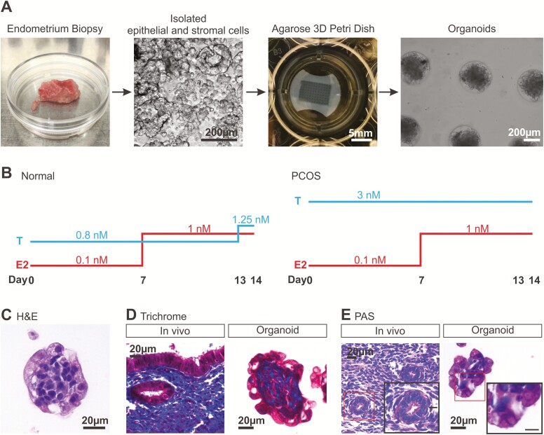
Objective: To study the effect of the characteristic increased androgen levels in PCOS on the endometrium, a novel scaffold-free multicellular endometrial organoid was established.

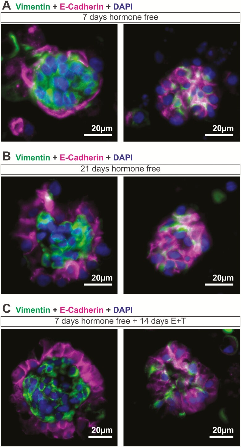
Design: Human endometrial organoids were constructed using primary endometrial epithelial and stromal cells from endometrial tissues. Organoids were treated for 14 days with physiologic levels of estradiol and testosterone to mimic a normal follicular phase or PCOS hormone profiles. Organoids were harvested for immunostaining and ribonucleic acid sequencing.

Setting: Academic institution.

Patients: Endometrial tissues from 10 premenopausal women undergoing hysterectomy for benign pathologies were obtained following written consent.

Main outcome measures: Organoid architecture, cell specific markers, functional markers, proliferation, and gene expression were measured.

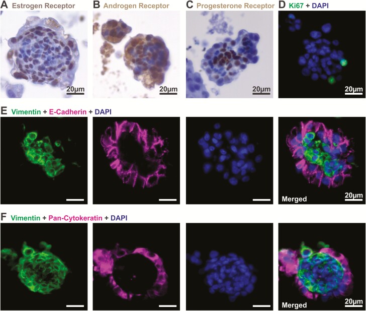
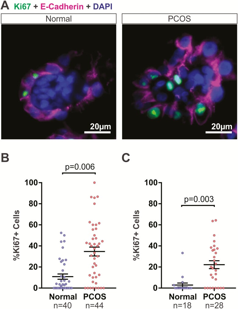
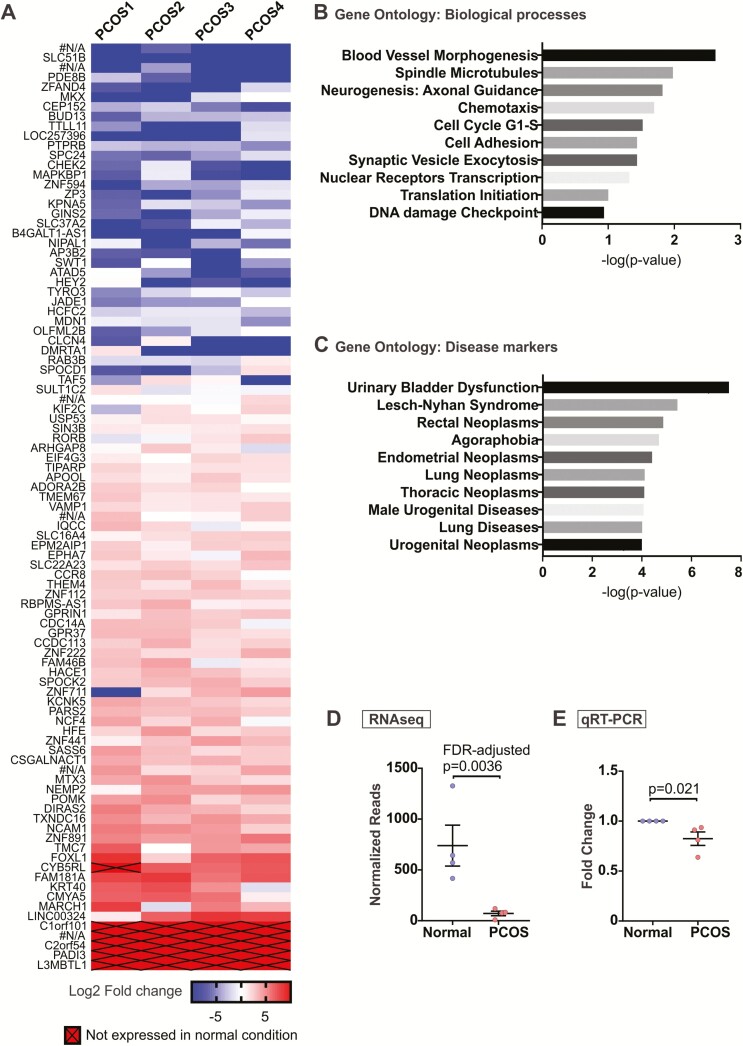
Results: A method to generate scaffold-free endometrial organoids containing epithelial and stromal cells was established. These organoids exhibited distinct organization with epithelial cells lining the outer surface and stromal cells in the center of the organoids. Epithelial cells were polarized, organoids expressed cell type specific and functional markers, as well as androgen, estrogen, and progesterone receptors. Treatment with PCOS hormones increased cell proliferation and dysregulated genes in endometrial organoids.

Conclusions: A new multicellular, scaffold-free endometrial organoid system was established that resembled physiology of the native endometrium. Excess androgens in PCOS promoted cell proliferation in endometrial organoids, revealing new mechanisms of PCOS-associated with risk of endometrial neoplasia.

Keywords: PCOS; endometrium; organoids; polycystic ovarian syndrome.


Figures

Figure 1.
Figure 1.
Generation of scaffold-free 3D endometrial organoids from human primary endometrial cells. (A) Endometrial epithelial and stromal cells were isolated from premenopausal endometrial tissues with benign pathology. Both stromal and epithelial cells were seeded into 1.5% agarose 3D Petri Dishes™ at a 1:3 ratio by volume and maintained in sex hormone-free medium for 7 days before downstream experiments. (B) Estradiol (E2) and testosterone (T) were added in a stepwise manner to the 3D cultures to mimic the levels of E2 and T during the follicular phase of a menstrual cycle. T levels were consistently higher (3 nM) in the polycystic ovarian syndrome hormone profile. After 14 days of normal hormone treatment, endometrial organoids were stained with (C) hematoxylin and eosin, (D) trichrome stain to detect collagen (blue), and (E) periodic acid-Schiff staining to stain mucosal substances (eg, mucins, glycoproteins; bright pink). Scale bar in inset of (E) is 10 µm.
Figure 2.
Figure 2.
Endometrial organoids express sex hormone receptors and exhibit structural organization. Organoids were observed after 14 days of stepwise hormone treatment mimicking the follicular phase of a normal menstrual cycle. Levels of (A) estrogen receptor, (B) androgen receptor, and (C) progesterone receptor were detected by immunohistochemical staining in endometrial organoids. (D) Proliferation was assessed in the organoids as indicated by Ki67 immunoreactivity. (E, F) Cell specific markers were assessed by immunofluorescent staining of E-cadherin, pan-cytokeratin (epithelial), and vimentin (stromal), which revealed structural organization of the cells in the organoids.
Figure 3.
Figure 3.
Hormonal effects of organoid architecture. Endometrial organoids were cultured in a (A, B) hormone free environment or (C) with the normal stepwise hormone treatment and immunofluorescent staining for vimentin (stromal) and E-cadherin (epithelial) was done at (A, C) 7 days postseeding and (B) 21 days postseeding. (A) At 7 days hormone-free, most organoids appeared to have stromal and epithelial cells intermingled within the organoids (left panel), with only 5/28 organoids (obtained from 3 patients) exhibited epithelial cells lining the outer surface of the organoid (right panel). (B) 8/24 organoids (obtained from 4 patients) established distinct structural organization when cultured for 21 days in a hormone free environment (right panel) while the remainder stayed intermixed with epithelial and stromal cells (left panel) (C) 27/34 organoids (obtained from 4 patients; left panel) exhibited structural organization with a clear distinction of epithelial and stromal layers when treated with normal levels of estradiol and testosterone for 14 days.
Figure 4.
Figure 4.
Excess androgens in polycystic ovary syndrome (PCOS) hormone condition increased proliferation of cells in endometrial organoids. (A) Endometrial organoids were cultured in normal (left) and PCOS (right) conditions as described in Fig. 1. Cell proliferation was detected by immunofluorescent staining of Ki67 in epithelial cells (dual stained with E-cadherin in pink). (B) Ki67+ cells were quantified in endometrial organoids treated with normal or PCOS hormones. Organoids were obtained from 4 patients. (C) Organoids were treated with normal or PCOS hormones using R1881 as the androgen and the Ki67+ cells were quantified. Organoids were obtained from 3 patients. Data were analyzed using unpaired Student’s t-test based on n = number of organoids.
Figure 5.
Figure 5.
Differential gene expression in polycystic ovary syndrome (PCOS) organoids. Endometrial organoids from normal versus PCOS hormone conditions were harvested at the end of 14 days. Ribonucleic acid (RNA) was isolated and RNA-sequencign (RNA-seq) was performed. (A) A total of 95 possible genes (false discovery rate–adjusted P-value < .05) were differentially expressed in PCOS organoids compared to those cultured in normal hormone condition based on RNA-seq analysis. Of these, 6 were unannotated genes with no known functions (#N/A). Genes with no expression in PCOS organoids were arbitrarily assigned a value of –10 on this log2 scale. Four separate sets of organoids obtained from n = 4 patients were used for RNA-seq. (B, C) Gene ontology analysis was performed using GeneGo to determine key pathways and biological processes (B) and disease markers (C) involving genes differentially expressed in PCOS organoids. (D) Normalized reads for BUD13 from the RNA-seq were plotted to compare with (E) quantitative reverse transcription polymerase chain reaction for relative BUD13 transcript levels in normal versus PCOS organoids obtained from 4 new patients, which was done for validation purposes.

References

    1. Ding T, Hardiman PJ, Petersen I, Wang FF, Qu F, Baio G. The prevalence of polycystic ovary syndrome in reproductive-aged women of different ethnicity: a systematic review and meta-analysis. Oncotarget. 2017;8(56):96351–96358. - PMC - PubMed
    1. Rotterdam ESHRE/ASRM-Sponsored PCOS Consensus Workshop Group. Revised 2003 consensus on diagnostic criteria and long-term health risks related to polycystic ovary syndrome. Fertil Steril. 2004;81(1):19–25. - PubMed
    1. Piltonen TT. Polycystic ovary syndrome: endometrial markers. Best Pract Res Clin Obstet Gynaecol. 2016;37:66–79. - PubMed
    1. Deng Y, Zhang Y, Li S, Zhou W, Ye L, Wang L, Tao T, Gu J, Yang Z, Zhao D, Gu W, Hong J, Ning G, Liu W, Wang W. Steroid hormone profiling in obese and nonobese women with polycystic ovary syndrome. Sci Rep. 2017;7(1):14156. - PMC - PubMed
    1. Stracquadanio M, Ciotta L, Palumbo MA. Relationship between serum anti-Mullerian hormone and intrafollicular AMH levels in PCOS women. Gynecol Endocrinol. 2018;34(3):223–228. - PubMed

Publication types

MeSH terms