. 2019 Oct 12;19(20):4424.
doi: 10.3390/s19204424.

An Enzyme- and Label-Free Fluorescence Aptasensor for Detection of Thrombin Based on Graphene Oxide and G-Quadruplex

Affiliations

An Enzyme- and Label-Free Fluorescence Aptasensor for Detection of Thrombin Based on Graphene Oxide and G-Quadruplex

Yani Wei et al. Sensors (Basel). .

Abstract

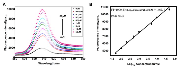
An enzyme- and label-free aptamer-based assay is described for the determination of thrombin. A DNA strand (S) consisting of two parts was designed, where the first (Sa) is the thrombin-binding aptamer and the second (Se) is a G-quadruplex. In the absence of thrombin, Sa is readily adsorbed by graphene oxide (GO), which has a preference for ss-DNA rather than for ds-DNA. Upon the addition of the N-methyl-mesoporphyrin IX (NMM), its fluorescence (with excitation/emission at 399/610 nm) is quenched by GO. In contrast, in the presence of thrombin, the aptamer will bind thrombin, and thus, be separated from GO. As a result, fluorescence will be enhanced. The increase is linear in the 0.37 µM to 50 µM thrombin concentration range, and the detection limit is 0.37 nM. The method is highly selective over other proteins, cost-effective, and simple. In our perception, it represents a universal detection scheme that may be applied to other targets according to the proper choice of the aptamer sequence and formation of a suitable aptamer-target pair.

Keywords: N-methyl-mesoporphyrin IX (NMM); aptamer; fluorescence; thrombin detection.


Conflict of interest statement

The authors declare no conflict of interest.

Figures

Scheme 1
Scheme 1
Schematic illustration of the universal, no-label, enzyme-free fluorescent detection of thrombin-based ongraphene oxide and G-quadruplex.
Figure 1
Figure 1
Fluorescence-emission spectra of the different samples: (a) S; (b) S + NMM; (c) S + Thrombin + GO + NMM; (d) S + GO + NMM; (e) NMM. The concentrations of S, thrombin, GO, and NMM are 0.25 μM, 0.2 μM, 30 µg·mL−1, and 1.5 μM, respectively.
Figure 2
Figure 2
(A) Effect of the length of the terminal signal carrier sequence (G-quadruplex) upon fluorescence emission spectra; (B) Effect of the concentration of S upon fluorescence emission spectra; (C) Effect of the concentration of GO upon fluorescence emission spectra; (D) Effect of the reaction time between G-quadruplex structure and NMM upon fluorescence emission spectra. The error bar was calculated in three independent experiments.
Figure 3
Figure 3
(A) Fluorescence emission spectra of the sensing method to different concentrations of thrombin. From a to i, the concentration of thrombin is 0, 0.05, 0.1, 0.2, 0.5, 1, 5, 10, 50 µM, respectively; (B) The linear region from 0.05 µM to 50 µM of thrombin, yielding a detection limit of 0.37 nM. The error bar was calculated in three independent experiments.
Figure 4
Figure 4
Selectivity evaluation of the biosensor for the detection of thrombin against other proteins of Lysozyme, Trypsase, lgG, BSA, and HSA. The concentration of the thrombin was 0.2 µM. However, the concentration of Lysozyme, Trypsase, lgG, BSA, and HSA was 2 µM. No thrombin or other proteins were used in the blank group. Inset: the changes in fluorescence intensity (F/F0), F and F0, are the fluorescence intensities in the presence and absence of protein, respectively. The error bar was calculated in three independent experiments.
Figure 5
Figure 5
Results obtained from the fluorescent tests of diluted serum samples spiked with different concentrations of thrombin and thrombin in buffer. The same reaction mixtures without thrombin were used as the blank group. The error bar was calculated from three independent experiments.

References

    1. Lequin R.M. Enzyme Immunoassay (EIA)/Enzyme-Linked Immunosorbent Assay (ELISA) Clin. Chem. 2005;51:2415–2418. doi: 10.1373/clinchem.2005.051532. - DOI - PubMed
    1. Lam M.T., Wan Q.H., Boulet C.A., Le X.C. Competitive immunoassay for staphylococcal enterotoxin a using capillary electrophoresis with laser-induced fluorescence detection. J. Chromatogr. A. 1999;853:545–553. doi: 10.1016/S0021-9673(99)00677-9. - DOI - PubMed
    1. Baker K.N., Rendall M.H., Patel A., Boyd P., James D.C. Rapid monitoring of recombinant protein products: A comparison of current technologies. Trends Biotechnol. 2002;20:149–156. doi: 10.1016/S0167-7799(01)01914-X. - DOI - PubMed
    1. Stoltenburg R., Reinemann C., Strehlitz B. Selex—A (r)evolutionary method to generate high-affinity nucleic acid ligands. Biomol. Eng. 2007;24:381–403. doi: 10.1016/j.bioeng.2007.06.001. - DOI - PubMed
    1. Birch C.M., Hou H.W., Han J., Niles J.C. Identification of malaria parasite-infected red blood cell surface aptamers by inertial microfluidic SELEX (I-SELEX) Sci. Rep. 2015;5:11347. doi: 10.1038/srep11347. - DOI - PMC - PubMed