Review
. 2019 Oct 12;12(20):3331.
doi: 10.3390/ma12203331.

Utilization of Finite Element Analysis for Articular Cartilage Tissue Engineering

Affiliations
Review

Utilization of Finite Element Analysis for Articular Cartilage Tissue Engineering

Chaudhry R Hassan et al. Materials (Basel). .

Abstract

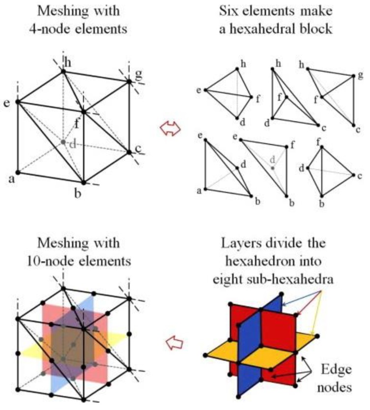
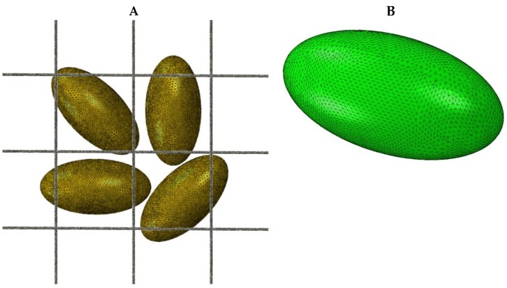
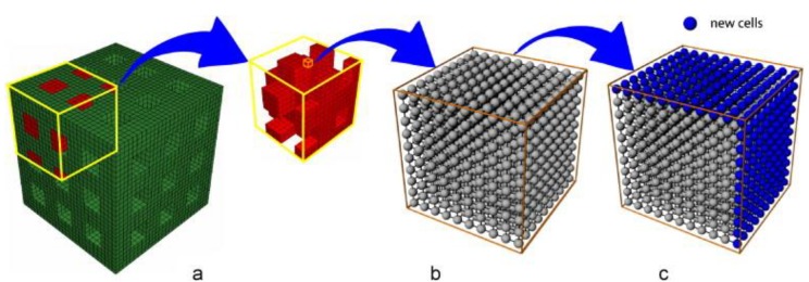
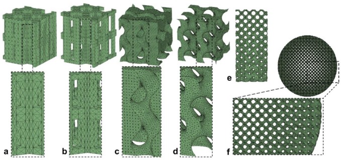
Scaffold design plays an essential role in tissue engineering of articular cartilage by providing the appropriate mechanical and biological environment for chondrocytes to proliferate and function. Optimization of scaffold design to generate tissue-engineered cartilage has traditionally been conducted using in-vitro and in-vivo models. Recent advances in computational analysis allow us to significantly decrease the time and cost of scaffold optimization using finite element analysis (FEA). FEA is an in-silico analysis technique that allows for scaffold design optimization by predicting mechanical responses of cells and scaffolds under applied loads. Finite element analyses can potentially mimic the morphology of cartilage using mesh elements (tetrahedral, hexahedral), material properties (elastic, hyperelastic, poroelastic, composite), physiological loads by applying loading conditions (static, dynamic), and constitutive stress-strain equations (linear, porous-elastic, biphasic). Furthermore, FEA can be applied to the study of the effects of dynamic loading, material properties cell differentiation, cell activity, scaffold structure optimization, and interstitial fluid flow, in isolated or combined multi-scale models. This review covers recent studies and trends in the use of FEA for cartilage tissue engineering and scaffold design.

Keywords: articular cartilage; finite element analysis; scaffold design; tissue engineering.


Conflict of interest statement

The authors declare no conflict of interest.

Figures

Figure 1
Figure 1
Tetrahedral (four-node) and hexahedral (eight-node and 20-node) elements are commonly used in the finite element mesh. The solid body is replaced with the selected type of elements and the stress–strain constitutive equations are solved at each node. (Figure taken from Kim, J., & Bathe, K. J. [15] and reprinted with permission).
Figure 2
Figure 2
(A) A representative model of a scaffold with embedded cells. The cell surface nodes are in contact with the scaffold’s rod-like structure. The force applied to the scaffold results in strain on cell surface nodes. (B) A detailed tetrahedral mesh of the ellipsoid shaped cell with a 100-µm major axis and a 50-µm minor axis that contains 6000 nodes.
Figure 3
Figure 3
(a) Finite element model of a scaffold with 50% porosity. The scaffold cavity is initially filled with granulation tissue (red). (b) Each element of the granulation tissue has a lattice for analysis of cellular activity. (c) The lattice expands by the addition of new cells (blue) to grow into the space of the dissolving scaffold material. (Figure is taken from Byrne et al. [47] and reprinted with permission).
Figure 4
Figure 4
Six scaffold designs with varying structure and porosity; hexagonal prisms with (a) 55% and (b) 70% porosity, gyroid with (c) 55% and (d) 70% porosity, (e) gyroid with height-based porosity gradient, and (f) gyroid with radial-based porosity gradient. (Figure is taken from Olivares et al. [24] and reprinted with permission).

References

    1. Goldring M.B. Chondrogenesis, chondrocyte differentiation, and articular cartilage metabolism in health and osteoarthritis. Ther. Adv. Musculoskelet. Dis. 2012;4:269–285. doi: 10.1177/1759720X12448454. - DOI - PMC - PubMed
    1. Rolauffs B., Williams J.M., Aurich M., Grodzinsky A.J., Kuettner K.E., Cole A.A. Proliferative remodeling of the spatial organization of human superficial chondrocytes distant from focal early osteoarthritis. Arthritis Rheum. 2010;62:489–498. doi: 10.1002/art.27217. - DOI - PMC - PubMed
    1. Bullough P., Goodfellow J. The significance of the fine structure of articular cartilage. J. Bone Jt. Surg. Br. Vol. 1968;50:852–857. doi: 10.1302/0301-620X.50B4.852. - DOI - PubMed
    1. Jeffery A., Blunn G., Archer C., Bentley G. Three-dimensional collagen architecture in bovine articular cartilage. J. Bone Jt. Surg. Br. Vol. 1991;73:795–801. doi: 10.1302/0301-620X.73B5.1894669. - DOI - PubMed
    1. Lee H.S., Millward-Sadler S.J., Wright M.O., Nuki G., Al-Jamal R., Salter D.M. Activation of Integrin—RACK1/PKCα signalling in human articular chondrocyte mechanotransduction. Osteoarthr. Cart. 2002;10:890–897. doi: 10.1053/joca.2002.0842. - DOI - PubMed

LinkOut - more resources