. 2019 Oct 13;20(20):5077.
doi: 10.3390/ijms20205077.

Graphene Oxide Enhances Chitosan-Based 3D Scaffold Properties for Bone Tissue Engineering

Affiliations

Graphene Oxide Enhances Chitosan-Based 3D Scaffold Properties for Bone Tissue Engineering

Sorina Dinescu et al. Int J Mol Sci. .

Abstract

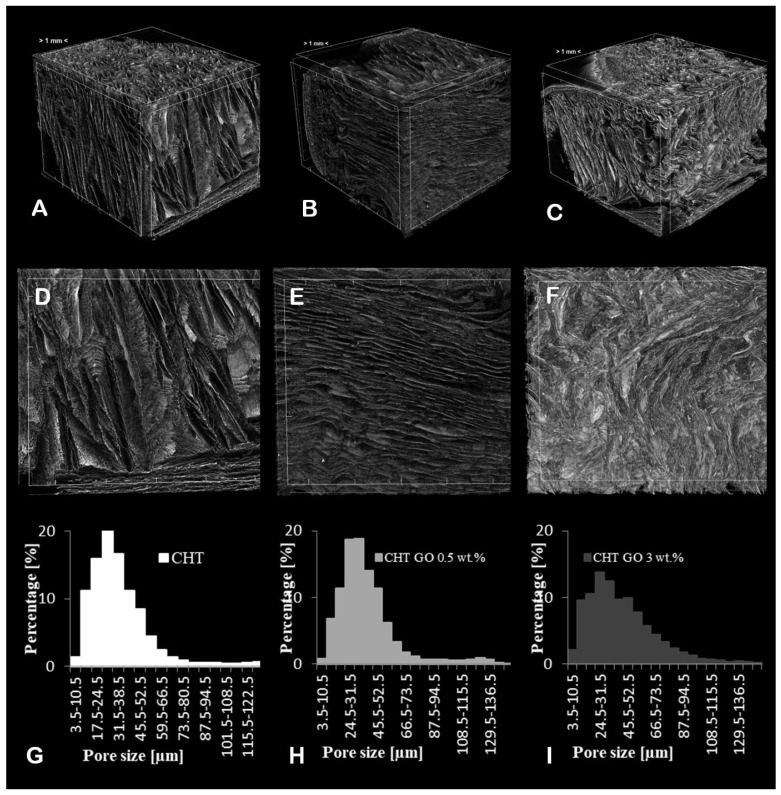
The main goal of bone tissue engineering (BTE) is to refine and repair major bone defects based on bioactive biomaterials with distinct properties that can induce and support bone tissue formation. Graphene and its derivatives, such as graphene oxide (GO), display optimal properties for BTE, being able to support cell growth and proliferation, cell attachment, and cytoskeleton development as well as the activation of osteogenesis and bone development pathways. Conversely, the presence of GO within a polymer matrix produces favorable changes to scaffold morphologies that facilitate cell attachment and migration i.e., more ordered morphologies, greater surface area, and higher total porosity. Therefore, there is a need to explore the potential of GO for tissue engineering applications and regenerative medicine. Here, we aim to promote one novel scaffold based on a natural compound of chitosan, improved with 3 wt.% GO, for BTE approaches, considering its good biocompatibility, remarkable 3D characteristics, and ability to support stem cell differentiation processes towards the bone lineage.

Keywords: biocompatibility; bone tissue engineering; graphene oxide; human adipose-derived stem cells; osteogenesis.


Conflict of interest statement

The authors declare no conflict of interest. The funders had no role in the design of the study; in the collection, analyses, or interpretation of data; in the writing of the manuscript, or in the decision to publish the results.

Figures

Figure 1
Figure 1
Cytocompatibility assessment of BC0.5–BC3 with human adipose-derived stem cells (hASCs). (a) Cell viability in contact with chitosan (CHT)/graphene oxide (GO) composites by 3-(4,5-dimethylthiazol-2-yl)-2,5-diphenyltetrazolium bromide (MTT) assay; (b) CHT/GO material levels of cytotoxicity on contact with hASC culture by lactate dehydrogenase (LDH) assay; (c) tridimensional reconstructions for BC0.5-BC3 and control showing live cells (green) and dead cells (red) after 7 days of culture resulted from Live/Dead assay and confocal microscopy analysis. */# p < 0.05; ** p < 0.01; ***/### p < 0.001.
Figure 2
Figure 2
Actin filaments developed by hASCs after 48 h of contact with CHT/GO (b–e) and the CHT reference (a), as shown by confocal microscopy. Scale bar 50 μm. Actin filaments are shown in green (phalloidin-fluorescein isothiocyanate (FITC)) and cell nuclei are shown in blue (DAPI).
Figure 3
Figure 3
CHT/GO material characterization by MicroCT. Three-dimensional renderings of CHT (A), CHT/0.5 wt.% GO (B), and CHT/3 wt.% GO (C), with complementary cross-sections (D,E,F) and pore size distributions (G,H,I). The overall scale bar is 1 mm.
Figure 4
Figure 4
Cell distribution and phenotype in BC0.5–BC3 systems. (a) hASC distribution and morphology in the 3D structure of BC0.5–BC3 and the hASC/CHT reference bioconstruct (BC) before and after 28 days of osteogenic differentiation, assessed by SEM; the red box marks the area enlarged below each image and the yellow arrows indicate mineralized deposits in the extracellular matrix (ECM) which was further characterized by EDAX; (b) the composition of extracellular matrix secreted by cells after 28 days of osteogenic differentiation, as revealed by energy dispersive X-ray analysis (EDAX) analysis.
Figure 5
Figure 5
Histological evaluation of osteogenic differentiation. Microtome sections of BC0.5, BC3, and BC stained with hematoxylin-eosin (for morphology) and with Alizarin Red S (to highlight mineral deposits). Scale bar 20 µm.
Figure 6
Figure 6
Gene expression of osteogenic specific markers—runx2, osx, opn, and ocn. (ad) runx2, osx, opn, and ocn profiles of gene expression obtained by qPCR in BC, BC0.5, and BC3 after 7, 14, and 28 days of differentiation of hASCs in pro-osteogenic conditions. */# p < 0.05; ** p < 0.01; ***/### p < 0.001. Fold change in qPCR data analysis was determined by 2−ΔΔCt method.
Figure 7
Figure 7
Protein expression of osteogenic specific markers Osx and Opn. (a) Osx and (b) Opn protein expression as revealed by confocal microscopy in BC, BC0.5, and BC3 up to 28 days of differentiation in pro-osteogenic conditions. The nuclei are labeled in blue with DAPI and Osx/Opn is labeled in red. The scale bar is 50 μm.
Figure 8
Figure 8
In vivo confirmation of osteogenesis activation. (a) Osx levels of protein expression in vivo after CHT/3 wt.% GO material implantation in mouse calvaria bone defect; (b) osx gene expression profile during 18 weeks post-implantation. ** p < 0.01; *** p < 0.001. Fold change in qPCR data analysis was determined by 2−ΔΔCt method.

References

    1. Nayak T.R., Andersen H., Makam V.S., Khaw C., Bae S., Xu X., Ee P.L., Ahn J.H., Hong B.H., Pastorin G., et al. Graphene for controlled and accelerated osteogenic differentiation of human mesenchymal stem cells. ACS Nano. 2011;5:4670–4678. doi: 10.1021/nn200500h. - DOI - PubMed
    1. Depan D., Misra R.D. The interplay between nanostructured carbon-grafted chitosan scaffolds and protein adsorption on the cellular response of osteoblasts: Structure function property relationship. Acta Biomater. 2013;9:6084–6094. doi: 10.1016/j.actbio.2012.12.019. - DOI - PubMed
    1. Gu M., Liu Y., Chen T., Du F., Zhao X., Xiong C., Zhou Y. Is graphene a promising nano-material for promoting surface modification of implants or scaffold materials in bone tissue engineering? Tissue Eng. Part B Rev. 2014;20:477–491. doi: 10.1089/ten.teb.2013.0638. - DOI - PMC - PubMed
    1. Wang Y., Xiao Y., Gao G., Chen J., Hou R., Wang Q., Liu L., Fu J. Conductive Graphene Oxide Hydrogels Reduced and Bridged by L-Cysteine to Support Cell Adhesion and Growth. J. Mater. Chem. B. 2018;5:511–516. doi: 10.1039/C6TB02333A. - DOI - PubMed
    1. Shen H., Zhang L., Liu M., Zhang Z. Biomedical applications of graphene. Theranostics. 2012;2:283–294. doi: 10.7150/thno.3642. - DOI - PMC - PubMed

LinkOut - more resources