

Mid-term outcomes of arthroscopic-assisted Core decompression of Precollapse osteonecrosis of femoral head-minimum of 5 year follow-up
- PMID: 31615502
- PMCID: PMC6794765
- DOI: 10.1186/s12891-019-2853-0
Mid-term outcomes of arthroscopic-assisted Core decompression of Precollapse osteonecrosis of femoral head-minimum of 5 year follow-up
Abstract
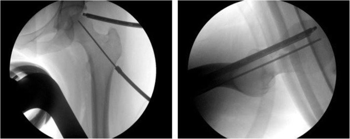
Background: Osteonecrosis of the femoral head (ONFH) is a progressive disease that leads to collapse and the development of secondary arthritis. The preferred management of ONFH remains controversial. Arthroscopic-assisted management of ONFH is a new and evolving approach for hip preservation. We hypothesis that arthroscopy is able to improve ONFH outcomes by achieving accurate and minimally invasive decompression while successfully addressing concomitant intraarticular pathologies resulting in reliable mid-term outcomes.
Methods: This was a retrospective cohort analysis. All patients had atraumatic ONFH with a precollapse lesion and a minimum follow-up of 5 years.
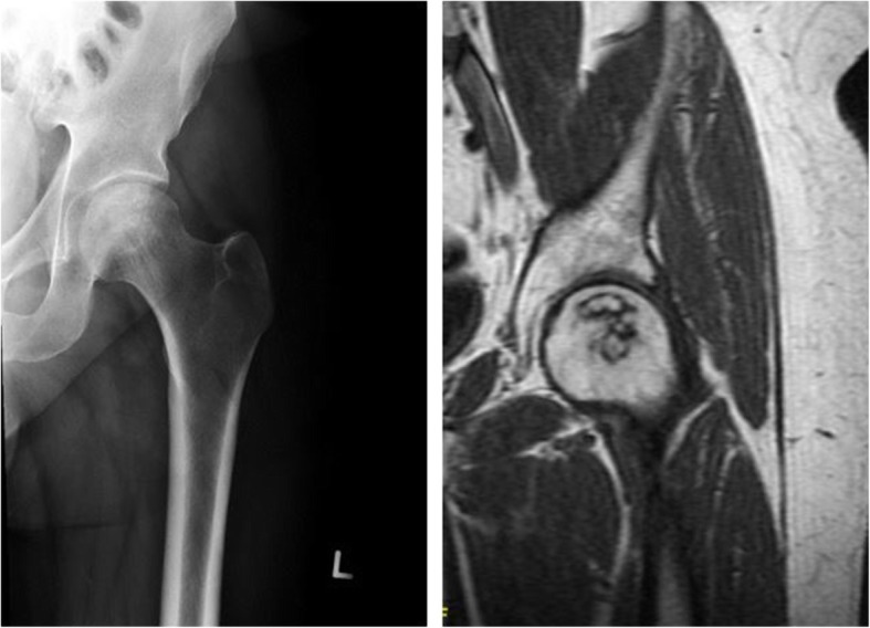
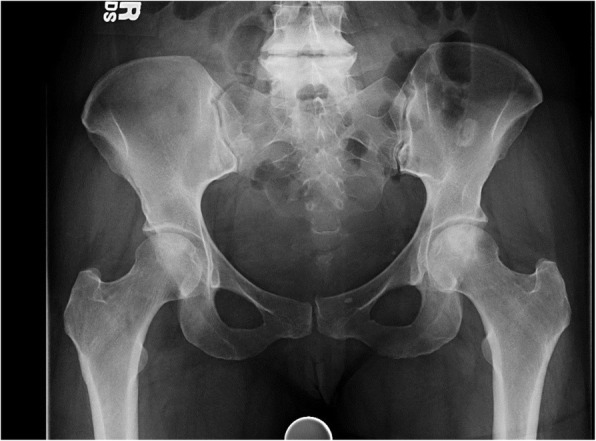
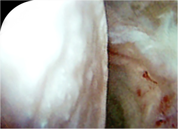
Results: A total cohort of 11 hips (8 patients) was identified. The mean patient follow-up was 7 years ±1.48 years (range, 64-118 months). The Ficat-Alret classification found on preoperative imaging was Stage I-3 (27.2%), IIa-4 (36.4%), and IIb-4 (36.4%) hips. Four (36.4%) hips experienced mechanical issues, including locking, catching, and buckling. The most common concomitant pathology addressed at the time of arthroscopy, was labral repair/debridement-8 (73%), followed by microfracture-7 (64%). At final follow-up, 6 hips (54.5%) had not converted to THA. Upon further stratification, Stage I-100%, Stage IIa-75%, for a combined 87%, had not converted to THA, in contrast, 100% of hips categorized as Stage IIb had converted to THA. Ficat-Alret staging, especially Stage IIb, was significantly associated with conversion to THA. (p-value = 0.015) There were 0% major or minor complications.
Conclusions: To our knowledge, this is the longest reported follow-up of arthroscopic-assisted management of ONFH. Arthroscopic-assisted management is a promising surgical approach that provides safe, accurate, and minimally invasive decompression, resulting in reliable results with an acceptable conversion rate to THA.
Level of evidence: Level IV, Case Series.
Keywords: Core decompression; Femoral head preservation; Femoral head preserving; Hip arthroscopy; Mechanical symptoms; Mid-term follow-up; Mid-term outcomes; ONFH; Osteonecrosis; Osteonecrosis of the femoral head.
Conflict of interest statement
The authors have no disclosures, conflicts of interest, or competing interests to make.
Figures
References
-
- Mont MA, Jones LC, Hungerford DS. Nontraumatic osteonecrosis of the femoral head: ten years later. J Bone Joint Surg Am. 2006;88:1117–1132. - PubMed
MeSH terms
LinkOut - more resources
Full Text Sources
Research Materials
