. 2019 Oct 22;8(10):bio045864.
doi: 10.1242/bio.045864.

Cellular response to bacterial infection in the grasshopper Oxya chinensis

Affiliations

Cellular response to bacterial infection in the grasshopper Oxya chinensis

Xiaomin Zhang et al. Biol Open. .

Abstract

Oxya chinensis is one of the most widespread grasshopper species found in China and one of the most common pests against rice. In view of the importance of haemocytes in insect immunity in general, and the lack of information on the haemocytes of O. chinensis, we examined the haemocytes of this species in detail. We challenged the cellular response of this grasshopper with the bacteria Escherichia coli, Staphylococcus aureus, and Bacillus subtilis Haemocyte morphology was observed using light, scanning electron and transmission electron microscopy, which revealed distinct morphological varieties of haemocytes. Granulocytes and plasmatocytes responded to the bacterial challenge by phagocytosis. Histochemical staining indicated the presence of acid phosphatase in plasmatocytes and granulocytes. We also observed non-phagocytic prohemocytes and vermicytes, but their functions in the circulation are unclear. Insect haemocytes play a crucial role in cellular immunity, and further research is needed for a comprehensive understanding.

Keywords: Haemocyte; Histochemistry; Morphology; Phagocytosis; Wright-Giemsa staining.


Conflict of interest statement

Competing interestsThe authors declare no competing or financial interests.

Figures
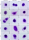
Fig. 1.
Fig. 1.
The phagocytic response of the grasshopper O. chinensis against E. coli, B. subtilis and S. aureus bacteria at 4, 8, 12, 24 and 48 h post-injection. The phagocytes shown are plasmatocytes (A–C,H–J,L–O) and granulocytes (D–G,K). Arrows point to bacteria. Scale bars: 5 μm.
Fig. 2.
Fig. 2.
Wright-Giemsa-stained haemocytes of O. chinensis under the light microscope. (A–C) Polymorphic granulocytes with purple cytoplasmic granules, which vary in density. (D) Oval-shaped plasmatocytes. (E,F) Plasmatocytes with extended pseudopodia (arrows). (G) A spheroid prohemocyte with limited cytoplasm. (H) A worm-shaped vermicyte with elongated cytoplasm and a nucleus containing some cytoplasmic granules (arrows). Scale bars: 5 μm.
Fig. 3.
Fig. 3.
Haemocytes of O. chinensis under the scanning electron microscope. (A) An irregularly shaped granulocyte with lumps on the membrane caused the presence of cytoplasmic granules. (B) An irregularly shaped plasmatocyte with a relatively smooth membrane. (C) A spheroid prohemocyte with smooth membrane. (D) A vermicyte with rough membrane and pseudopodia. Scale bars: 7 μm (A,D), 5 μm (B), 4 μm (D) .
Fig. 4.
Fig. 4.
Granulocytes of O. chinensis under the transmission electron microscope. The slides depict sections of granulocytes with visible granules, indicated by arrows. (A,B) Electron-dense (red arrows) and electron-lucent (black arrows) granules in the cytoplasm are shown. (C) Section of a granulocyte with two clusters of small bright inclusions (arrows). (D) Arrows indicate electron-lucent granules. The nucleus (n), mitochondria (mi) and endoplasmic reticulum (er) are found in the cytoplasm. Scale bars: 0.5 μm (A), 0.8 μm (B), 0.6 μm (C), 1 μm (D).
Fig. 5.
Fig. 5.
Plasmatocytes of O. chinensis under the transmission electron microscope. (A) Many elongated pseudopodia (p) extended outward from the plasmatocytes. (B) Arrows indicate exocytosis-like activity. (C,D) Nuclei (n), mitochondria (mi), endoplasmic reticulum (er), Golgi apparatus (go) and vacuole (va) were observed in the cytoplasm. Scale bars: 1 μm (A), 0.25 μm (B), 0.5 μm (C,D).
Fig. 6.
Fig. 6.
A prohemocyte and vermicytes of O. chinensis under the transmission electron microscope. (A–C) Prohemocytes (A). B and C are enlarged sections of A (red rectangle). Golgi apparatus (go), nuclei (n), mitochondria (mi), endoplasmic reticulum (er) and vacuole (va) are visible in the cytoplasm. (D–F) Vermicytes of O. chinensis under the transmission electron microscope. (D) Two worm-shaped vermicytes with elongated cytoplasm and nuclei. (E) Arrows indicate some of the vermicytes' cytoplasmic inclusions. (F) An enlarged section of E (red rectangle). Nuclei, mitochondria, endoplasmic reticulum, vacuole, and additional prohemocytes (PR) and plasmatocytes (PL) were also observed. Scale bars: 1 μm (A), 0.5 μm (B,C,E,F), 2.5 μm (D).
Fig. 7.
Fig. 7.
Histochemically stained haemocytes of O. chineneis. (A–C) Purplish-red colouration indicates a positive reaction in haemocytes stained with ACP. (A) Two ACP-positive plasmatocytes, one of which contains an unstained vacuole (arrow). (B) Two weakly ACP-positive plasmatocytes (black arrows) and two granulocytes with one containing an ACP-positive red granule (red arrow). (C) An ACP-positive plasmatocyte and an ACP-negative granulocyte. The two red arrows point to different sized ACP-positive red granules. (D–F) Haemocytes stained with CAE reaction. The haemocytes are CAE-negative, while the background is positively reacted and red in colour. Plasmatocytes are shown in D and E. (F) A naked nucleus with no conspicuous cytoplasm. (G–I) Haemocytes stained with ORO. (G) Two weakly ORO-positive plasmatocytes (red arrows). (H,L) Granulocyte with clear granules, indicating negative ORO and PAS reactions. (I) A weakly ORO-positive plasmatocyte (arrow) and three nuclei with no defined cytoplasm. (J–L) Haemocytes stained in PAS reaction. (J) One of the two plasmatocytes has PAS-positive purplish-red substances (arrow). (K) Two negatively reacted plasmatocytes. Scale bars: 5 μm.
Fig. 8.
Fig. 8.
Haemocyte counts from the Wright-Giemsa-stained preparations of normal adults (A), and phagocyte proportion of adults injected with (B) Bacillus subtilis, (C) Escherichia coli and (D) Staphylococcus aureus. (A) Prohemocyte (PR), plasmatocyte (PL), granulocyte (GR) and vermicyte (VE). (B-D) The percentages of plasmatocytes and granulocytes with and without attachment or engulfment of injected bacteria are shown on the y-axes.

References

    1. Akai H. and Sato S. (1979). Surface and internal ultrastructure of hemocytes of some insects. In Insect Hemocytes, Development, Forms, Functions and Techniques (ed. Gupta A. P.), pp. 129-154. Cambridge, London: Cambridge University Press.
    1. Anggraeni T., Melanie T. and Putra R. E. (2011). Cellular and humoral immune defenses of Oxya japonica (Orthoptera: Acrididae) to entomopathogenic fungi Metarhizium anisopliae. Entomol. Res. 41, 1-6. 10.1111/j.1748-5967.2010.00311.x - DOI
    1. Arp A. P., Martini X. and Pelz-Stelinski K. S. (2017). Innate immune system capabilities of the Asian citrus psyllid, Diaphorina citri. J. Invertebr. Pathol. 148, 94-101. 10.1016/j.jip.2017.06.002 - DOI - PubMed
    1. Blandin S. A. and Levashina E. A. (2007). Phagocytosis in mosquito immune responses. Immunol. Rev. 219, 8-16. 10.1111/j.1600-065X.2007.00553.x - DOI - PubMed
    1. Brehélin M., Zachary D. and Hoffmann J. A. (1978). A comparative ultrastructural study of blood cells from nine insect orders. Cell Tissue Res. 195, 45-57. 10.1007/BF00233676 - DOI - PubMed