. 2019 Nov 15;294(46):17463-17470.
doi: 10.1074/jbc.RA119.011255. Epub 2019 Oct 15.

Kinetic and structural evidence that Asp-678 plays multiple roles in catalysis by the quinoprotein glycine oxidase

Affiliations

Kinetic and structural evidence that Asp-678 plays multiple roles in catalysis by the quinoprotein glycine oxidase

Kyle J Mamounis et al. J Biol Chem. .

Abstract

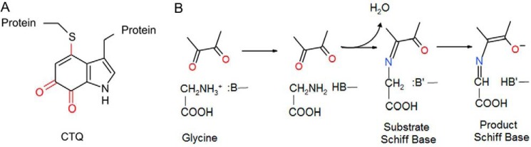
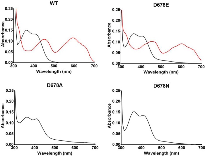
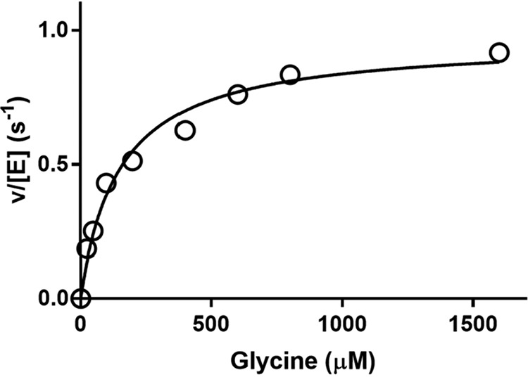
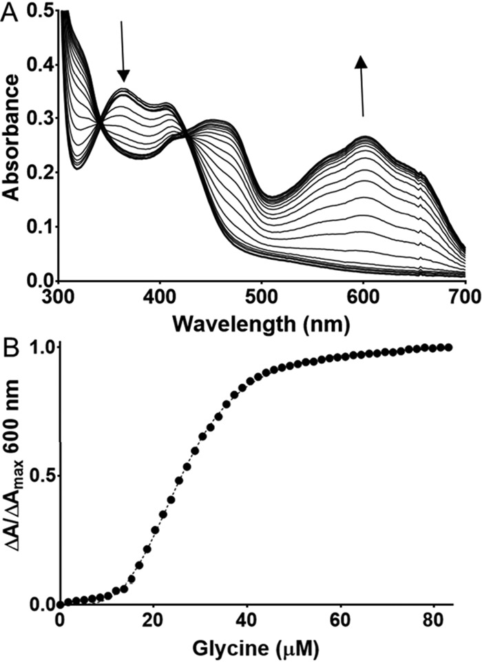
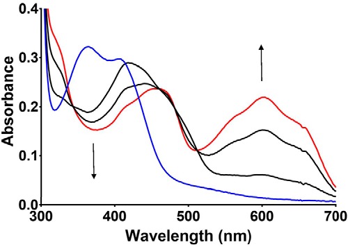
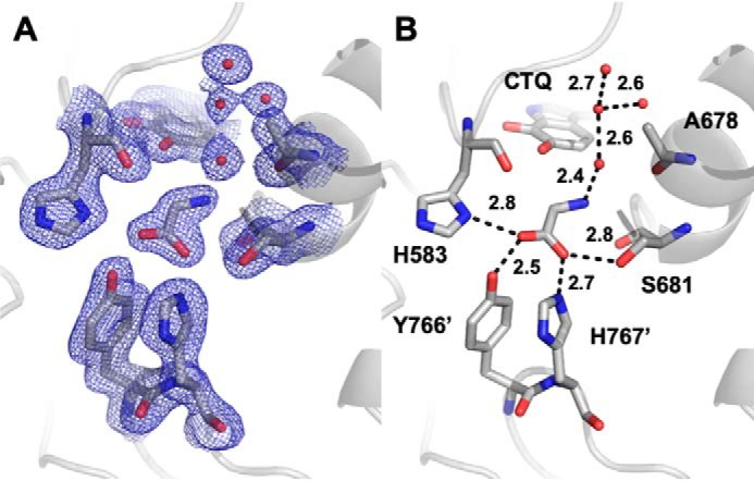
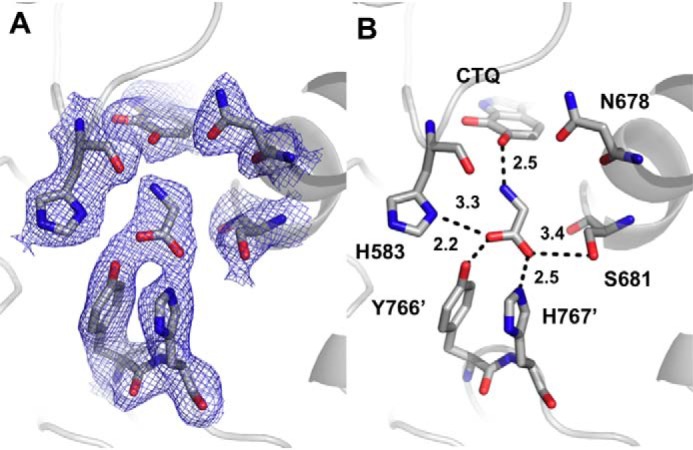
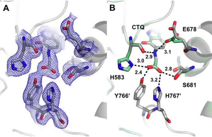
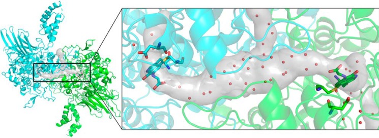
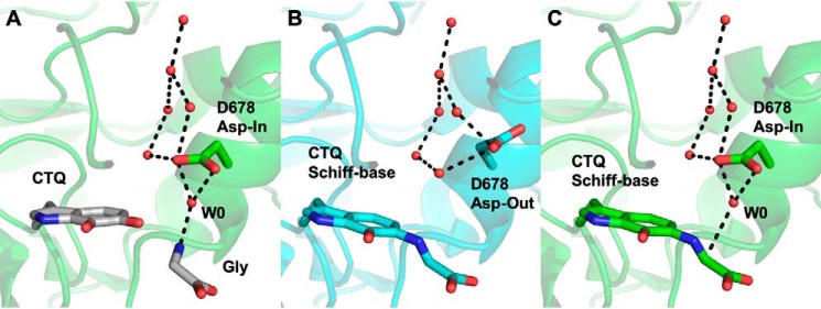
PlGoxA from Pseudoalteromonas luteoviolacea is a glycine oxidase that utilizes a protein-derived cysteine tryptophylquinone (CTQ) cofactor. A notable feature of its catalytic mechanism is that it forms a stable product-reduced CTQ adduct that is not hydrolyzed in the absence of O2 Asp-678 resides near the quinone moiety of PlGoxA, and an Asp is structurally conserved in this position in all tryptophylquinone enzymes. In those other enzymes, mutation of that Asp results in no or negligible CTQ formation. In this study, mutation of Asp-678 in PlGoxA did not abolish CTQ formation. This allowed, for the first time, studying the role of this residue in catalysis. D678A and D678N substitutions yielded enzyme variants with CTQ, which did not react with glycine, although glycine was present in the crystal structures in the active site. D678E PlGoxA was active but exhibited a much slower kcat This mutation altered the kinetic mechanism of the reductive half-reaction such that one could observe a previously undetected reactive intermediate, an initial substrate-oxidized CTQ adduct, which converted to the product-reduced CTQ adduct. These results indicate that Asp-678 is involved in the initial deprotonation of the amino group of glycine, enabling nucleophilic attack of CTQ, as well as the deprotonation of the substrate-oxidized CTQ adduct, which is coupled to CTQ reduction. The structures also suggest that Asp-678 is acting as a proton relay that directs these protons to a water channel that connects the active sites on the subunits of this homotetrameric enzyme.

Keywords: GoxA; LodA-like protein; acid-base catalysis; allostery; cysteine tryptophylquinone (CTQ); enzyme kinetics; enzyme mechanism; glycine oxidase; oxidase; protein structure; proton transfer; proton transport; quinone; quinoprotein; water channel.


Conflict of interest statement

The authors declare that they have no conflicts of interest with the contents of this article

Figures

Figure 1.
Figure 1.
A, cysteine tryptophylquinone. B, reductive half-reaction of PlGoxA. Only the quinone portion of CTQ is shown.
Figure 2.
Figure 2.
Absorbance spectra of WT and variant PlGoxA proteins. The spectra of the oxidized proteins are black. After the addition of 5 mm glycine to WT and D678E PlGoxA, the red spectrum is immediately observed. After the glycine addition to D678A and D678N PlGoxA, there is no change in the spectrum.
Figure 3.
Figure 3.
Steady-state kinetic analysis of glycine oxidase activity of D678E PlGoxA. The line is a fit of the data by Equation 1.
Figure 4.
Figure 4.
Titration of glycine binding to CTQ in D678E PlGoxA. A, changes in the spectrum observed on incremental additions of glycine. B, the line is a fit of the data by Equation 3.
Figure 5.
Figure 5.
Changes in absorbance of D678E PlGoxA immediately after the addition of glycine. The reaction was performed at 15 °C. The initial spectrum of the oxidized enzyme is blue. The initial species observed after the addition of glycine is the black spectrum, which transitions to the red spectrum characteristic of the product-reduced CTQ Schiff base. The time between scans was ∼1.5 s.
Figure 6.
Figure 6.
The active site of D678A PlGoxA at pH 5.5 showing 2FoFc (A) electron density contoured at 1σ and hydrogen bond interactions and distances between glycine, active site residues, and water molecules (B).
Figure 7.
Figure 7.
The active site of D678N GoxA soaked with glycine at pH 5.5 showing 2FoFc electron density contoured at 1σ (A) and hydrogen bond interactions and distances between glycine and active site residues (B).
Figure 8.
Figure 8.
The active site of D678E PlGoxA soaked with glycine at pH 5.5 showing 2FoFc electron density contoured at 1σ (A) and hydrogen bond interactions and distances between glycine and active-site residues (B). Chain B (green) is overlaid on chain A (gray) to show the two conformations of Glu-678.
Figure 9.
Figure 9.
Substrate tunnels in the native PlGoxA (Protein Data Bank entry 6BYW) as determined by MOLEonline (18) are shown as a gray surface. The structure of substrate-reduced PlGoxA dimer is shown in a cartoon colored according to chain. CTQ cofactors and Asp-678 are shown as sticks colored according to element, and waters within the tunnels and in the active site are shown as red spheres.
Figure 10.
Figure 10.
Comparison of structures suggesting a proton relay in PlGoxA. A, superimposition of a water molecule (W0), CTQ, and glycine from the D678A PlGoxA structure (gray sticks) on the “Asp-in” conformation of WT PlGoxA soaked in Gly (6EER, green). B, the “Asp-out” conformation of WT PlGoxA soaked in Gly (6EER, blue). C, the “Asp-in” conformation of WT PlGoxA soaked in Gly (6EER, green) superimposed with W0, suggesting the next step in the catalytic cycle as W0 approaches the α-carbon, indicated by a dotted line in C. All other dotted lines indicate putative hydrogen bonds.

References

    1. Andreo-Vidal A., Mamounis K. J., Sehanobish E., Avalos D., Campillo-Brocal J. C., Sanchez-Amat A., Yukl E. T., and Davidson V. L. (2018) Structure and enzymatic properties of an unusual cysteine tryptophylquinone-dependent glycine oxidase from Pseudoalteromonas luteoviolacea. Biochemistry 57, 1155–1165 10.1021/acs.biochem.8b00009 - DOI - PMC - PubMed
    1. Gómez D., Lucas-Elío P., Sanchez-Amat A., and Solano F. (2006) A novel type of lysine oxidase: l-lysine-epsilon-oxidase. Biochim. Biophys. Acta 1764, 1577–1585 10.1016/j.bbapap.2006.08.014 - DOI - PubMed
    1. Okazaki S., Nakano S., Matsui D., Akaji S., Inagaki K., and Asano Y. (2013) X-ray crystallographic evidence for the presence of the cysteine tryptophylquinone cofactor in l-lysine ϵ-oxidase from Marinomonas mediterranea. J. Biochem. 154, 233–236 10.1093/jb/mvt070 - DOI - PubMed
    1. Chacón-Verdú M. D., Campillo-Brocal J. C., Lucas-Elío P., Davidson V. L., and Sánchez-Amat A. (2015) Characterization of recombinant biosynthetic precursors of the cysteine tryptophylquinone cofactors of l-lysine-ϵ-oxidase and glycine oxidase from Marinomonas mediterranea. Biochim. Biophys. Acta 1854, 1123–1131 10.1016/j.bbapap.2014.12.018 - DOI - PubMed
    1. Chacón-Verdú M. D., Gómez D., Solano F., Lucas-Elío P., and Sánchez-Amat A. (2014) LodB is required for the recombinant synthesis of the quinoprotein l-lysine-ϵ-oxidase from Marinomonas mediterranea. Appl. Microbiol. Biotechnol. 98, 2981–2989 10.1007/s00253-013-5168-3 - DOI - PubMed

Publication types

Supplementary concepts

Associated data

LinkOut - more resources