. 2019 Sep 10:10:997.
doi: 10.3389/fphar.2019.00997. eCollection 2019.

Skeletal Muscle Atrophy Was Alleviated by Salidroside Through Suppressing Oxidative Stress and Inflammation During Denervation

Affiliations

Skeletal Muscle Atrophy Was Alleviated by Salidroside Through Suppressing Oxidative Stress and Inflammation During Denervation

Ziwei Huang et al. Front Pharmacol. .

Abstract

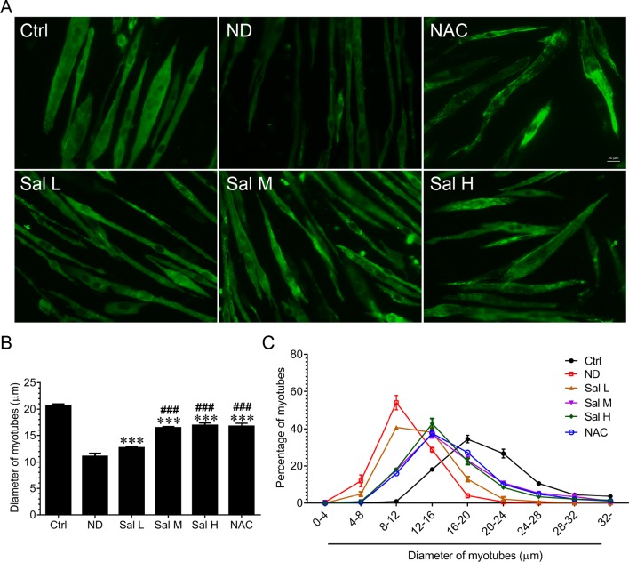
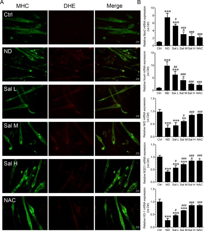
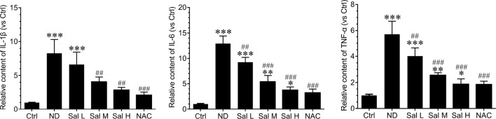
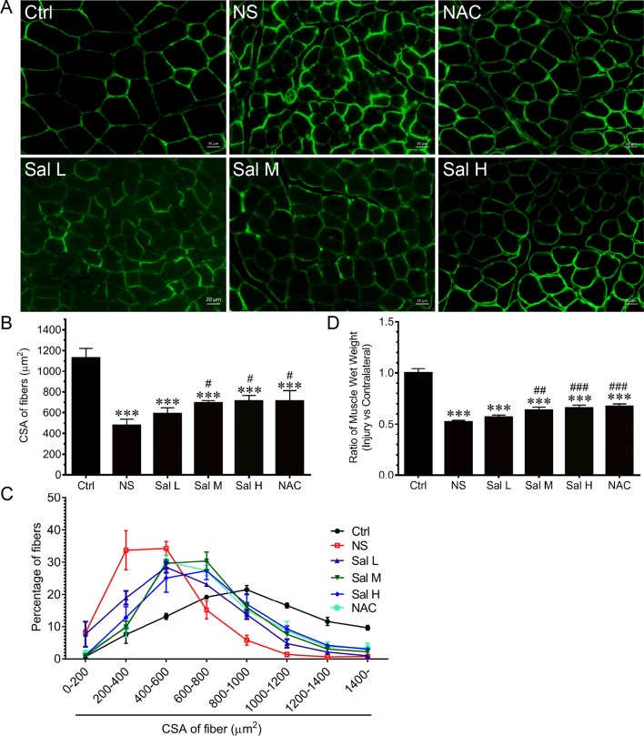
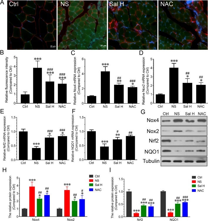
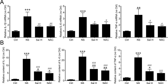
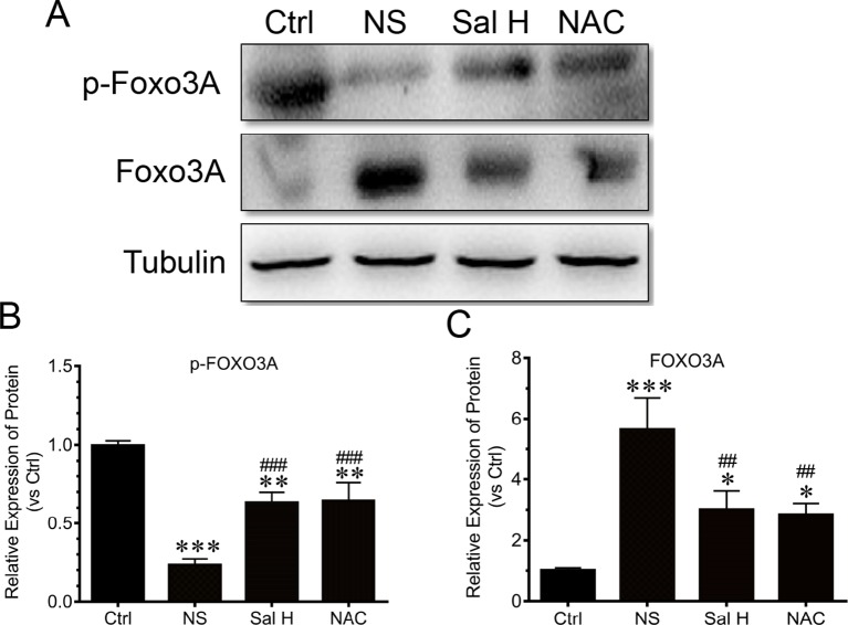
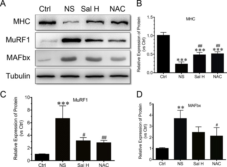
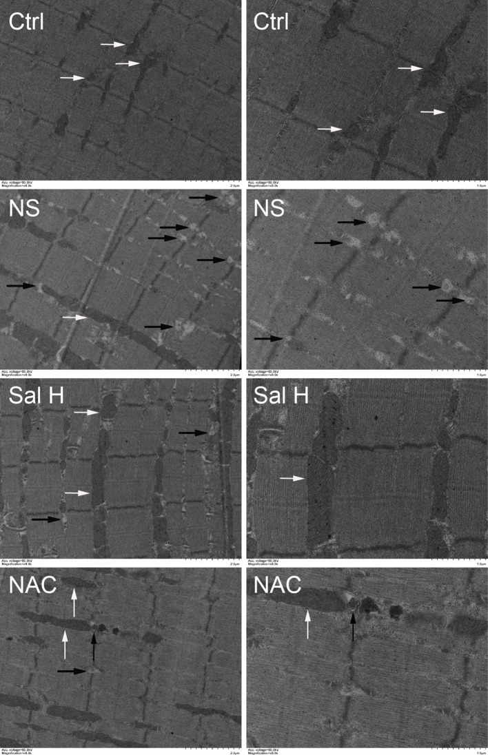
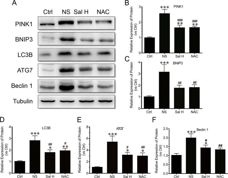
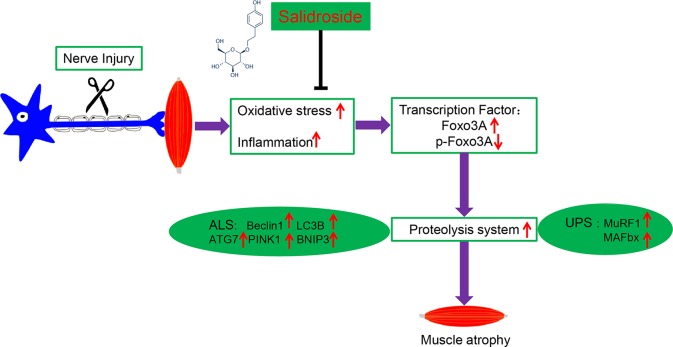
Skeletal muscle atrophy is a common and debilitating condition that lacks an effective therapy. Oxidative stress and inflammation are two main molecular mechanisms involved in muscle atrophy. In the current study, we want to explore whether and how salidroside, with antioxidant and anti-inflammatory properties, protects against skeletal muscle atrophy induced by denervation. First, oxidative stress and inflammatory response were examined during myotube atrophy induced by nutrition deprivation. The results demonstrated that oxidative stress and inflammatory response were induced in cultured myotubes suffered from nutrition deprivation, and salidroside not only inhibited oxidative stress and inflammatory response but also attenuated nutrition deprivation-induced myotube atrophy, as evidenced by an increased myotube diameter. The antioxidant, anti-inflammatory, and antiatrophic properties of salidroside in cultured myotubes were confirmed in denervated mouse models. The mice treated with salidroside showed less oxidative stress and less inflammatory cytokines, as well as higher skeletal muscle wet weight ratio and larger average cross sectional areas of myofibers compared with those treated with saline only during denervation-induced skeletal muscle atrophy. Moreover, salidroside treatment of denervated mice resulted in an inhibition of the activation of mitophagy in skeletal muscle. Furthermore, salidroside reduced the expression of atrophic genes, including MuRF1 and MAFbx, autophagy genes, including PINK1, BNIP3, LC3B, ATG7, and Beclin1, and transcription factor forkhead box O3 A (Foxo3A), and improved the expression of myosin heavy chain and transcriptional factor phosphorylated Foxo3A. Taken together, these results suggested that salidroside alleviated denervation-induced muscle atrophy by suppressing oxidative stress and inflammation.

Keywords: inflammation; muscle atrophy; nerve injury; oxidative stress; salidroside.


Figures

Figure 1
Figure 1
Salidroside alleviated nutrition deprivation-induced myotube atrophy. (A) Representative images of myosin heavy chain (MHC)-stained myotubes at 12 h following treatment with Hank’s balanced salt solution (HBSS) with or without the presence of salidroside (Sal L: 40 μM, Sal M: 80 μM, Sal H: 160 μM) or NAC (5 mM). Green indicates MHC staining. Scale bar, 20 μm. (B) Quantification of myotube size. At least 100 myotubes were analyzed for each group. Mean ± SD of diameter of myotubes. (C) Frequency distribution of diameter of myotubes. Data are expressed as mean ± SD. ***p < 0.001 versus the normal control (Ctrl) group. ###p < 0.001 versus the (nutrition deprivation) ND group.
Figure 2
Figure 2
Salidroside inhibited oxidative stress in cultured myotubes suffered from nutrition deprivation. (A) Representative images of dihydroethidium (DHE)-stained myotubes at 12 h following treatment with HBSS with or without the presence of salidroside (Sal L: 40 μM, Sal M: 80 μM, Sal H: 160 μM) or NAC (5 mM).Green and red indicate MHC staining and DHE staining, respectively. Scale bar, 20 μm. (B) The qRT-PCR analyses of the expression of oxidative stress-related genes Nox2, Nox4, Nrf2, NQO1, and HO-1 mRNA in myotubes. Data are expressed as mean ± SD. *p < 0.05, **p < 0.01, and ***p < 0.001 versus the normal control (Ctrl) group. #p < 0.05, ##p < 0.01, and ###p < 0.001 versus the (nutrition deprivation) ND group.
Figure 3
Figure 3
Salidroside inhibited inflammatory cytokines in cultured myotubes suffered from nutrition deprivation. The content of inflammatory cytokines, including interleukin 1 beta (IL-1β), interleukin-6 (IL-6), and tumor necrosis factor alpha (TNF-α), were determined by enzyme-linked immunosorbent assay (ELISA) in the myotubes at 12 h following treatment with HBSS with or without the presence of salidroside (Sal L: 40 μM, Sal M: 80 μM, Sal H: 160 μM), or N-acetyl-cysteine (NAC) (5 mM). Data are expressed as mean ± SD.*p < 0.05, **p < 0.01, and ***p < 0.001 versus the normal control (Ctrl) group. ##p < 0.01, and ###p < 0.001 versus the (nutrition deprivation) ND group.
Figure 4
Figure 4
Salidroside alleviated denervation-induced skeletal muscle atrophy. After denervation, mice were injected intraperitoneally with saline vehicle plus salidroside (Sal L: 5 mg/kg/day, Sal M: 10 mg/kg/day, Sal H: 20 mg/kg/day), saline vehicle plus NAC (20 mg/kg/day; NAC), or saline vehicle only (NS) for 14 days. After sham operation, mice were injected intraperitoneally with saline vehicle (Ctrl) for 14 days. Then the tibialis anterior (TA) muscles were harvested to undergo laminin staining analysis. (A) Representative images of laminin-stained TA muscle cross-sections. Green indicates laminin staining. Scale bar, 20 μm. (B) Mean ± SD of CSA in TA fibers from each group. (C) Frequency distribution of CSA of TA muscles. (D) The ratio of TA muscles wet weight. Data are expressed as mean ± SD. ***p < 0.001 versus Ctrl. #p < 0.05, ##p < 0.01, and ###p < 0.001 versus NS. n = 6 mice in each group.
Figure 5
Figure 5
Salidroside relieved oxidative stress in skeletal muscles suffered from sciatic nerve transection. After denervation, mice were injected intraperitoneally with saline vehicle plus salidroside (Sal H: 20 mg/kg/day), saline vehicle plus NAC (20 mg/kg/day; NAC), or saline vehicle only (NS) for 14 days. After sham operation, mice were injected with saline vehicle (Ctrl) for 14 days. Then, the TA muscles were harvested to undergo DCF staining analysis. (A) Representative images of DCF-stained TA muscles cross-sections. Green and red indicate DCF staining and laminin staining, respectively. Scale bar, 20 μm. (B) Quantification of relative DCF fluorescence intensity. (CF) The quantitative real-time polymerase chain reaction (qRT-PCR) analyses of the expression of oxidative stress-related genes Nox2, Nox4, Nrf2, and NQO1 mRNA in TA muscles. (G) The expression of Nox2, Nox4, Nrf2, and NQO1 in TA muscles was determined by Western blotting. (H, I) Protein levels of Nox2, Nox4, Nrf2, and NQO1 in TA muscles. Data are expressed as mean ± SD. *p < 0.05, **p < 0.01, and ***p < 0.001 versus Ctrl. #p < 0.05, ##p < 0.01, and ###p < 0.001 versus NS.
Figure 6
Figure 6
Salidroside suppressed inflammatory cytokines in skeletal muscles suffered from sciatic nerve transection. After denervation, mice were injected intraperitoneally with saline vehicle plus salidroside (Sal H: 20 mg/kg/day), saline vehicle plus NAC (20 mg/kg/day; NAC), or saline vehicle only (NS) for 14 days. After sham operation, mice were injected intraperitoneally with saline vehicle (Ctrl) for 14 days. Then, the inflammatory cytokines were determined through ELISA and qRT-PCR analyses. (A) The qRT-PCR analyses of the expression of inflammatory cytokines IL-1β, IL-6, and TNF-α in TA muscles. (B) The content of inflammatory cytokines, including IL-1β, IL-6, and TNF-α, were determined by ELISA. Data are expressed as mean ± SD. *p < 0.05, **p < 0.01, and ***p < 0.001 versus Ctrl. #p < 0.05, ##p < 0.01, and ###p < 0.001 versus NS.
Figure 7
Figure 7
Salidroside improved the expression of transcripitional factor forkhead box O3 A (Foxo3A) and phosphorylated Foxo3A in TA muscles of mice suffered from sciatic nerve transection. After denervation, mice were injected intraperitoneally with saline vehicle plus salidroside (Sal H: 20 mg/kg/day), saline vehicle plus NAC (20 mg/kg/day; NAC), or saline vehicle only (NS) for 14 days. After sham operation, mice were injected intraperitoneally with saline vehicle (Ctrl) for 14 days. Then the TA muscles were harvested to undergo Western blotting analysis. (A) Representative Western blot images were shown for Foxo3A and phosphorylated Foxo3A. (B) Histograms comparing phosphorylated Foxo3A levels among different muscle samples. (C) Histograms comparing Foxo3A levels among different muscle samples. Data are expressed as mean ± SD. *p < 0.05, **p < 0.01, and ***p < 0.001 versus Ctrl. ##p < 0.01 and ###p < 0.001 versus NS.
Figure 8
Figure 8
Salidroside inhibited the expression of ubiquitin–proteasome pathway—especially E3 ubiquitin ligases muscle-specific F-box (MAFbx) and muscle ring-finger protein-1 (MuRF1), and improved the expression of MHC in TA muscles of mice suffered from sciatic nerve transection. (A) Representative blots of MHC, MAFbx, and MuRF1 in TA muscles. (B) Relative expression of MHC in TA muscles. (C) Relative expression of MuRF1 in TA muscles. (D) Relative expression of MAFbx in TA muscles. Data are expressed as mean ± SD. **p < 0.01 and ***p < 0.001 versus Ctrl. #p < 0.05 and ##p < 0.01 versus NS.
Figure 9
Figure 9
Salidroside inhibited mitophagy in the TA muscles following sciatic nerve transection. After denervation, mice were injected intraperitoneally with saline vehicle plus salidroside (Sal H: 20 mg/kg/day), saline vehicle plus NAC (20 mg/kg/day; NAC), or saline vehicle only (NS) for 14 days. After sham operation, mice were injected intraperitoneally with saline vehicle (Ctrl) for 14 days. Then the TA muscles were harvested to undergo transmission electron microscopy (TEM) analysis. Representative TEM micrographs of TA muscles. The right is a local enlargement of the left. White arrows indicate normal mitochondria. Black arrows indicate mitophagy.
Figure 10
Figure 10
Salidroside inhibited the expression of autophagy genes, including PTEN-induced putative kinase 1 (PINK1), Bcl2/adenovirus E1B 19-kDa protein-interacting protein 3 (BNIP3), microtubule-associated protein 1 light chain 3 beta (LC3B), autophagy-related protein 7 (ATG7), and Beclin1, in TA muscles of mice suffered from sciatic nerve transection. (A) Representative blots of PINK1, BNIP3, LC3B, ATG7, and Beclin1 in TA muscles. (B) Relative expression of PINK1 in TA muscles. (C) Relative expression of BNIP3 in TA muscles. (D) Relative expression of LC3B in TA muscles. (E) Relative expression of ATG7 in TA muscles. (F) Relative expression of Beclin1 in TA muscles. Data are expressed as mean ± SD. *p < 0.05, **p < 0.01, and ***p < 0.001 versus Ctrl. #p < 0.05, ##p < 0.01, and ###p < 0.001 versus NS.
Figure 11
Figure 11
A schematic diagram illustrating the proposed mechanism by which salidroside attenuates denervation-induced skeletal muscle atrophy. Denervation-induced skeletal muscle atrophy is associated with elevated oxidative stress and inflammation, elevated Foxo3A, and decreased p-Foxo3A, greater activation of proteolysis and mitophagy in this study. Interestingly, salidroside improved skeletal muscle atrophy by inhibiting oxidative stress, inflammation and mitophagy, and giving rise to reduced proteolysis in denervated skeletal muscle.

References

    1. Abrigo J., Rivera J. C., Simon F., Cabrera D., Cabello-Verrugio C. (2016). Transforming growth factor type beta (TGF-beta) requires reactive oxygen species to induce skeletal muscle atrophy. Cell Signal 28 (5), 366–376. 10.1016/j.cellsig.2016.01.010 - DOI - PubMed
    1. Anderson L. J., Liu H., Garcia J. M. (2017). Sex differences in muscle wasting. Adv. Exp. Med. Biol. 1043, 153–197. 10.1007/978-3-319-70178-3_9 - DOI - PubMed
    1. Cai X., Yuan Y., Liao Z., Xing K., Zhu C., Xu Y., et al. (2018). Alpha-Ketoglutarate prevents skeletal muscle protein degradation and muscle atrophy through PHD3/ADRB2 pathway. FASEB J. 32 (1), 488–499. 10.1096/fj.201700670R - DOI - PMC - PubMed
    1. Ceelen J. J. M., Schols A., van Hoof S. J., de Theije C. C., Verhaegen F., Langen R. C. J. (2017). Differential regulation of muscle protein turnover in response to emphysema and acute pulmonary inflammation. Respir. Res. 18 (1), 75. 10.1186/s12931-017-0531-z - DOI - PMC - PubMed
    1. Chal J., Pourquie O. (2017). Making muscle: skeletal myogenesis in vivo and in vitro. Development 144 (12), 2104–2122. 10.1242/dev.151035 - DOI - PubMed