. 2019 Sep 20:10:1013.
doi: 10.3389/fphar.2019.01013. eCollection 2019.

Panax Notoginseng Saponins Protect Cardiac Myocytes Against Endoplasmic Reticulum Stress and Associated Apoptosis Through Mediation of Intracellular Calcium Homeostasis

Affiliations

Panax Notoginseng Saponins Protect Cardiac Myocytes Against Endoplasmic Reticulum Stress and Associated Apoptosis Through Mediation of Intracellular Calcium Homeostasis

Jun Chen et al. Front Pharmacol. .

Abstract

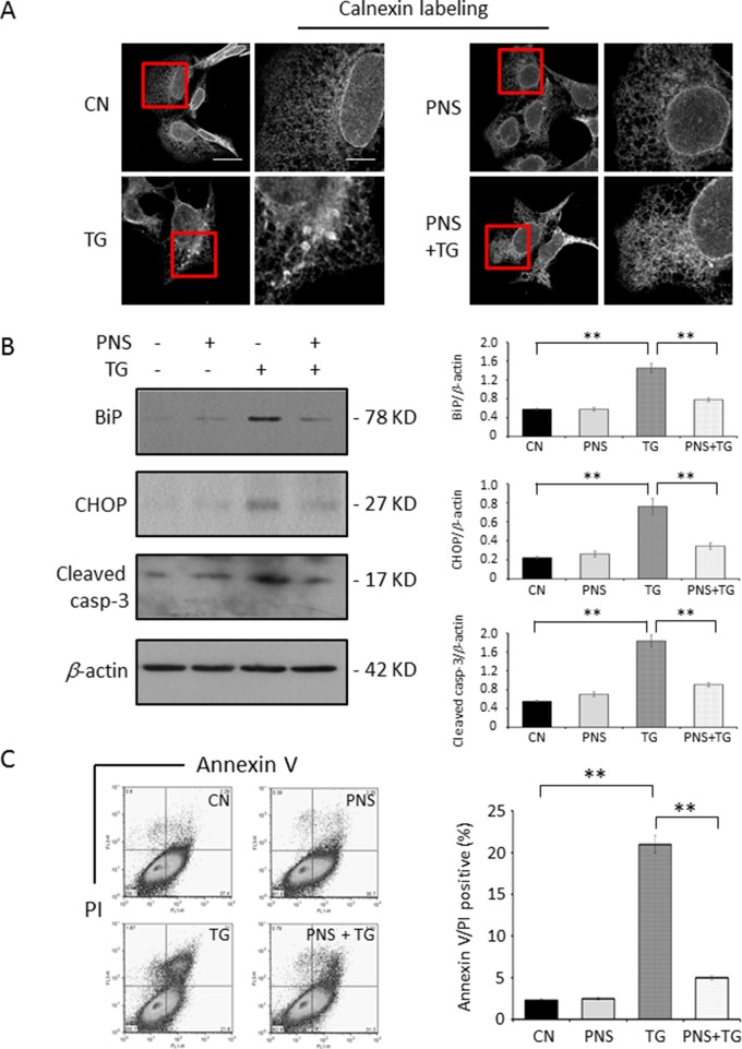
Endoplasmic reticulum (ER) stress has been demonstrated to play important roles in the pathogenesis of various cardiovascular diseases. The ER stress pathway is therefore a promising therapeutic target in cardiovascular disease. Although Panax notoginseng saponins (PNS) are one of the patent medicines that are traditionally used to treat cardiovascular disorders, their effects on ER stress in cardiac myocytes remain unexploited so far. This study investigates the effects of PNS on ER stress and its associated cell apoptosis along with the related mechanism in cardiac myocytes. PNS compounds were identified via high-performance liquid chromatograph (HPLC) assay. PNS-pretreated H9c2 cells, HL-1 cells, and primary cultured neonatal rat cardiomyocytes were stimulated with thapsigargin (TG) to induce ER stress response and apoptosis. ER stress response was tested by immunofluorescence or immunoblot of the ER protein chaperones-calnexin, binding immunoglobulin protein (BiP) and the C/EBP homologous protein (CHOP). Cell viability was tested by methyl thiazolyl tetrazolium (MTT) assay. Cell apoptosis was detected by immunoblot of Cleaved caspase-3 and flow cytometry analysis of Annexin V/propidium iodide (PI) staining. Cytosolic, mitochondrial, and ER calcium dynamics were investigated by calcium imaging. Moreover, a ryanodine receptor type-2 (RyR2) overexpression stable cell line was generated to verify the mechanism of RyR2 involved in PNS in the inhibition of ER stress and cell apoptosis. We demonstrate here that PNS protected cardiac myocytes from ER stress response and associated cell death in a concentration-dependent manner. Importantly, PNS reduced the elevation of cytosolic calcium, mitochondria calcium, as well as ER calcium in response to either TG or histamine treatment. PNS protection in ER stress was regulated by RyR2 expression. In summary, PNS protection against TG-induced ER stress response and its associated cell apoptosis in cardiac myocytes is calcium dependent. Through the regulation of ER calcium release mediated by RyR2, a novel mechanism for PNS in the prevention of cardiovascular diseases is thereby identified.

Keywords: Panax notoginseng saponins; apoptosis; endoplasmic reticulum stress; intracellular calcium homeostasis; ryanodine receptor.


Figures

Figure 1
Figure 1
PNS compound identification and cell viability test. (A) HPLC profiles of PNS compounds. P1: notoginsenoside R1; P2: ginsenoside Rg1; P3: ginsenoside Re; P4: ginsenoside Rb1, and P5: ginsenoside Rd. (B) PNS compound identification: formula, molecular weight, and structure. (C) Cell viability tests of H9c2 cells treated with various concentrations of PNS (0, 20, 40, 80, 100 μg/ml) either with or without 1 μM TG (dark, −TG group; grey, +TG group). Bar graph shows percentage of viability compared with the untreated cells. (D) H9c2 cells treated as indicated in C were immunoblotted with antibodies to BiP and β-actin. Bands were quantified relative to β-actin by densitometry. (Mean ± SEM; NS, not significant; *P < 0.05, **P < 0.01 relative to CN group or indicated group.) PNS, Panax notoginseng saponin; HPLC, high-performance liquid chromatograph; TG, thapsigargin; BiP, binding immunoglobulin protein; SEM, the standard error of the mean; CN, control.
Figure 2
Figure 2
PNS prevents TG-induced ER stress response and cell apoptosis. (A) H9c2 cells, either untreated (CN group) or pretreated with 40 μg/ml PNS for 12 h (PNS group), before addition of 1 μM TG (TG group or PNS plus TG group) for 12 h were immunofluorescenced with primary anti-calnexin antibody. Scale bar, 30 μm; in box: 10 μm. (B) H9c2 cells treated as in A were immunoblotted with antibodies to BiP, CHOP, Cleaved caspase-3, and β-actin. Bands were quantified relative to β-actin by densitometry. (C) H9c2 cells treated as in A were double-stained with Annexin V/PI and analyzed by flow cytometry. Bar graph shows percentage of Annexin V/PI double-positive cells. (Mean ± SEM; **P < 0.01 relative to CN group or indicated group.) ER, endoplasmic reticulum; PI, propidium iodide; CHOP, the C/EBP homologous protein.
Figure 3
Figure 3
PNS suppresses cytosolic Ca2+ transients evoked by TG and histamine. (A) Representative recordings of TG-evoked cytosolic Ca2+ transients recorded by Fura-2 ratios (F 340/F 380) in H9c2 cells with (grey, PNS group) or without (dark, CN group) PNS pretreatment (40 μg/ml; 12 h), as indicated. Bar graphs show cytosolic Ca2+ peak amplitude, area under the curve (AUC), as well as time decay of Ca2+ transient response to TG stimulation. (B) Representative recordings of histamine-evoked cytosolic Ca2+ transients recorded by Fura-2 ratios (F 340/F 380) in H9c2 cells with (grey) or without (dark) PNS pretreatment (40 μg/ml; 12 h), as indicated. Bar graphs show cytosolic Ca2+ peak amplitude, AUC, as well as time decay of Ca2+ transient response to histamine stimulation. (Mean ± SEM; 60–80 responding cells; NS, not significant; *P < 0.05, **P < 0.01 relative to CN group.)
Figure 4
Figure 4
PNS suppresses mitochondrial Ca2+ uptake and ER Ca2+ release induced by TG. (A) Confocal microscope images of Rhod-2 AM loaded H9c2 cells counterstained with anti-Tom20 mitochondrial antibody. Scale bar, 10 μm; in box: 3 μm. (B) Confocal microscope images of H9c2 cells loaded with D1ER cameleon and anti-derlin antibody. Scale bar, 10 μm; in box: 3 μm. (C) Representative recordings of TG-evoked mitochondrial Ca2+ elevation recorded by Rhod-2 fluorescence (F/F 0) in H9c2 cells with (grey, PNS group) or without (dark, CN group) PNS pretreatment (40 μg/ml; 12 h), as indicated. Bar graphs show mitochondrial Ca2+ peak amplitude and AUC in response to TG stimulation. (Mean ± SEM; 60–80 responding cells; *P < 0.05, **P < 0.01 relative to CN group.) (D) Representative recordings of TG-induced ER Ca2+ dynamics were recorded by the FRET-to-CFP emission ratio (FRET/CFP) in H9c2 cells with (grey, PNS group) or without (dark, CN group) PNS pretreatment (40 μg/ml; 12 h), as indicated. Bar graphs show ER Ca2+ peak amplitude and AUC in response to TG stimulation. (Mean ± SEM; 20–30 responding cells; *P < 0.05, **P < 0.01 relative to CN group.) FRET, fluorescence resonance energy transfer; AM, acetoxymethyl; D1ER, ER-targeted cameleon; CFP, cyan fluorescent protein.
Figure 5
Figure 5
PNS prevention of TG-induced ER stress and apoptosis is Ca2+ dependent. H9c2 cells were either untreated (CN group) or pretreated with 40 μg/ml PNS for 12 h (PNS group) before addition of 1 μM TG for 12 h in the presence of 1 mM Ca2+ plus 1 μM ionomycin (Iono; A) or 40 μM Bapta-AM (Bapta; B), as indicated. Treated cells were immunoblotted with antibodies to BiP, Cleaved caspase-3, as well as β-actin, and bands were quantified relative to β-actin by densitometry. (Mean ± SEM; NS, not significant; **P < 0.01 relative to CN group or indicated group.)
Figure 6
Figure 6
PNS prevention of TG-induced ER stress and apoptosis is regulated by RyR2. (A) H9c2 cells, either untreated (CN group) or pretreated with 40 μg/ml PNS for 12 h (PNS group) before addition of 1 μM TG (TG group or PNS plus TG group) for 12 h, were immunoblotted to antibodies to RyR2 and SERCA2 as well as β-actin. Bands were quantified relative to β-actin by densitometry. (B) Non-target control (NTC group) or RyR2-mfGFP (RyR2-mfGFP group) inducible stable HL-1 cell lines were identified by immunofluorescence and immunoblot to anti-GFP antibody. Scale bar, 50 μm. (C) Non-target control (NTC group) or RyR2-mfGFP transfected (RyR2-mfGFP group) HL-1 cells either untreated (CN group) or pretreated with 40 μg/ml PNS for 12 h before addition of 1 μM TG for 12 h (TG group or PNS plus TG group) were immunoblotted with antibodies to BiP, CHOP, and Cleaved caspase-3 as well as β-actin. Bands were quantified relative to β-actin by densitometry. (D) NTC or RyR2-mfGFP HL-1 cells treated as indicated in (C) were double-stained with Annexin V and PI, and analyzed by flow cytometry. Bar graph shows percentage of Annexin V/PI double-positive cells. (Mean ± SEM; NS, not significant; **P < 0.01 relative to CN group or indicated group.) RyR2, ryanodine receptor type-2. SERCA2, sarco/endoplasmic reticulum Ca2+-ATPase; mfGFP; multifunctional GFP.
Figure 7
Figure 7
PNS prevents TG-induced ER stress response and cell apoptosis in primary cultured cardiomyocytes. (A) Primary cultured cardiomyocytes were immunofluorescenced with primary anti-α-actin antibody. Scale bar, 50 μm. (B) Primary cultured cardiomyocytes either untreated (CN group) or pretreated with 40 μg/ml PNS for 12 h (PNS group), before addition of 1 μM TG (TG group or PNS plus TG group), were immunoblotted with antibodies to BiP and Cleaved caspase-3 and β-actin. Bands were quantified relative to β-actin by densitometry. (C) Primary cultured cardiomyocytes treated as in (B) were double-stained with Annexin V/PI and analyzed by flow cytometry. Bar graph shows percentage of Annexin V/PI double-positive cells. (D) Primary cultured cardiomyocytes treated as in (B) were immunoblotted with antibodies to RyR2 and β-actin. Bands were quantified relative to β-actin by densitometry. (Mean ± SEM; **P < 0.01 relative to CN group or indicated group.)
Figure 8
Figure 8
Mechanism of PNS protects ER stress–induced cell death. TG inhibits Ca2+ re-uptake from ER to cytosol through SERCA2 and thereby elevates intracellular concentration and mitochondrial calcium uptake. TG disturbs the calcium homeostasis and causes protein-folding dysfunction, and thus, the accumulated misfolded/unfolded proteins (e.g. BiP and CHOP) induce ER stress. Prolonged TG treatment initiates the intrinsic apoptotic pathway by permeabilizing the mitochondrial membrane and thus releasing cytochrome c and AIF to cytosol, resulting in apoptosome formation and leading to activation of caspase-3. PNS pretreatment significantly reduced upregulation of BiP, CHOP, and Cleaved caspase-3 induced by TG. PNS reduced the elevation of cytosolic calcium transients, mitochondrial calcium uptake, as well as ER calcium release in response to either TG or histamine. PNS decreases the RyR2 expression, and PNS prevention of ER stress and associated apoptosis is mediated by RyR2 expression. These results suggest that by the intermediary through regulation of intracellular calcium homeostasis, especially suppression of ER calcium release mediated by RyR2 and thus inhibition of the cytosolic and mitochondrial calcium overload, PNS therefore protected against ER stress–induced cell death. AIF, apoptosis inducing factor.

References

    1. Belevych A. E., Radwanski P. B., Carnes C. A., Gyorke S. (2013). ‘Ryanopathy’: causes and manifestations of RyR2 dysfunction in heart failure. Cardiovasc. Res. 98 (2), 240–247. 10.1093/cvr/cvt024 - DOI - PMC - PubMed
    1. Benkusky N. A., Farrell E. F., Valdivia H. H. (2004). Ryanodine receptor channelopathies. Biochem. Biophys. Res. Commun. 322 (4), 1280–1285. 10.1016/j.bbrc.2004.08.033 - DOI - PubMed
    1. Bernardi P. (1999). Mitochondrial transport of cations: channels, exchangers, and permeability transition. Physiol. Rev. 79 (4), 1127–1155. 10.1152/physrev.1999.79.4.1127 - DOI - PubMed
    1. Berridge M. J., Lipp P., Bootman M. D. (2000). The versatility and universality of calcium signalling. Nat. Rev. Mol. Cell Biol. 1 (1), 11–21. 10.1038/35036035 - DOI - PubMed
    1. Bers D. M. (2002). Cardiac excitation–contraction coupling. Nature 415 (6868), 198–205. 10.1038/415198a - DOI - PubMed