. 2019 Oct 9;8(5):22.
doi: 10.1167/tvst.8.5.22. eCollection 2019 Sep.

Imaging Distal Aqueous Outflow Pathways in a Spontaneous Model of Congenital Glaucoma

Affiliations

Imaging Distal Aqueous Outflow Pathways in a Spontaneous Model of Congenital Glaucoma

Kevin C Snyder et al. Transl Vis Sci Technol. .

Abstract

Purpose: To validate the use of aqueous angiography (AA) in characterizing distal aqueous outflow pathways in normal and glaucomatous cats.

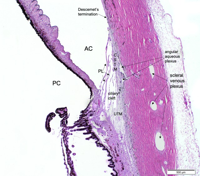
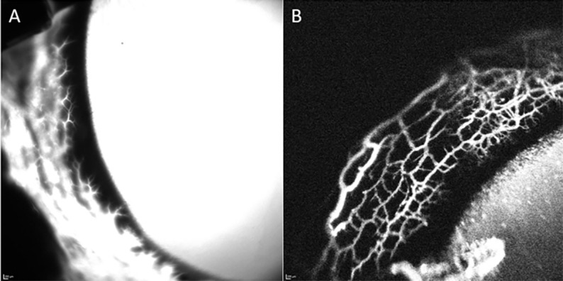
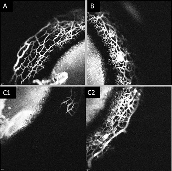
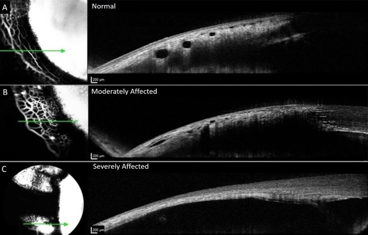
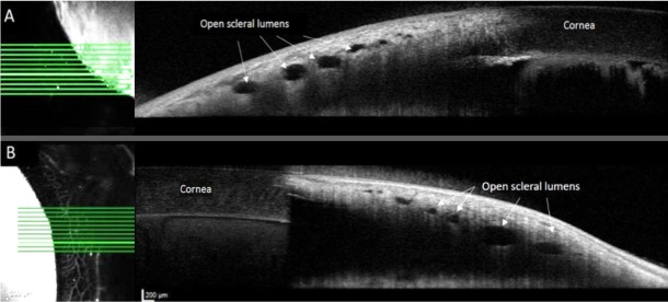
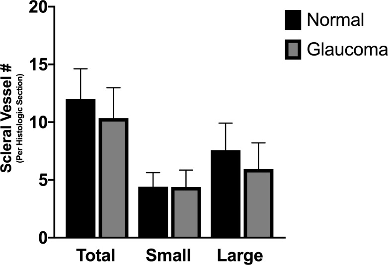
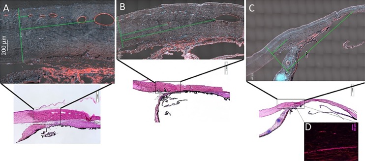
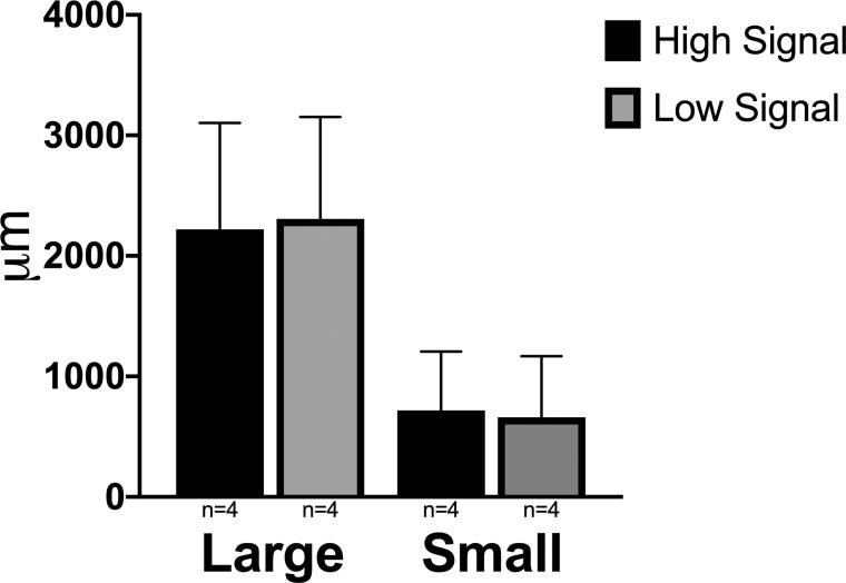
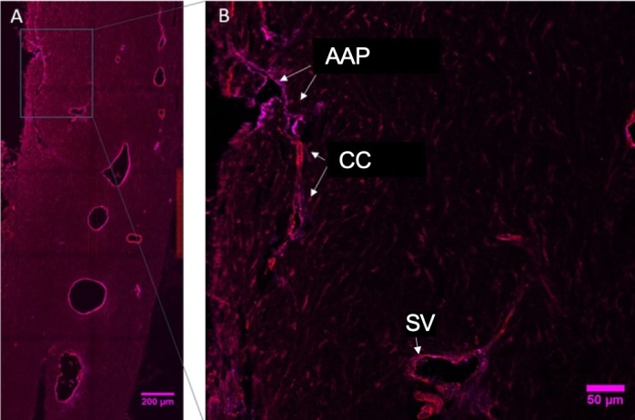
Methods: Ex vivo AA and optical coherence tomography (OCT) were performed in nine adult cat eyes (5 feline congenital glaucoma [FCG] and 4 normal), following intracameral infusion of 2.5% fluorescein and/or 0.4% indocyanine green (ICG) at physiologic intraocular pressure (IOP). Scleral OCT line scans were acquired in areas of high- and low-angiographic signal. Tissues dissected in regions of high- and low-AA signal, were sectioned and hematoxylin and eosin (H&E)-stained or immunolabeled (IF) for vascular endothelial and perivascular cell markers. Outflow vessel numbers and locations were compared between groups by Student's t-test.

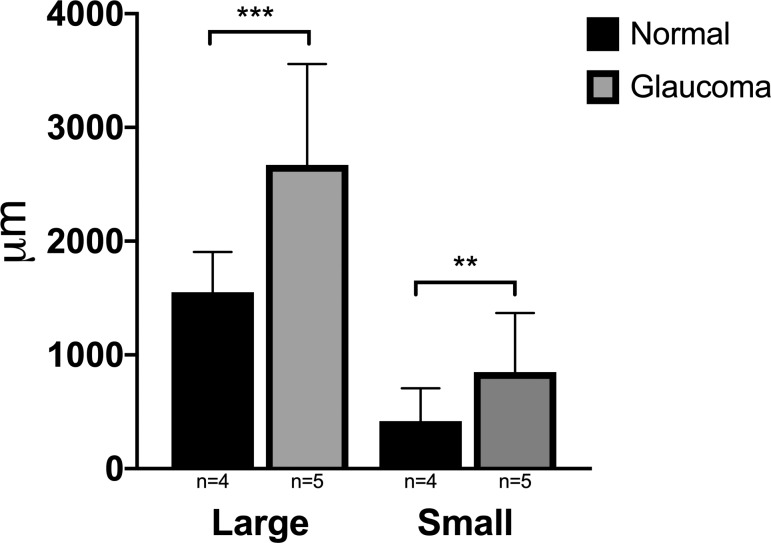
Results: AA yielded circumferential, high-quality images of distal aqueous outflow pathways in normal and FCG eyes. No AA signal or scleral lumens were appreciated in one buphthalmic FCG eye, though collapsed vascular profiles were identified on IF. The remaining eight of nine eyes all showed segmental AA signal, distinguished by differences in time of signal onset. AA signal always corresponded with lumens seen on OCT. Numbers of intrascleral vessels were not significantly different between groups, but scleral vessels were significantly more posteriorly located relative to the limbus in FCG.

Conclusions: A capacity for distal aqueous humor outflow was confirmed by AA in FCG eyes ex vivo but with significant posterior displacement of intrascleral vessels relative to the limbus in FCG compared with normal eyes.

Translational relevance: This report provides histopathologic correlates of advanced diagnostic imaging findings in a spontaneous model of congenital glaucoma.

Keywords: aqueous angiography; glaucoma anterior segment; imaging; optical coherence tomography.


Figures

Figure 1
Figure 1
Photomicrograph of a normal adult feline iridocorneal angle stained with H&E, illustrating important structures of the feline conventional aqueous outflow pathway. Anterior chamber (AC), posterior chamber (PC), pectinate ligaments (PL), corneoscleral trabecular meshwork (CSTM), Descemet's membrane termination, ciliary cleft, uveoscleral trabecular meshwork (UTM), angular aqueous plexus, and scleral venous plexus are labeled for reference.
Figure 2
Figure 2
ICG (0.4%) yielded higher resolution images for longer imaging time versus 2.5% fluorescein as a tracer. (A) Representative image of angiographic signal in a normal feline eye with 2.5% fluorescein 10 minutes after intracameral infusion. Note the diffuse loss of detail and lack of outflow architecture due to fluorescein diffusion. (B) Representative image of angiographic signal in a normal feline eye with 0.4% ICG approximately 12 minutes after injection. Note improved resolution and detail of outflow architecture.
Figure 3
Figure 3
Segmental outflow in a normal feline eye. Images of three quadrants (A–C) from the same eye taken at approximately 7 minutes post 0.4% ICG injection. (A, B) Depict high-angiographic signal and (C1) depicts no signal. (C2) depicts the same quadrant as in (C1), imaged at 20 minutes postinjection of tracer, which now exhibits an angiographic signal.
Figure 4
Figure 4
Representative OCT images from normal and glaucomatous feline eyes. Images on the left represent en face confocal laser scanning ophthalmoscopy images of AA signal and images on the right are corresponding, OCT line scans registered to these images *Green arrow corresponds to precise location of OCT line scan. (A) Depicts normal eye; (B) depicts globe affected by glaucoma. OCT lumens visible but appear attenuated. (C) Depicts severely affected glaucoma eye with no vascular outflow signal, or scleral lumens visible on the corresponding OCT image. The sclera is thin and no scleral vessel lumens are visible. Angiographic signal on the left represents combination of diffusion into overlying conjunctival tissues after extended period of time and possibly signal visible from within, through extremely thin sclera.
Figure 5
Figure 5
Open scleral lumens were observed on OCT images (right), both in regions of low-AA signal (A), and of high-AA signal (B) in different quadrants of the same representative normal feline eye taken consecutively at approximately 9 minutes postinjection of 0.4% iCG. Brightest horizontal lines on en face AA images (left) correspond to location of respective OCT line scans on right.
Figure 6
Figure 6
Number of scleral vessels identified on IF histologic sections did not differ between normal cats and cats with feline congenital glaucoma. Total number of scleral vessels, number of large scleral vessels (>500 μm in circumference), and number of small scleral vessels (<500 μm in circumference) were not significantly different between glaucoma affected and normal eyes. Error bars represent standard deviation.
Figure 7
Figure 7
Representative, tiled sections from feline anterior segment tissues IF labeled for vascular endothelial cells with vWF (red) and DAPI nuclear counterstain (blue) include differential interference contrast (DIC) signal for visualization of scleral tissue (A–C). (A) Normal eye, (B) moderately affected eye, and (C, D) severely affected eye, in which collapsed scleral vessel lumens were observed with IF (D) that were not visible on OCT. *Green line on left of each image is a line drawn perpendicularly to sclera corresponding to the termination of Descemet's membrane and two perpendicular green lines depict distance measured to first small scleral vessel lumen and first large vessel lumen.
Figure 8
Figure 8
Mean distance in micrometers from the termination of Descemet's membrane to first large and small vessel lumen, comparing normal and glaucoma groups from histologic sections. These distances were significantly greater in eyes with glaucoma than in normal eyes (P ≤ 0.0001 and P = 0.001, respectively). Error bars represent standard deviation.
Figure 9
Figure 9
Mean distances in micrometers from the termination of Descemet's membrane to either the first large or first small vessel lumen were not significantly different between histologic sections from high AA signal and low AA signal regions. Error bars represent standard deviation.
Figure 10
Figure 10
(A) Immunofluorescent labelled section through the perilimbal region of a representative normal feline eye (image is intentionally overexposed to allow visualization of tissue morphology) with region delineated by a rectangle shown at higher magnification in (B), which depicts the location of the normal AAP labeled positively with vWF (endothelial cells; magenta) while the collector channels (CC) and scleral vessels (SV) are positively labeled for both vWF and alpha-smooth muscle actin (perivascular cells; red).

References

    1. Thylefors B, Négrel AD. The global impact of glaucoma. Bull World Health Organ. 1994;72:323–326. - PMC - PubMed
    1. Steinkuller PG, Du L, Gilbert C, Foster A, Collins ML, Coats DK. Childhood blindness. J AAPOS. 1999;3:26–32. - PubMed
    1. Rutz-Mendicino MM, Snella EM, Jens JK, et al. Removal of potentially confounding phenotypes from a Siamese-derived feline glaucoma breeding colony. Comp Med. 2011;61:251–257. - PMC - PubMed
    1. Kuehn MH, Lipsett KA, Menotti-Raymond M, et al. A mutation in LTBP2 causes congenital glaucoma in domestic cats (Felis catus) PLoS One. 2016;11:e0154412. - PMC - PubMed
    1. Ali M, McKibbin M, Booth A, et al. Null mutations in LTBP2 cause primary congenital glaucoma. Am J Hum Genet. 2009;84:664–671. - PMC - PubMed