. 2019 Oct 2;5(10):eaax0059.
doi: 10.1126/sciadv.aax0059. eCollection 2019 Oct.

MMOD-induced structural changes of hydroxylase in soluble methane monooxygenase

Affiliations

MMOD-induced structural changes of hydroxylase in soluble methane monooxygenase

Hanseong Kim et al. Sci Adv. .

Abstract

Soluble methane monooxygenase in methanotrophs converts methane to methanol under ambient conditions. The maximum catalytic activity of hydroxylase (MMOH) is achieved through the interplay of its regulatory protein (MMOB) and reductase. An additional auxiliary protein, MMOD, functions as an inhibitor of MMOH; however, its inhibitory mechanism remains unknown. Here, we report the crystal structure of the MMOH-MMOD complex from Methylosinus sporium strain 5 (2.6 Å). Its structure illustrates that MMOD associates with the canyon region of MMOH where MMOB binds. Although MMOD and MMOB recognize the same binding site, each binding component triggers different conformational changes toward MMOH, which then respectively lead to the inhibition and activation of MMOH. Particularly, MMOD binding perturbs the di-iron geometry by inducing two major MMOH conformational changes, i.e., MMOH β subunit disorganization and subsequent His147 dissociation with Fe1 coordination. Furthermore, 1,6-hexanediol, a mimic of the products of sMMO, reveals the substrate access route.


Figures

Fig. 1
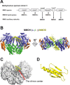
Fig. 1. Crystal structure of the MMOH-MMOD complex.
(A) Schematic overview of the sMMO operon in M. sporium strain 5. (B) Front and top views of the MMOH-MMOD complex. MMOH is shown using a cartoon model (α subunit, blue; β subunit, green; and γ subunit, orange); MMOD is colored yellow, and the orange balls represent di-irons. (C) Front view of the MMOH-MMOD complex. MMOH is shown using a surface representation model (white), and MMOD (yellow) is shown in cartoon form. Red dot marks the location of the di-iron center in MMOH. (D) MMOD contains a ββββα-fold, which is shown in cartoon form. Illustrations of the protein structure used in all figures were generated with PyMOL (DeLano Scientific LLC).
Fig. 2
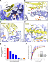
Fig. 2. Key residues participate in both the MMOH-MMOD binding and the inhibitory activity of MMOD.
(A) MMOD β4, loop, and helix α (yellow) directly participate in recognizing the canyon region of MMOH. MMOD residues that do not interact with MMOH are colored gray. The two iron atoms are labeled and colored orange. (B to D) Detailed molecular interactions in the MMOH-MMOD complex. (E) Enzymatic activities (n = 3) of MMOH in the presence of MMOB, MMOR, and/or MMOD, as determined on the basis of the conversion from propylene to propylene oxide in the presence of NADH. Condition H + B + R (red) indicates the absence of MMOD; blue indicates the addition of MMOD. Conditions H + B (green) and H + R (orange) represent the absence of MMOR and MMOB, respectively (n = 3, average ± SEM). (F) Binding of MMOB, MMOD, and truncated MMODs to MMOH as detected by fluorescence spectroscopy (n = 3). All error bars represent SEMs. Quenching of intrinsic fluorescence of MMOH was monitored by titration of MMOB (blue), MMOD (red), 1 to 74 MMOD (green), 12 to 111 MMOD (purple), and 12 to 74 MMOD (orange). The dissociation constant (Kd1 and Kd2) values were determined through nonlinear curve fitting for the two binding sites of MMOH (0.32 μM).
Fig. 3
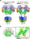
Fig. 3. Structural comparison of MMOH (PDB ID: 1MHY) (20) and the MMOH-MMOD complex.
(A) Front views of MMOH and MMOH-MMOD. Red box indicates the region with the major structural differences. The schematic model shows that MMOHβ-NT functions as a latch to tightly lock the MMOHα helix bundles. MMOD binding unlocks the latch and relaxes the overall helix architecture of MMOHα. (B) M. sporium strain 5 MMOD structures are overlaid onto M. trichosporium OB3b MMOH. The N terminus of MMOHβ is displayed in green. The MMOD C-terminal long helix is shown in yellow. Dashed yellow lines indicate the extended, disordered C-terminal region of MMOD. The red arrow indicates the viewing angle of the inset. The inset shows the clash between MMOHβ-NT and the C-terminal helix of MMOD.
Fig. 4
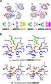
Fig. 4. Structural comparison of the di-iron center of MMOH (PDB ID: 1MHY) (20), MMOH-MMOB (PDB ID: 4GAM) (14), and MMOH-MMOD.
(A) Conformational changes in the MMOH four-helix bundle (helices B, C, E, and F) upon MMOB and MMOD binding. Six residues that coordinate to the two iron atoms are on helices B, C, E, and F. Red arrows indicate the translation and rotation of the indicated helices upon MMOD and MMOB binding. (B) Stereo views of the di-iron center geometry in MMOH-MMOD and MMOH. The two Fe atoms (orange) are surrounded and coordinated by four glutamate and two histidine residues. Water molecules (H2O) are displayed as red spheres.
Fig. 5
Fig. 5. Product analog 1,6-hexanediol at the substrate access cavity near the di-iron center and the cavity opening in the MMOH-MMOD complex.
(A) Composite omit map [1.5σ contour, calculated using the PHENIX software package (38)] of MMOH-MMOD near the di-iron center. The electron density map is shown as a mesh (gray), and 1,6-hexanediol and formate are colored green. (B) Substrate access cavity in the M. trichosporium OB3b MMOH (gray, PDB ID: 1MHY) (20) and the M. sporium strain 5 MMOH-MMOD complex (yellow). The two gate-forming residues (Leu110 and Phe188) are shown as red spheres, and 1,6-hexanediol is shown in green. The cavities calculated using PyMOL are shown as surface models. (C) Schematic model showing the association of MMOH with its auxiliary proteins MMOB and MMOD, which compete for the canyon region of MMOH.

References

    1. Hanson R. S., Hanson T. E., Methanotrophic bacteria. Microbiol. Rev. 60, 439–471 (1996). - PMC - PubMed
    1. Higgins I. J., Best D. J., Hammond R. C., New findings in methane-utilizing bacteria highlight their importance in the biosphere and their commercial potential. Nature 286, 561–564 (1980). - PubMed
    1. Wang V. C.-C., Maji S., Chen P. P.-Y., Lee H. K., Yu S. S.-F., Chan S. I., Alkane oxidation: Methane monooxygenases, related enzymes, and their biomimetics. Chem. Rev. 117, 8574–8621 (2017). - PubMed
    1. Tinberg C. E., Lippard S. J., Dioxygen activation in soluble methane monooxygenase. Acc. Chem. Res. 44, 280–288 (2011). - PMC - PubMed
    1. Sirajuddin S., Rosenzweig A. C., Enzymatic oxidation of methane. Biochemistry 54, 2283–2294 (2015). - PMC - PubMed

Publication types

Supplementary concepts

LinkOut - more resources