. 2020 Jan;139(1):63-77.
doi: 10.1007/s00401-019-02080-2. Epub 2019 Oct 16.

4-Repeat tau seeds and templating subtypes as brain and CSF biomarkers of frontotemporal lobar degeneration

Affiliations

4-Repeat tau seeds and templating subtypes as brain and CSF biomarkers of frontotemporal lobar degeneration

Eri Saijo et al. Acta Neuropathol. 2020 Jan.

Erratum in

Abstract

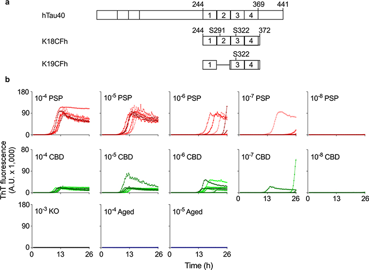
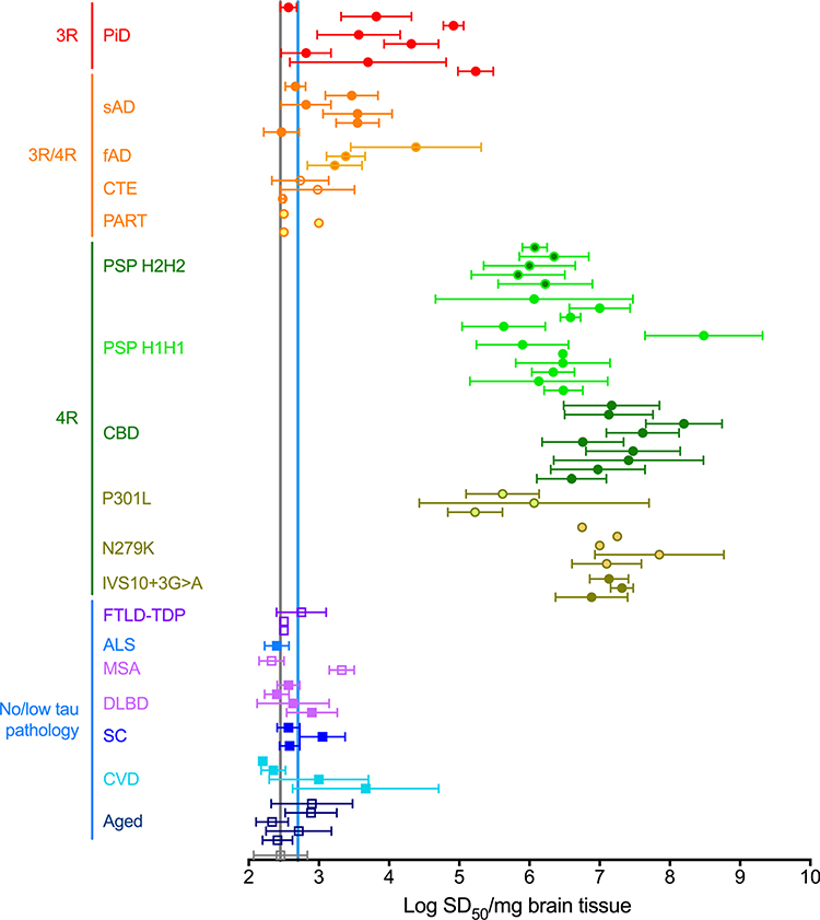
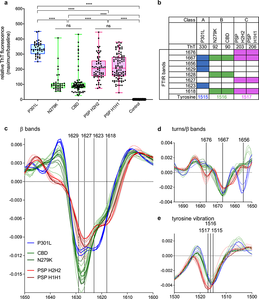
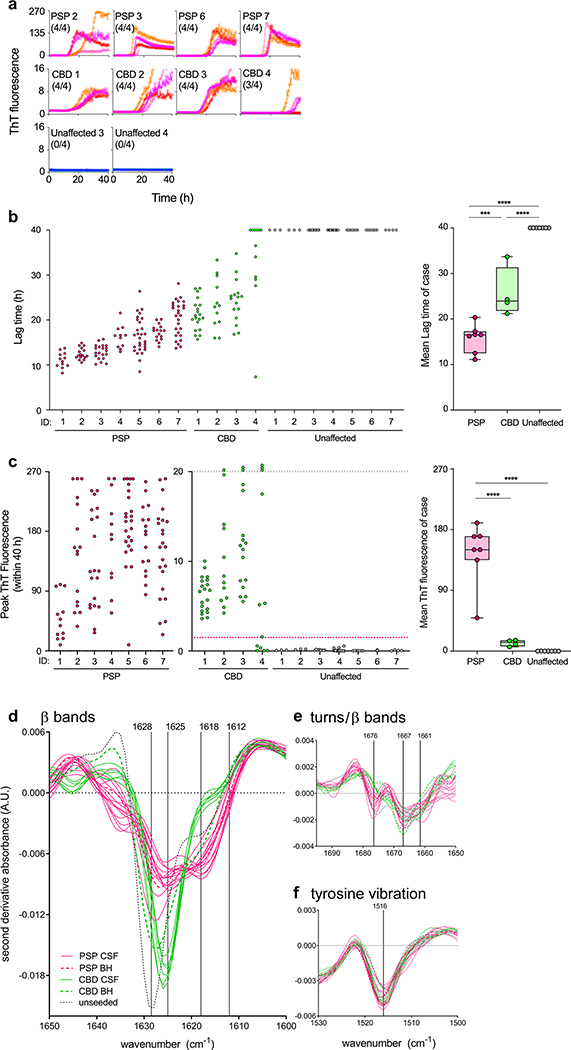
To address the need for more meaningful biomarkers of tauopathies, we have developed an ultrasensitive tau seed amplification assay (4R RT-QuIC) for the 4-repeat (4R) tau aggregates of progressive supranuclear palsy (PSP), corticobasal degeneration (CBD), and other diseases with 4R tauopathy. The assay detected seeds in 106-109-fold dilutions of 4R tauopathy brain tissue but was orders of magnitude less responsive to brain with other types of tauopathy, such as from Alzheimer's disease cases. The analytical sensitivity for synthetic 4R tau fibrils was ~ 50 fM or 2 fg/sample. A novel dimension of this tau RT-QuIC testing was the identification of three disease-associated classes of 4R tau seeds; these classes were revealed by conformational variations in the in vitro amplified tau fibrils as detected by thioflavin T fluorescence amplitudes and FTIR spectroscopy. Tau seeds were detected in postmortem cerebrospinal fluid (CSF) from all neuropathologically confirmed PSP and CBD cases but not in controls. CSF from living subjects had weaker seeding activities; however, mean assay responses for cases clinically diagnosed as PSP and CBD/corticobasal syndrome were significantly higher than those from control cases. Altogether, 4R RT-QuIC provides a practical cell-free method of detecting and subtyping pathologic 4R tau aggregates as biomarkers.

Keywords: Biomarker; Corticobasal degeneration; Diagnosis; Progressive supranuclear palsy; Strain; Tau.


Figures

Fig. 1
Fig. 1
a Schematics of truncated and mutated tau RT-QuIC substrates relative to full-length human tau isoform (hTau40). The poly-histidine tags on K18CFh and K19CFh are not depicted. b End-point dilution 4R RT-QuIC analysis of PSP and CBD brain tissue. The designated dilutions of brain tissue from a human PSP (#12, top), CBD (#1, middle), and non-demented aged individuals (#2, bottom) were tested. Also shown was simultaneous testing of brain from tau-free KO mouse (bottom). Individual traces from eight replicate reactions are shown in each panel
Fig. 2
Fig. 2
Concentrations of seeding units (SD50s) in 4R tauopathy and other brain homogenates using 4R RT-QuIC analysis. Data points represent mean ± SD of multiple independent log SD50/mg brain determinations for individual cases with the designated diagnoses. Vertical grey and blue lines mark the average values from brains of tau KO mice and humans lacking immunohistochemical evidence of tau pathology, respectively. Case abbreviations: PiD pick disease, sAD sporadic AD, fAD familial AD, CTE chronic traumatic encephalopathy, PART primary age-related tauopathy, PSP progressive supranuclear palsy, CBD corticobasal degeneration, P301L frontotemporal dementia with Parkinsonism linked to chromosome 17 MAPT (FTDP-17 MAPT) with P301L mutation, N279K FTDP-17 MAPT with N279K mutation, IVS10 + 3G > A FTDP-17 MAPT with IVS10 + 3G > A mutation, FTLD-TDP frontotemporal lobar degeneration with TDP-43, ALS amyotrophic lateral sclerosis, MSA multiple system atrophy, DLBD diffuse Lewy body disease, SC senile changes (non-tau-associated), CVD cerebrovascular disease
Fig. 3
Fig. 3
ThT maxima and FTIR reveal three distinct RT-QuIC products from 4R tauopathy subtypes. a Points represent maximum relative ThT fluorescence (rfu) values (maximum/baseline) for 4R tauopathy seeded RT-QuIC reactions. Boxes represent interquartile range around the median and whiskers represent the maximum and minimum values; ****p < 0.0001, ns non-significant. b Summarized FTIR banding patterns from RT-QuIC products in panel a, filled boxes represent the presence of a band. ce Second derivative FTIR spectra of RT-QuIC products from individual subject brain homogenates (individual traces); traces of same color indicate cases of the same type of tauopathy. Each trace represents 4–8 pooled RT-QuIC reaction wells seeded with an individual brain homogenate at 10−4 dilution for the specified 4R tauopathy
Fig. 4
Fig. 4
4R seeding activities detected in postmortem PSP and CBD CSF by 4R RT-QuIC. a CSF samples (12 μL) from PSP (n = 4), CBD (n = 4), and control (n = 2) cases, identified by their ID numbers from Online Resource Table 3, were analyzed by 4R RT-QuIC. Traces from individual quadruplicate reactions are plotted with ThT rfu indicated in thousands. The fraction of the quadruplicate reactions with rfu exceeding 1500 rfu is shown in parentheses. b Lag times (time to 1500 rfu within 40 h) for each reaction from all samples tested is shown in the dot plot. Right graph: Data points show average lag times for each case with the designated diagnosis. Boxes indicate the median and interquartile range, and whiskers indicate the maximum and minimum case averages. Significance of differences between means by one-way ANOVA followed by uncorrected Fisher’s LSD test: ***p < 0.001, ****p < 0.0001. c Baseline-subtracted maximum rfu values. CBD and unaffected control cases were plotted in the middle with a smaller y-axis. Off-scale values exceeding 20,000 rfu are stacked above the grey dotted line. Magenta dotted line: 1500 rfu threshold. Box plots as described in b, but with peak rfu instead of lag times. df Second derivative ATR-FTIR analysis of 4R RT-QuIC products seeded with individual PSP (magenta), CBD (greens), and control (black) CSF samples. Dashed lines are typical brain homogenate (BH) RT-QuIC product spectra reported in Fig. 3c for comparison
Fig. 5
Fig. 5
4R seeding activities detected in intra-vitam PSP and CBD CSF by 4R RT-QuIC. a Representative CSF samples (12 μL) from PSP, CBD, AD, and non-neurological (normal) control cases, identified by their ID numbers from Online Resource Table 3, were analyzed by 4R RT-QuIC. Traces from individual quadruplicate reactions are plotted with ThT rfu indicated in thousands. The fraction of the quadruplicate reactions exceeding the 1500 rfu threshold is shown in parentheses. b Lag times (times to 1500 rfu within 40 h) for individual reactions seeded with CSF from the designated PSP, CBS/CBD, MSA, AD, MCI mild cognitive impairment, RPD rapidly progressive dementia, PPA primary progressive aphasia, PD Parkinson disease, dementia with Lewy bodies/Lewy body dementia (DLB/LBD), carcinoma and normal control cases (ID numbers on x-axis) were shown in the dot plot. Right graph: data points show average lag times for each case with the designated diagnosis. Boxes indicate the median and interquartile range, and whiskers indicate the maximum and minimum case averages. Significance of differences between means by one-way ANOVA followed by uncorrected Fisher’s LSD test: *p < 0.05, **p < 0.001, ****p < 0.0001. c Baseline-subtracted rfu values within 36 h. Off-scale values exceeding 30,000 rfu are stacked above the grey dotted line. Magenta dotted line:1500 rfu threshold. Box plots as described in b, but for peak rfu values instead of lag times

References

    1. Alster P, Nieciecki M, Koziorowski DM, Cacko A, Charzynska I, Krolicki L et al. (2019) Thalamic and cerebellar hypoperfusion in single photon emission computed tomography may differentiate multiple system atrophy and progressive supranuclear palsy. Medicine (Baltimore) 98:e16603 10.1097/MD.0000000000016603 - DOI - PMC - PubMed
    1. Atarashi R, Satoh K, Sano K, Fuse T, Yamaguchi N, Ishibashi D et al. (2011) Ultrasensitive human prion detection in cerebrospinal fluid by real-time quaking-induced conversion. Nat Med 17:175–178. 10.1038/nm.2294 - DOI - PubMed
    1. Baron GS, Hughson AG, Raymond GJ, Offerdahl DK, Barton KA, Raymond LD et al. (2011) Effect of glycans and the glycophosphatidylinositol anchor on strain dependent conformations of scrapie prion protein: improved purifications and infrared spectra. Biochemistry 50:4479–4490. 10.1021/bi2003907 - DOI - PMC - PubMed
    1. Bessen RA, Kocisko DA, Raymond GJ, Nandan S, Lansbury PT Jr, Caughey B (1995) Nongenetic propagation of strain-specific phenotypes of scrapie prion protein. Nature 375:698–700 - PubMed
    1. Bongianni M, Orrù CD, Groveman BR, Sacchetto L, Fiorini M, Tonoli G et al. (2017) Diagnosis of human prion disease using real-time quaking-induced conversion testing of olfactory mucosa and cerebrospinal fluid samples. JAMA Neurol 74:1–8. 10.1001/jamaneurol.2016.4614 - DOI - PubMed

Publication types

MeSH terms

LinkOut - more resources