

Review
doi: 10.1007/s10858-019-00280-2.
Epub 2019 Oct 15.
Perspective: the essential role of NMR in the discovery and characterization of intrinsically disordered proteins
Affiliations
- PMID: 31617035
- PMCID: PMC7043288
- DOI: 10.1007/s10858-019-00280-2
Item in Clipboard
Review
Perspective: the essential role of NMR in the discovery and characterization of intrinsically disordered proteins
J Biomol NMR.
2019 Dec.
Abstract
The 2019 ISMAR Prize recognized NMR studies of disordered proteins. Here we provide a highly personal perspective on the discovery of intrinsically disordered proteins and the development and application of NMR methods to characterize their conformational ensembles, dynamics, and interactions.
Keywords: Immunogenic peptides; Intrinsically disordered proteins; Protein folding; Relaxation dispersion.
Figures

1H–15N HSQC spectra of disordered partners for a the DNA-binding domain of LEF-1 (Love et al. 1995, 2004), black—free LEF-1, red, LEF-1 bound to a 15 base-pair cognate DNA sequence. Selected resonances shifted between the free and bound spectra are connected and labeled. Resonances that are absent from the spectrum of the free protein but appear in the bound spectrum are labeled in green. b p21 residues 9–85 bound to the cyclin-dependent kinase Cdk2 (Kriwacki et al. 1996), green—resonances found at the same chemical shift in the free protein and the Cdk2 complex; red, new cross peaks that appear in the complex. c free pKID (top) and pKID bound to the KIX domain (bottom) (Radhakrishnan et al. 1997). Significantly shifted resonances are labeled. The intense, narrow resonances that appear between 7.8 and 8.5 ppm arise from residues in the unstructured terminal regions of pKID in the complex. The box at lower right shows (aliased) arginine side chain resonances. b, c reproduced with permission from references (Kriwacki et al. 1996; Radhakrishnan et al. 1997)

Structure of a HIF-1α interaction domain fragment (residues 798–805) bound to two targets, a TAZ1 domain of CBP (Dames et al. 2002), and b asparagine hydroxylase FIH (Elkins et al. 2003). The backbone of each partner protein is shown as a gray ribbon, the backbone of the HIF-1α fragment in green and side chains in yellow. The asparagine residue that functions as a redox switch is highlighted. Figure adapted from reference (Dyson and Wright 2005) with permission
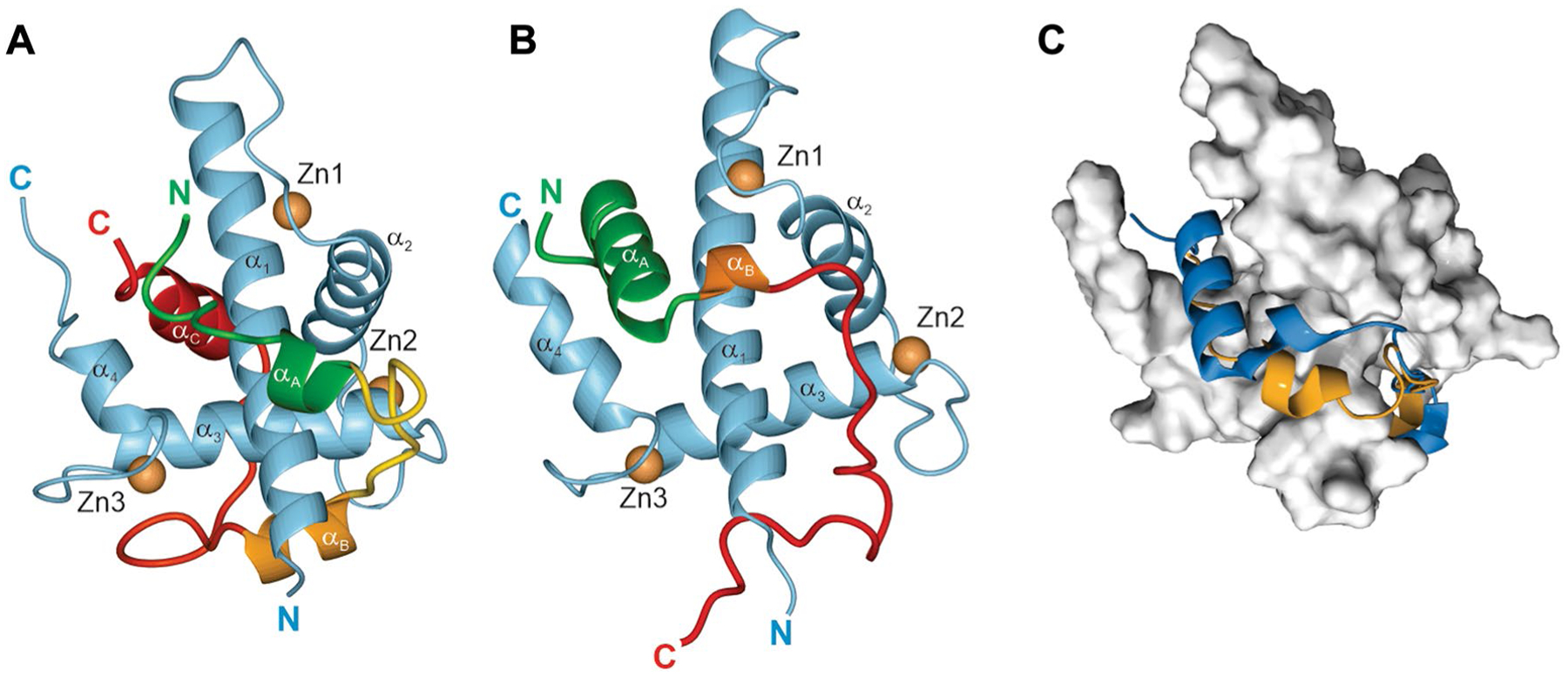
Single NMR structures of TAZ1 in complex with HIF-1α and CITED2. a TAZ1:HIF-1α complex (Dames et al. 2002). b TAZ1:CITED2 complex (De Guzman et al. 2004). The HIF-1α and CITED2 sequences are color-coded from N (green) to C (red). c Superposition of the two structures on the TAZ1 structure (Berlow et al. 2017). The gray surface shows TAZ1, with HIF-1α in gold and CITED2 in blue

1H–15N HSQC spectra of TAZ1 and complexes (Berlow et al. 2017). a Superimposed spectra of 15N-labeled TAZ1 alone (black), and in complex with HIF-1α (gold) and CITED2 (blue). Assignments are shown for selected cross peaks. The tryptophan indole resonance is shown as an inset in the bottom left corner. b Superimposed spectra of 15N-labeled TAZ1 in complex with HIF-1α (gold) and with CITED2 (blue). The red spectrum (with fewer contours displayed for visibility) was obtained from a solution containing equimolar amounts of 15N-labeled TAZ1, HIF-1α and CITED2. Adapted from reference (Berlow et al. 2017), with permission

Schematic mechanism for displacement of HIF-1α (gold) by CITED2 (blue) from TAZ1 (gray) (Berlow et al. 2017). ΔgC and ΔgH represent thermodynamic coupling between the αA and LPEL motifs of CITED2 and between the LPQL-αB and αC motifs of HIF-1α, respectively. Reproduced from reference (Berlow et al. 2017), with permission
References
Publication types
MeSH terms
Substances
Grants and funding
- NS14069/NS/NINDS NIH HHS/United States
- R01 DK034909/DK/NIDDK NIH HHS/United States
- P01 CA027489/CA/NCI NIH HHS/United States
- P01 GM071862/GM/NIGMS NIH HHS/United States
- R01 GM057374/GM/NIGMS NIH HHS/United States
- CA96865/CA/NCI NIH HHS/United States
- CA27489/CA/NCI NIH HHS/United States
- R01 CA214054/CA/NCI NIH HHS/United States
- GM075995/GM/NIGMS NIH HHS/United States
- R01 GM127807/GM/NIGMS NIH HHS/United States
- P01 GM056879/GM/NIGMS NIH HHS/United States
- CA229652/CA/NCI NIH HHS/United States
- AG21601/NIAging
- GM113251/GM/NIGMS NIH HHS/United States
- P01 AG002132/AG/NIA NIH HHS/United States
- DK34909/DK/NIDDK NIH HHS/United States
- R01 CA096865/CA/NCI NIH HHS/United States
- R35 GM131693/GM/NIGMS NIH HHS/United States
- R37 DK034909/DK/NIDDK NIH HHS/United States
- P01 AG021601/AG/NIA NIH HHS/United States
- R01 GM075995/GM/NIGMS NIH HHS/United States
- R01 GM113251/GM/NIGMS NIH HHS/United States
- GM56879/GM/NIGMS NIH HHS/United States
- R01 CA229652/CA/NCI NIH HHS/United States
- CA214054/CA/NCI NIH HHS/United States
- GM38794/GM/NIGMS NIH HHS/United States
- GM57374/GM/NIGMS NIH HHS/United States
- AI19499/National Institute of Allergy and Infectious Diseases
LinkOut - more resources
Full Text Sources
