. 2019 Mar;16(1):6-14.
doi: 10.14245/ns.1836322.161. Epub 2019 Mar 31.

Evolution of Spinal Endoscopic Surgery

Affiliations

Evolution of Spinal Endoscopic Surgery

Manyoung Kim et al. Neurospine. 2019 Mar.

Abstract

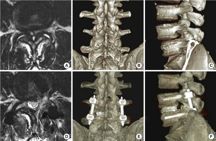
Innovations in the development of endoscopic spinal surgery were classified into different generations and reviewed. Future developments and directions for endoscopic spinal surgery were discussed. Surgical therapy for spinal disease has been gradually changing from traditional open surgery to minimally invasive spinal surgery. Recently, endoscopic spinal surgery, which initially was limited to the treatment of soft tissue lesions, has expanded to include other aspects of spinal disease and good clinical results have been reported. As the paradigm of spinal surgery shifts from open surgery to endoscopic surgery, we discussed the evolution of endoscopic spine surgery in our literature review. Through this description, we presented possibilities of future developments and directions in endoscopic spine surgery.

Keywords: Endoscopic spinal surgery; Interlaminar approach; Percutaneous spinal surgery; Transforaminal approach.


Conflict of interest statement

The authors have nothing to disclose.

Figures

Fig. 1.
Fig. 1.
Illustration of 3 anatomical barriers. Sympathetic trunk and ganglia (a), exiting nerve and ganglia (b), traversing nerve (c), and sinuvertebral nerves (d).
Fig. 2.
Fig. 2.
Illustrations of mobile inside-out technique of percutaneous endoscopic transforamial discectomy. (A) Preparation for approach with bone trimming. Levering the cannula against the ventral facet to direct the cannula trajectory to the ventral (B) or dorsal (C) disc cavity.
Fig. 3.
Fig. 3.
Illustrations of mobile outside-in technique of percutaneous endoscopic transforamial discectomy. (A) This panel shows the initial placement of the cannula in the transforaminal approach. (B) This panel shows the levering of the working cannula. (C) This panel shows the suprapedicular route. (D) This panel shows the intervertebral route. (E) This panel shows the foraminal route. (F) This panel shows the round cannula placement for the far lateral disc.
Fig. 4.
Fig. 4.
Illustrations of annular sealing during percutaneous endoscopic interlaminar discectomy. The change in an annular defect can be seen before (A), during (B), and after (C) annular sealing.
Fig. 5.
Fig. 5.
In the C-arm image, it shows the approach from ipsilateral to contralateral foraminal to extraforaminal.
Fig. 6.
Fig. 6.
(A) A schematic diagram of spinal stenosis with combined lesions. (B) A schematic diagram comparing the transforaminal and interlaminar approaches.
Fig. 7.
Fig. 7.
(A) Preoperative magnetic resonance imaging (MRI) showing the severe spinal stenosis combined with lumbar herniated nucleus pulposus. (B) Postoperative MRI taken 1 day after operation showing the sufficient decompression and adequate resection of lumbar herniated disk.
Fig. 8.
Fig. 8.
Intraoperative simple X-ray and endoscopic images obtained during endoscopic lumbar interbody fusion.
Fig. 9.
Fig. 9.
Preoperative magnetic resonance imaging (MRI) (A), 3-dimensional computed tomography (3D CT) posterior (B) and lateral (C) views showing the severe spinal stenosis combined with facet cyst and degenerative spondylolisthesis. The patient received endoscopic transforaminal lumbar interbody fusion. Postoperative MRI (D), 3D CT posterior (E) and lateral (F) views demonstrating the adequate decompression of stenosis and reduction of spondylolisthesis.

References

    1. Ishimoto Y, Yoshimura N, Muraki S, et al. Prevalence of symptomatic lumbar spinal stenosis and its association with physical performance in a population-based cohort in Japan: the Wakayama Spine Study. Osteoarthritis Cartilage. 2012;20:1103–8. - PubMed
    1. Ahn Y. Percutaneous endoscopic decompression for lumbar spinal stenosis. Expert Rev Med Devices. 2014;11:605–16. - PubMed
    1. Kim JH, Kim HS, Kapoor A, et al. Feasibility of full endoscopic spine surgery in patients over the age of 70 years with degenerative lumbar spine disease. Neurospine. 2018;15:131–7. - PMC - PubMed
    1. Gibson JN, Cowie JG, Iprenburg M. Transforaminal endoscopic spinal surgery: the future 'gold standard' for discectomy? - A review. Surgeon. 2012;10:290–6. - PubMed
    1. Lee DY, Shim CS, Ahn Y, et al. Comparison of percutaneous endoscopic lumbar discectomy and open lumbar microdiscectomy for recurrent disc herniation. J Korean Neurosurg Soc. 2009;46:515–21. - PMC - PubMed