. 2020 Jan;18(1):46-56.
doi: 10.1158/1541-7786.MCR-19-0359. Epub 2019 Oct 16.

A TFAP2C Gene Signature Is Predictive of Outcome in HER2-Positive Breast Cancer

Affiliations

A TFAP2C Gene Signature Is Predictive of Outcome in HER2-Positive Breast Cancer

Vincent T Wu et al. Mol Cancer Res. 2020 Jan.

Abstract

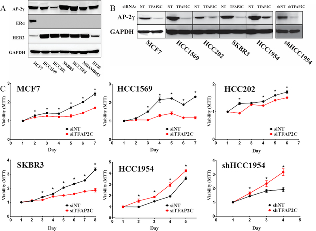
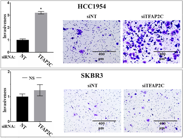
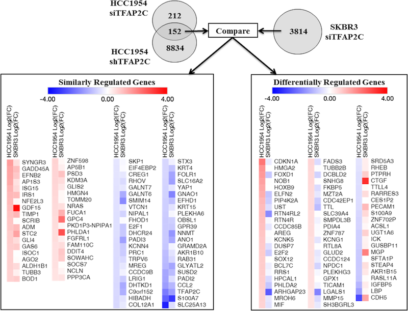
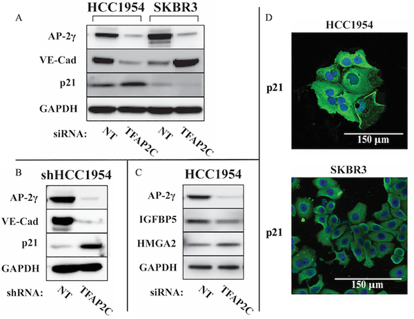
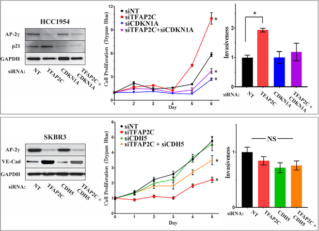
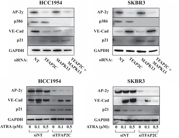
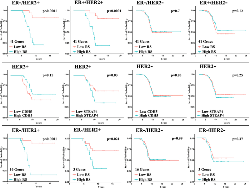
The AP-2γ transcription factor, encoded by the TFAP2C gene, regulates the expression of estrogen receptor-alpha (ERα) and other genes associated with hormone response in luminal breast cancer. Little is known about the role of AP-2γ in other breast cancer subtypes. A subset of HER2+ breast cancers with amplification of the TFAP2C gene locus becomes addicted to AP-2γ. Herein, we sought to define AP-2γ gene targets in HER2+ breast cancer and identify genes accounting for physiologic effects of growth and invasiveness regulated by AP-2γ. Comparing HER2+ cell lines that demonstrated differential response to growth and invasiveness with knockdown of TFAP2C, we identified a set of 68 differentially expressed target genes. CDH5 and CDKN1A were among the genes differentially regulated by AP-2γ and that contributed to growth and invasiveness. Pathway analysis implicated the MAPK13/p38δ and retinoic acid regulatory nodes, which were confirmed to display divergent responses in different HER2+ cancer lines. To confirm the clinical relevance of the genes identified, the AP-2γ gene signature was found to be highly predictive of outcome in patients with HER2+ breast cancer. We conclude that AP-2γ regulates a set of genes in HER2+ breast cancer that drive cancer growth and invasiveness. The AP-2γ gene signature predicts outcome of patients with HER2+ breast cancer and pathway analysis predicts that subsets of patients will respond to drugs that target the MAPK or retinoic acid pathways. IMPLICATIONS: A set of genes regulated by AP-2γ in HER2+ breast cancer that drive proliferation and invasion were identified and provided a gene signature that is predictive of outcome in HER2+ breast cancer.


Conflict of interest statement

Conflict of Interest: The authors state no conflicts of interest.

Figures

Figure 1.
Figure 1.. Proliferative Response with Knockdown of TFAP2C.
A. A panel of breast cancer cell lines was screened for expression of AP-2γ, ERα and HER2. B. Knockdown of TFAP2C with siRNA and stable knockdown of TFAP2C with shRNA in HCC1954 cells. C. Proliferative response to knockdown of TFAP2C using MTT assay in cell lines indicated.
Figure 2.
Figure 2.. Invasion Assay in HCC1954 and SKBR3 with TFAP2C Knockdown.
Graph showing relative cell invasiveness after knockdown of TFAP2C compared to non-targeting (NT) siRNA in HCC1954 (top) and SKBR3 (bottom). Panels to right show examples of cell invasion assay with knockdown of NT and TFAP2C.
Figure 3.
Figure 3.. Summary of RNA-seq Data with Knockdown of TFAP2C.
Significant changes in gene expression were first compared in HCC1954 with siRNA or shRNA stable knockdown. The 152 genes with significant and consistent changes were compared to knockdown of TFAP2C in SKBR3 cells with siRNA. Similarly regulated genes (on left) and differentially regulated genes (on right) are summarized with intensity of color indicating direction and magnitude of gene expression changes.
Figure 4.
Figure 4.. Expression of Proteins of Differentially Regulated AP-2γ-Responsive Genes.
A. Western blots for AP-2γ, VE-cadherin and p21 in HCC1954 and SKBR3 with knockdown of TFAP2C compared to NT. B. Western blots for protein expression in HCC1954 with stable knockdown comparing shRNA for NT vs. TFAP2C. C. Select Western blots of additional AP-2γ-responsive genes in HCC1954. D. Immunofluorescence staining for p21 in HCC1954 and SKBR3 demonstrates cytoplasmic localization.
Figure 5.
Figure 5.. Response of Proliferation and Invasiveness to CDKN1A and CDH5.
HCC1954 cells (top panels) with knockdown of TFAP2C, CDKN1A or both; western blots confirm knockdown of proteins (left panel); proliferation determined by counting viable cells (trypan blue) (middle panel); bar graph showing relative invasiveness in HCC1954 with knockdown of TFAP2C with or without knockdown of CDKN1A (right panel). Parallel experiments in SKBR3 cells (bottom panels) with knockdown of TFAP2C, CDH5 or both; western blots confirm knockdown (left panel); proliferation measured by viable cell counts (trypan blue) (middle panel); bar graph shows relative cell invasiveness (right panel).
Figure 6.
Figure 6.. Expression of VE-cadherin and p21 in Response to MAPK13 and ATRA.
Western blots for AP-2γ, p38δ, VE-cadherin, p21 and GAPDH showing response in HCC1954 and SKBR3 cells with knockdown of TFAP2C with or without co-knockdown of MAPK13 (top panels). Western blots showing changes of expression of AP-2γ, VE-cadherin, p21 and GAPDH in HCC1954 and SKBR3 with knockdown using NT vs. TFAP2C siRNA with increasing concentration of ATRA at 0, 0.1 and 0.5 μM (bottom panels).
Figure 7.
Figure 7.. Kaplan-Meier Plots for DMFS Based on Recurrence Score Model.
Top Row: Using the model with the 41-gene panel, Kaplan-Meier plots for distant metastasis-free survival (DMFS) for patients with each tumor subtypes as shown. For ER-/HER2+ n=37, ER+/HER2+ n=32, ER-/HER2- n=119, ER+/HER2- n=266. Middle Row: Examples of Kaplan-Meier plots for DMFS for individual genes, CDH5 and STEAP4, for HER2+ and HER2- tumors (with any ER status). Bottom Row: Kaplan-Meier plots for DMFS using the model with 16-gene and 3-gene panels for ER-/HER2+ and ER-/HER2- patients, as indicated.

References

    1. Weigel RJ, deConinck EC. Transcriptional control of estrogen receptor in estrogen receptor-negative breast carcinoma. Cancer research 1993;53(15):3472–4. - PubMed
    1. deConinck EC, McPherson LA, Weigel RJ. Transcriptional regulation of estrogen receptor in breast carcinomas. Mol Cell Biol 1995;15(4):2191–6. - PMC - PubMed
    1. McPherson LA, Baichwal VR, Weigel RJ. Identification of ERF-1 as a member of the AP2 transcription factor family. Proceedings of the National Academy of Sciences of the United States of America 1997;94(9):4342–7. - PMC - PubMed
    1. Woodfield GW, Horan AD, Chen Y, Weigel RJ. TFAP2C controls hormone response in breast cancer cells through multiple pathways of estrogen signaling. Cancer research 2007;67(18):8439–43. - PubMed
    1. Kang HJ, Lee MH, Kang HL, Kim SH, Ahn JR, Na H, et al. Differential Regulation of Estrogen Receptor alpha Expression in Breast Cancer Cells by Metastasis-Associated Protein 1. Cancer research 2014. doi 10.1158/0008-5472.CAN-13-2020. - DOI - PubMed

Publication types