. 2019 Oct 16;11(514):eaau6870.
doi: 10.1126/scitranslmed.aau6870.

A modulator of wild-type glucocerebrosidase improves pathogenic phenotypes in dopaminergic neuronal models of Parkinson's disease

Affiliations

A modulator of wild-type glucocerebrosidase improves pathogenic phenotypes in dopaminergic neuronal models of Parkinson's disease

Lena F Burbulla et al. Sci Transl Med. .

Abstract

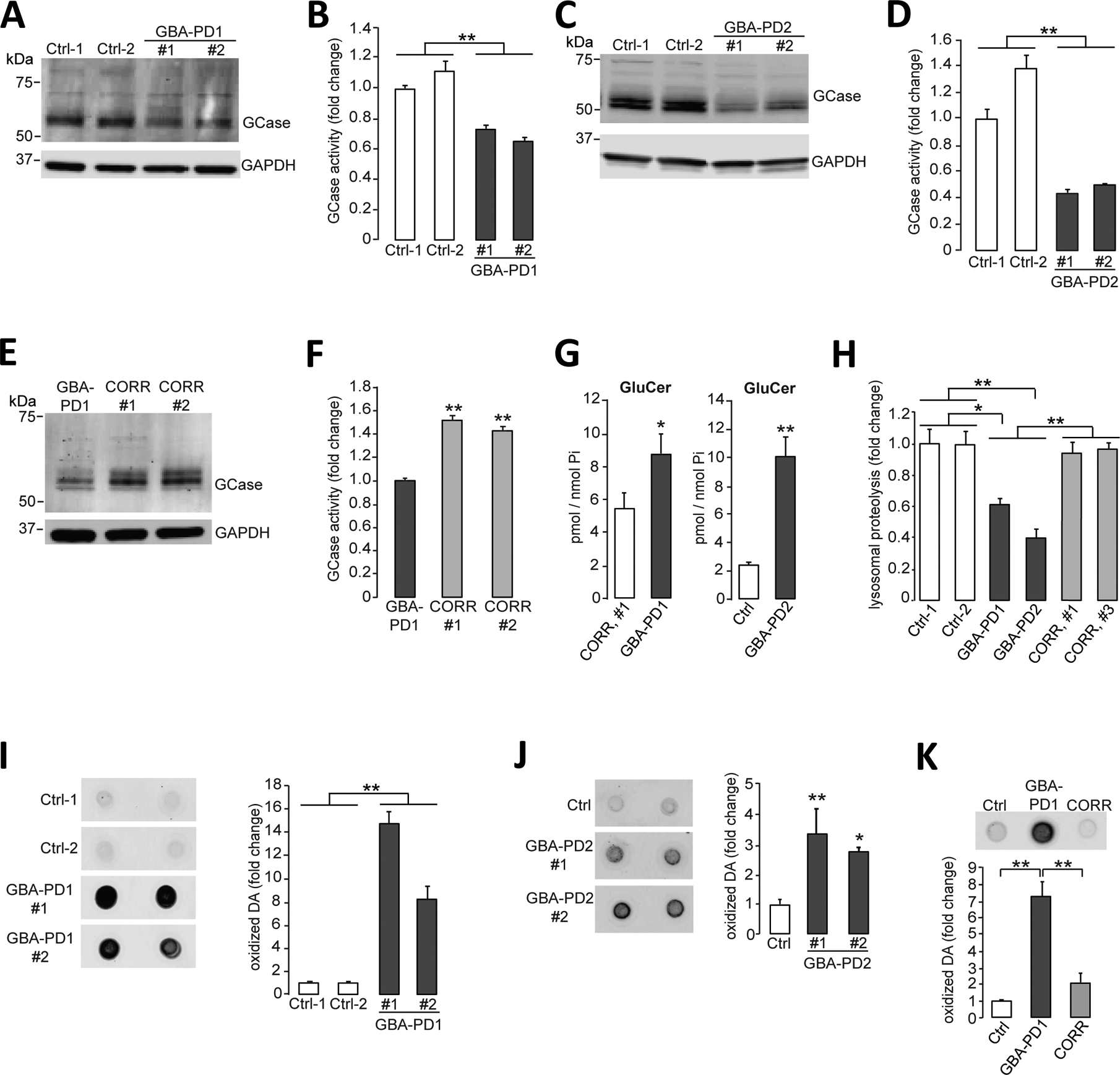
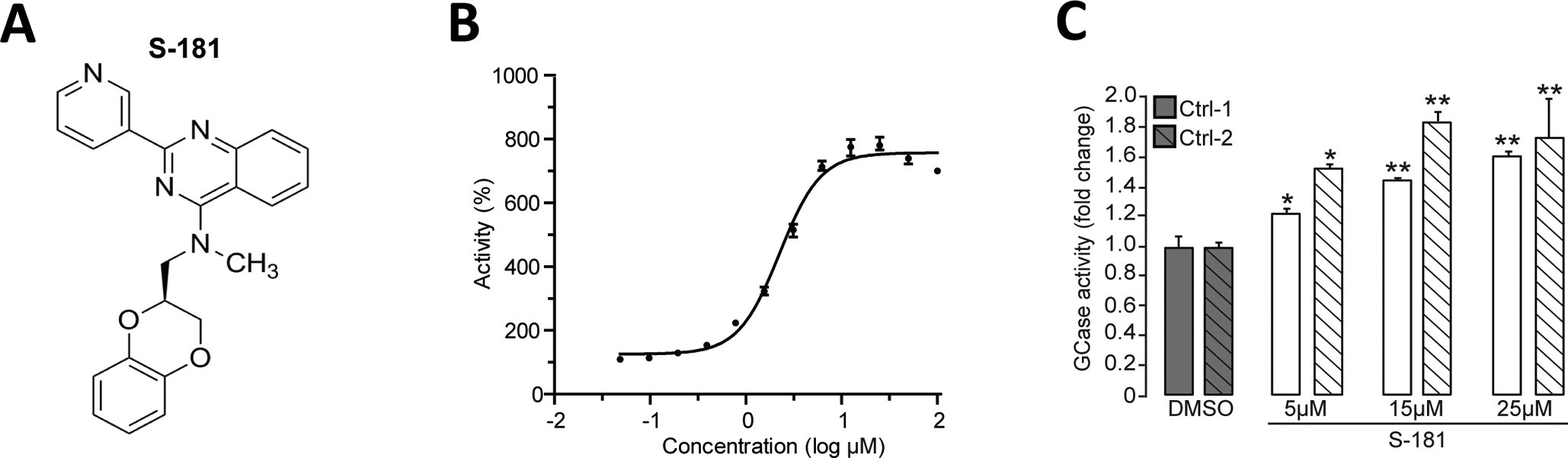
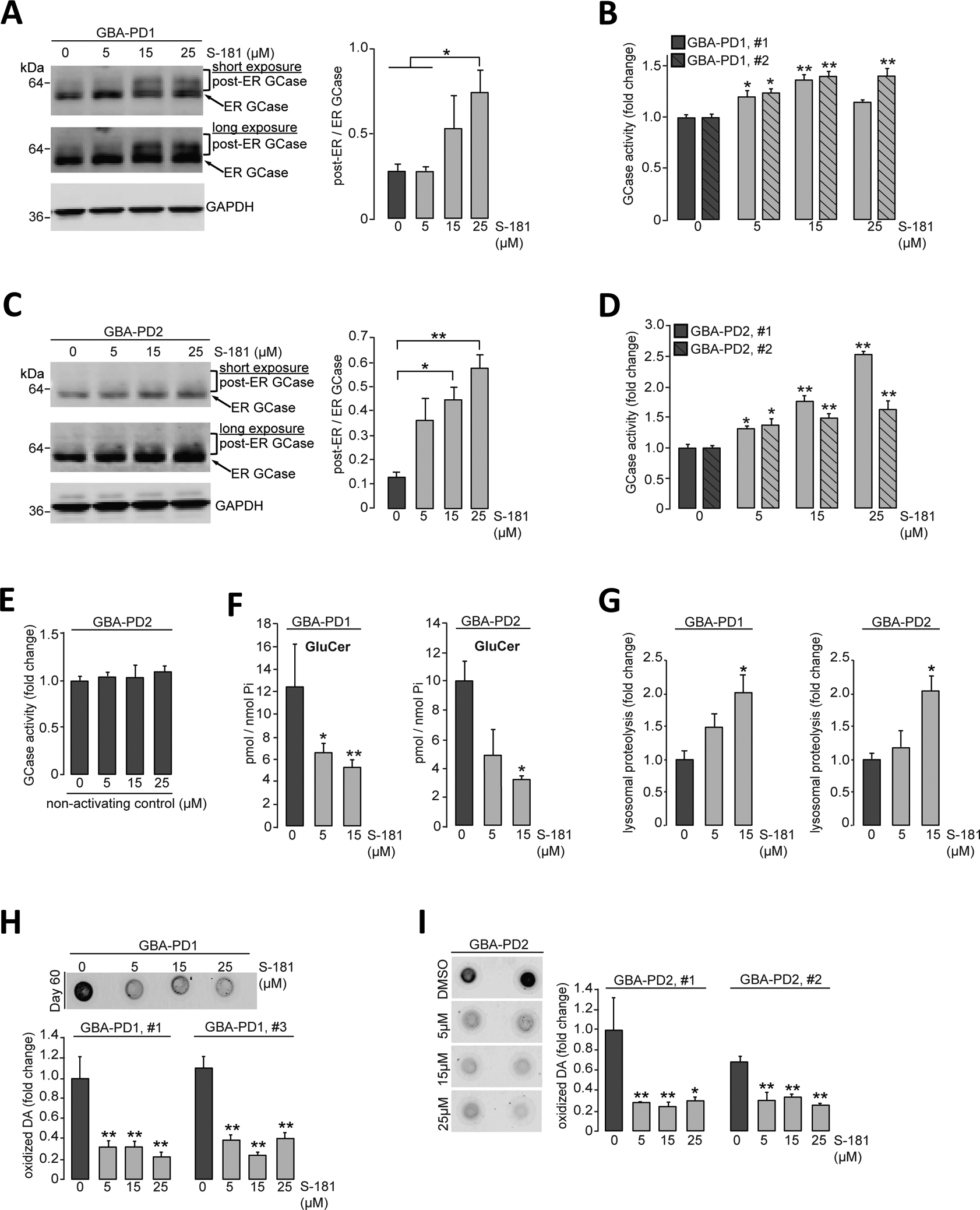
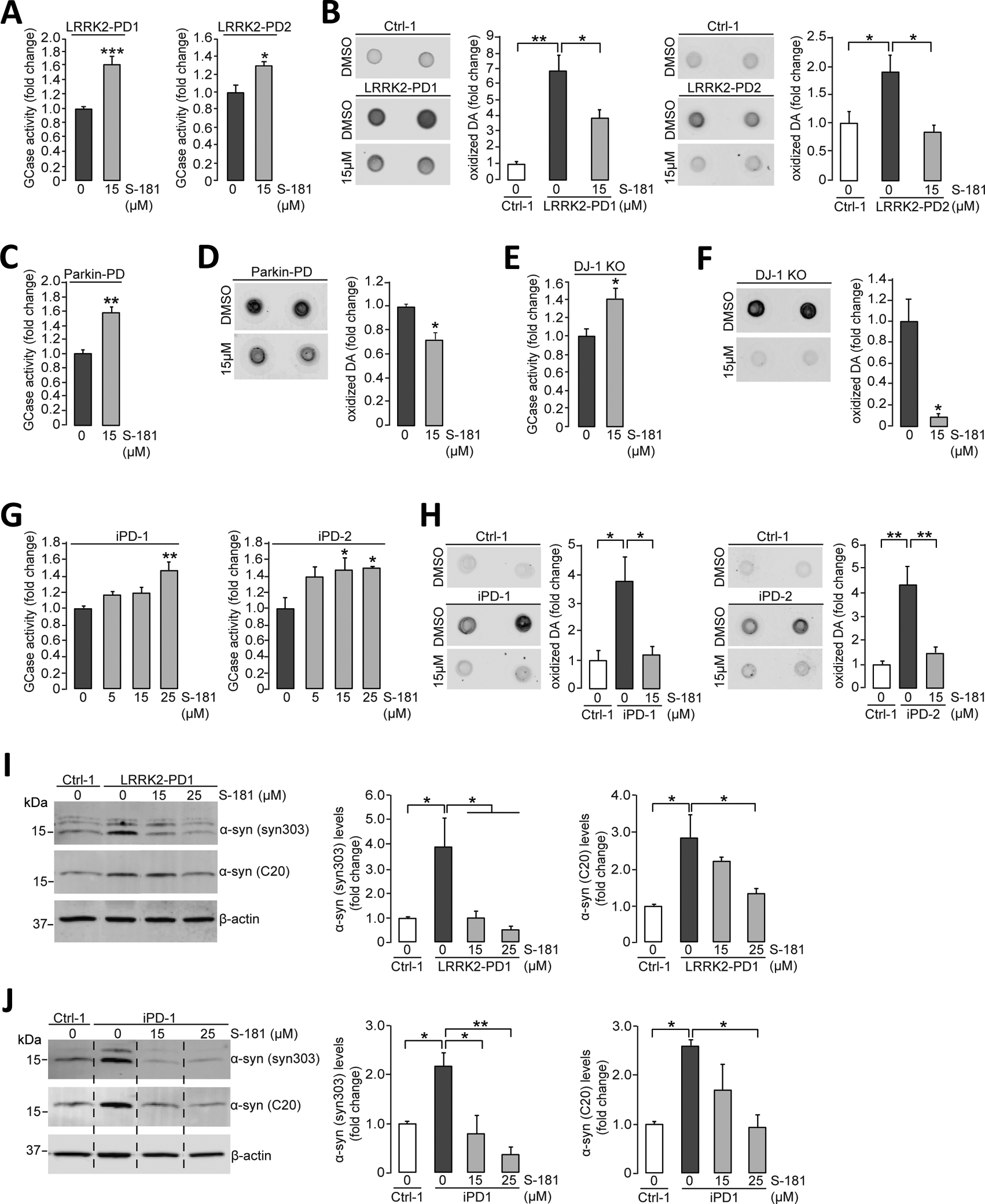
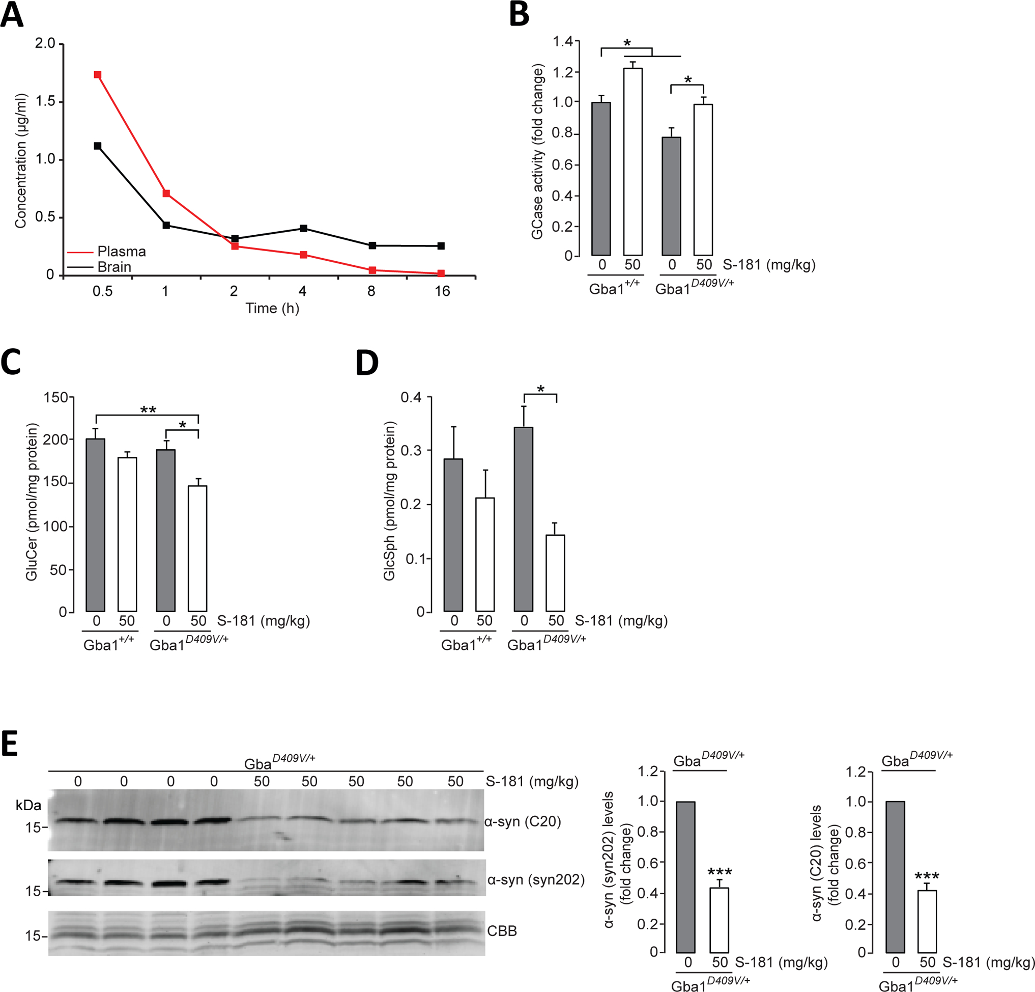
Mutations in the GBA1 gene encoding the lysosomal enzyme β-glucocerebrosidase (GCase) represent the most common risk factor for Parkinson's disease (PD). GCase has been identified as a potential therapeutic target for PD and current efforts are focused on chemical chaperones to translocate mutant GCase into lysosomes. However, for several GBA1-linked forms of PD and PD associated with mutations in LRRK2, DJ-1, and PARKIN, activating wild-type GCase represents an alternative approach. We developed a new small-molecule modulator of GCase called S-181 that increased wild-type GCase activity in iPSC-derived dopaminergic neurons from sporadic PD patients, as well as patients carrying the 84GG mutation in GBA1, or mutations in LRRK2, DJ-1, or PARKIN who had decreased GCase activity. S-181 treatment of these PD iPSC-derived dopaminergic neurons partially restored lysosomal function and lowered accumulation of oxidized dopamine, glucosylceramide and α-synuclein. Moreover, S-181 treatment of mice heterozygous for the D409V GBA1 mutation (Gba1D409V/+ ) resulted in activation of wild-type GCase and consequent reduction of GCase lipid substrates and α-synuclein in mouse brain tissue. Our findings point to activation of wild-type GCase by small-molecule modulators as a potential therapeutic approach for treating familial and sporadic forms of PD that exhibit decreased GCase activity.


Conflict of interest statement

Competing interests: D.K is Founder and Scientific Advisory Board Chair of Lysosomal Therapeutics Inc. D.K. serves on the scientific advisory boards of The Silverstein Foundation, Intellia Therapeutics, and Prevail Therapeutics and is a Venture Partner at OrbiMed. D.K., R.B.S. and J.Z. are coinventors on patent no. PCT/US2018/043703 entitled “Substituted fused pyrimidine compounds and uses thereof”. All other authors declare that they have no competing interests.

Figures

Figure 1.
Figure 1.. Lysosomal dysfunction and oxidized dopamine accumulation in PD patient iPSC-derived dopaminergic neurons.
(A and B) Triton-soluble lysates of iPSC-derived dopaminergic neurons obtained from fibroblasts from healthy control individuals or patients with PD carrying the heterozygous 84GG GBA1 mutation (GBA-PD1, clone #1 and clone #2) were prepared at day 80 of differentiation. Neuronal lysates were analyzed for (A) GCase protein amounts by immunoblotting and (B) GCase activity by in vitro enzyme activity assay (N=3 independent experiments). (C and D) Triton-soluble lysates of iPSC-derived dopaminergic neurons obtained from fibroblasts from healthy control individuals or patients with PD carrying the heterozygous 84GG GBA1 mutation (GBA-PD2, clone #1 and #2) were prepared at day 60 of differentiation. Neuronal lysates were analyzed for (C) GCase protein amounts by immunoblotting and (D) GCase activity by in vitro enzyme activity assay (N=3 independent experiments). (E and F) Triton-soluble lysates from GBA-PD1 iPSC-derived dopaminergic neurons and isogenic control iPSC-derived dopaminergic neurons with the GBA1 mutation corrected by CRISPR-Cas9 gene editing (CORR, clone #1 and #2) were prepared at day 60 of differentiation. Neuronal lysates were analyzed for (E) GCase protein amounts by immunoblotting (representative blot shown) and (F) GCase activity by in vitro enzyme activity assay (N=3 independent experiments). (G) Quantification of intracellular glucosylceramide (GluCer) by mass spectrometry normalized to internal phosphate (Pi) was performed in GBA-PD1 mutant iPSC-derived dopaminergic neurons and CRISPR-Cas9-corrected GBA1 isogenic control iPSC-derived dopaminergic neurons (CORR, clone #1) (left, N=3 to 4 independent experiments). Quantification of intracellular glucosylceramide (GluCer) by mass spectrometry normalized to internal phosphate (Pi) was performed in GBA-PD2 mutant and healthy control iPSC-derived dopaminergic neurons (Ctrl) (right, N=3 independent experiments) at day 70 of differentiation. (H) Quantification of lysosomal proteolysis of long-lived proteins was measured by radioactive pulse-chase at day 80 of differentiation in healthy control (Ctrl), in GBA-PD1 and GBA-PD2 iPSC-derived dopaminergic neurons, and in CRISPR-Cas9 gene-edited GBA1 isogenic control iPSC-derived dopaminergic neurons (CORR, clone #1 and #3) (N=4 independent experiments). (I to K) Detection and quantification of oxidized dopamine (DA) by near-infrared fluorescence assay was performed (I) in healthy control iPSC-derived dopaminergic neurons (Ctrl) and GBA-PD1 mutant iPSC-derived dopaminergic neurons (clone #1 and #2) at day 80 of differentiation (N=3 independent experiments), (J) in healthy control iPSC-derived dopaminergic neurons (Ctrl) and GBA-PD2 mutant iPSC-derived dopaminergic neurons (clone #1 and #2) at day 60 of differentiation (N=5 to 6 independent experiments), and (K) in healthy control (Ctrl) and GBA-PD1 mutant iPSC-derived dopaminergic neurons and CRISPR-Cas9 gene-edited GBA1 isogenic control iPSC-derived dopaminergic neurons (CORR) at day 50 to130 of differentiation (N=3 independent experiments). Error bars, mean ± SEM. *P<0.05 and **P<0.01, Student’s t test (G) or one-way ANOVA with Tukey post hoc test (B, D, F, and H to K).
Figure 2.
Figure 2.. Activation of wild-type GCase by the small molecule S-181.
(A) Structure of the GCase modulator S-181. (B) Shown is the cell-free in vitro enzyme activity of GCase with S-181 with a concentration of half-maximal activity (AC50) of 1.49μM. (C) Measurement of GCase activity in Triton-soluble lysates of iPSC-derived dopaminergic neurons in vitro (derived from two healthy control subjects) treated with either dimethyl sulfoxide (DMSO) (vehicle) or S-181 (5, 15, and 25μM) for 10 days (N=3–5 independent experiments). Error bars, mean ± SEM. *P<0.05 and **P<0.01, one-way ANOVA with Tukey post hoc test (C).
Figure 3.
Figure 3.. Activation of wild-type GCase with S-181 partially rescues pathogenic phenotypes in GBA-PD iPSC-derived dopaminergic neurons.
(A and B) Heterozygous 84GG GBA1 mutant iPSC-derived dopaminergic neurons (GBA-PD1 clone #1 and #2) were treated with either DMSO (vehicle) or S-181 (5, 15, and 25μM) for 10 days. Triton-soluble lysates were obtained at day 60 of differentiation and analyzed for (A) GCase protein amount by immunoblotting with quantification of post-ER/ER GCase (N=3 independent experiments) and (B) GCase activity assessed by in vitro enzyme activity assay (N=3 independent experiments). (C and D) Heterozygous 84GG GBA1 mutant iPSC-derived dopaminergic neurons (GBA-PD2, clone #1 and #2) were treated with either DMSO (vehicle) or S-181 (5, 15, and 25μM) for 10 days. Triton-soluble lysates were analyzed at day 60 of differentiation for (C) GCase protein amounts by immunoblotting with quantification of post-ER/ER GCase (N=3 independent experiments) and (D) GCase activity assessed by in vitro enzyme activity assay (N=3 to 6 independent experiments). (E) Heterozygous 84GG GBA1 mutant iPSC-derived dopaminergic neurons (GBA-PD2) were treated with either DMSO (vehicle) or a nonactivating control compound (5, 15, and 25μM) for 10 days. GCase activity was analyzed in Triton-soluble lysates at day 60 of differentiation (N=3 independent experiments). (F) Quantification of intracellular glucosylceramide (GluCer) by mass spectrometry normalized to internal phosphate (Pi) was performed at day 70 of differentiation in heterozygous 84GG GBA1 mutant iPSC-derived dopaminergic neurons [GBA-PD1 (left), N=3 to 4 independent experiments and GBA-PD2 (right), N=3 independent experiments] treated with either DMSO (vehicle) or S-181 (5 and 15μM) for 10 days. (G) Lysosomal proteolysis of long-lived proteins was quantified at day 80 of differentiation by radioactive pulse-chase of heterozygous 84GG GBA1 mutant iPSC-derived dopaminergic neurons [GBA-PD1 (left), N=3 independent experiments and GBA-PD2, N=3 to 5 independent experiments] treated with either DMSO (vehicle) or S-181 (5 and 15μM) for 10 days. (H and I) Detection and quantification of oxidized dopamine (DA) was performed by near-infrared fluorescence assay at day 60 of differentiation of heterozygous 84GG GBA1 mutant iPSC-derived dopaminergic neurons (H) GBA-PD1 clone #1 and #3 (N=3 to 4 independent experiments), (I) GBA-PD2 clone #1 and #2 (N=4 to 6 independent experiments) treated with either DMSO (vehicle) or S-181 (5, 15, and 25μM) for 10 days. Error bars, mean ± SEM. *P<0.05 and **P<0.01, one-way ANOVA with Tukey post hoc test.
Figure 4.
Figure 4.. Activation of wild-type GCase by S-181 lowers oxidized dopamine and α-synuclein in non-GBA1-linked PD patient iPSC-derived dopaminergic neurons.
(A) Mutant iPSC-derived dopaminergic neurons carrying the LRRK2 R1441C mutation (LRRK2-PD1) or G2019S mutation (LRRK2-PD2) were treated with either DMSO (vehicle) or S-181 (15μM) for 10 days. Cell lysates were then analyzed at day 100 of differentiation for GCase activity assessed by in vitro enzyme activity assay (N=6 to 9 independent experiments for LRRK2-PD1; N=3 independent experiments for LRRK2-PD2). (B) Mutant iPSC-derived dopaminergic neurons (LRRK2-PD1 and LRRK2-PD2) and healthy control iPSC-derived dopaminergic neurons were treated with either DMSO (vehicle) or S-181 (15μM) for 10 days. Cell lysates were analyzed at day 100 of differentiation for oxidized dopamine (DA) by near-infrared fluorescence assay (N=7 independent experiments for LRRK2-PD1; N= 4–5 independent experiments for LRRK2-PD2). (C and D) Mutant iPSC-derived dopaminergic neurons generated from fibroblasts of a patient with a compound heterozygous PARKIN mutation were treated with either DMSO (vehicle) or S-181 (15μM) for 10 days. Cell lysates were analyzed at day 150 of differentiation for (C) GCase activity assessed by in vitro enzyme activity assay (N=3 independent experiments) and (D) oxidized DA measured by near-infrared fluorescence assay (N=3 independent experiments). (E and F) Mutant iPSC-derived dopaminergic neurons in which DJ-1 was deleted by CRISPR-Cas9 gene editing (DJ-1 KO) were treated with either DMSO (vehicle) or S-181 (15μM) for 10 days. Cell lysates were analyzed at day 160 of differentiation for (E) GCase activity by in vitro enzyme activity assay (N=3 independent experiments) and (F) oxidized DA measured by near-infrared fluorescence assay (N=3 independent experiments). (G) iPSC-derived dopaminergic neurons generated from fibroblasts from two patients with idiopathic PD (iPD-1 and iPD-2) were treated with either DMSO (vehicle) or S-181 (5, 15, and 25μM) for 10 days. Cell lysates were analyzed at day 130 of differentiation for GCase activity (N=4 to 5 independent experiments for iPD1; N=3 to 5 independent experiments for iPD2). (H) iPSC-derived dopaminergic neurons generated from fibroblasts from two patients with idiopathic PD (iPD-1 and iPD-2) and one healthy control (Ctrl-1) were treated with either DMSO (vehicle) or S-181 (15μM) for 10 days. Cell lysates were analyzed at day 130 of differentiation for oxidized DA measured by near-infrared fluorescence assay (N=3 to 4 independent experiments for iPD1; N= 3 to 6 independent experiments for iPD2). (I) Mutant iPSC-derived dopaminergic neurons carrying the LRRK2 G2019S mutation (LRRK2-PD1) and healthy control iPSC-derived dopaminergic neurons (Ctrl-1) were treated with either DMSO (vehicle) or S-181 (15 and 25μM) for 10 days. Triton-soluble lysates were analyzed at day 100 of differentiation for the amount of α-synuclein by immunoblotting with C20 and syn303 anti-α-synuclein antibodies. β-Actin was used as a loading control (N=3 to 4 independent experiments). (J) iPSC-derived dopaminergic neurons generated from fibroblasts from a patient with idiopathic PD (iPD-1) and a healthy control (Ctrl-1) were treated with either DMSO (vehicle) or S-181 (15 and 25μM) for 10 days. Triton-soluble lysates were analyzed at day 100 of differentiation for the amount of α-synuclein by immunoblotting with C20 and syn303 anti-α-synuclein antibodies. β-Actin was used as a loading control (N=3 to 4 independent experiments). Error bars, mean ± SEM. *P<0.05, **P<0.01, and ***P<0.001, Student’s t test (A and C to F) or one-way ANOVA with Tukey post hoc test (B and G to J).
Figure 5.
Figure 5.. Activation of wild-type GCase by S-181 reduces lipid substrates and α-synuclein in mice.
(A) Mean concentration-time profiles for S-181 in plasma and brain tissue after administration of intraperitoneal single ​dose (50 mg/kg) to C57BL/6 wild-type mice (n=3 mice per time point). (B) GCase activity was measured by in vitro enzyme activity assay in lysates of hippocampal tissue from wild-type Gba1+/+ mice and from Gba1D409V/+ heterozygous mutant mice that had been treated with 5% dextrose (vehicle) (n=8 mice per group) or S-181 (50mg/kg; n=5 mice per group) intraperitoneally twice daily for 15 days. (C and D) Quantification of intracellular (C) glucosylceramide (GluCer) and (D) glucosylsphingosine (GluSph) by mass spectrometry in hippocampal tissue from wild-type Gba1+/+ and Gba1D409V/+ heterozygous mutant mice treated with 5% dextrose (vehicle) (n=8 mice per group) or S-181 (50mg/kg; n=5 to 6 mice per group) intraperitoneally twice daily for 15 days. (E) Immunoblot analysis of α-synuclein (using C20 and syn202 anti-α-synuclein antibodies) in Triton-insoluble lysates of hippocampal tissue from Gba1D409V/+ heterozygous mutant mice treated with 5% dextrose (vehicle) (n=8 mice per group) or S-181 (50mg/kg; n=5 mice per group) intraperitoneally twice daily for 15 days. Coomassie Brilliant Blue (CBB) was used as a loading control. Mice (mixed group of males and females) were between 13 and 15 months of age. Error bars, mean ± SEM. *P<0.05, **P<0.01, and ***P<0.001, Student’s t test (E) or one-way ANOVA with Tukey post hoc test (B to D).

References

    1. Poewe W, Seppi K, Tanner CM, Halliday GM, Brundin P, Volkmann J, Schrag AE, Lang AE, Parkinson disease. Nature reviews. Disease primers 3, 17013 (2017); published online EpubMar 23 (10.1038/nrdp.2017.13). - DOI - PubMed
    1. Surmeier DJ, Obeso JA, Halliday GM, Parkinson’s Disease Is Not Simply a Prion Disorder. The Journal of neuroscience : the official journal of the Society for Neuroscience 37, 9799–9807 (2017); published online EpubOct 11 (10.1523/JNEUROSCI.1787-16.2017). - DOI - PMC - PubMed
    1. Spillantini MG, Schmidt ML, Lee VM, Trojanowski JQ, Jakes R, Goedert M, Alpha-synuclein in Lewy bodies. Nature 388, 839–840 (1997); published online EpubAug 28 (10.1038/42166). - DOI - PubMed
    1. Sidransky E, Nalls MA, Aasly JO, Aharon-Peretz J, Annesi G, Barbosa ER, Bar-Shira A, Berg D, Bras J, Brice A, Chen CM, Clark LN, Condroyer C, De Marco EV, Durr A, Eblan MJ, Fahn S, Farrer MJ, Fung HC, Gan-Or Z, Gasser T, Gershoni-Baruch R, Giladi N, Griffith A, Gurevich T, Januario C, Kropp P, Lang AE, Lee-Chen GJ, Lesage S, Marder K, Mata IF, Mirelman A, Mitsui J, Mizuta I, Nicoletti G, Oliveira C, Ottman R, Orr-Urtreger A, Pereira LV, Quattrone A, Rogaeva E, Rolfs A, Rosenbaum H, Rozenberg R, Samii A, Samaddar T, Schulte C, Sharma M, Singleton A, Spitz M, Tan EK, Tayebi N, Toda T, Troiano AR, Tsuji S, Wittstock M, Wolfsberg TG, Wu YR, Zabetian CP, Zhao Y, Ziegler SG, Multicenter analysis of glucocerebrosidase mutations in Parkinson’s disease. N Engl J Med 361, 1651–1661 (2009); published online EpubOct 22 (10.1056/NEJMoa0901281). - DOI - PMC - PubMed
    1. Mazzulli JR, Xu YH, Sun Y, Knight AL, McLean PJ, Caldwell GA, Sidransky E, Grabowski GA, Krainc D, Gaucher disease glucocerebrosidase and alpha-synuclein form a bidirectional pathogenic loop in synucleinopathies. Cell 146, 37–52 (2011); published online EpubJul 8 (10.1016/j.cell.2011.06.001). - DOI - PMC - PubMed

Publication types

Substances