. 2019 Oct 16;9(1):14870.
doi: 10.1038/s41598-019-51006-1.

Structural and biochemical characterisation of a novel alginate lyase from Paenibacillus sp. str. FPU-7

Affiliations

Structural and biochemical characterisation of a novel alginate lyase from Paenibacillus sp. str. FPU-7

Takafumi Itoh et al. Sci Rep. .

Abstract

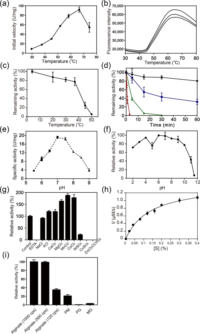
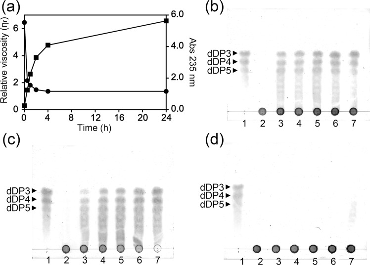
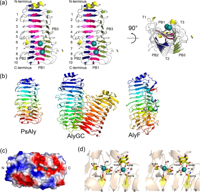
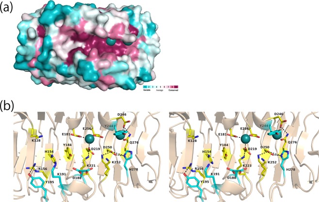
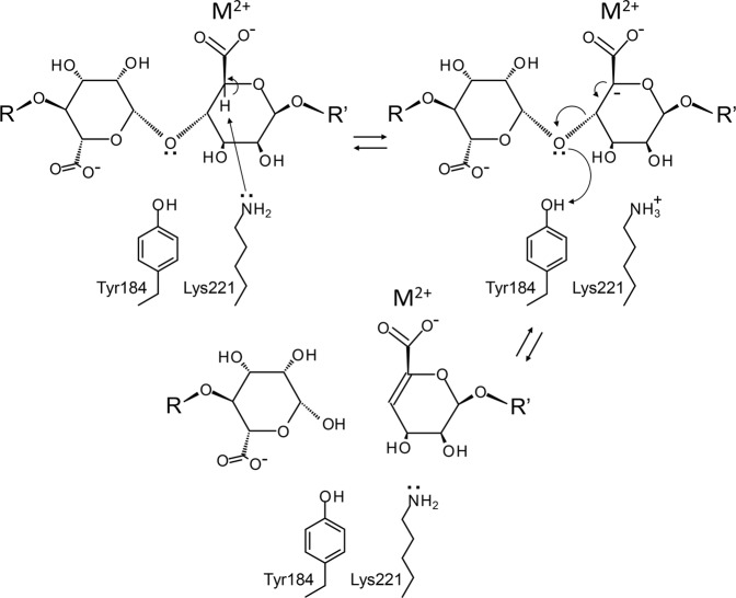
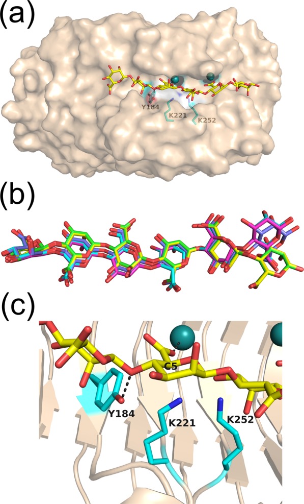
A novel alginate lyase, PsAly, with a molecular mass of 33 kDa and whose amino acid sequence shares no significant similarity to other known proteins, was biochemically and structurally characterised from Paenibacillus sp. str. FPU-7. The maximum PsAly activity was obtained at 65 °C, with an optimum pH of pH 7-7.5. The activity was enhanced by divalent cations, such as Mg2+, Mn2+, or Co2+, and inhibited by a metal chelator, ethylenediaminetetraacetic acid. The reaction products indicated that PsAly is an endolytic enzyme with a preference for polymannuronate. Herein, we report a detailed crystal structure of PsAly at a resolution of 0.89 Å, which possesses a β-helix fold that creates a long cleft. The catalytic site was different from that of other polysaccharide lyases. Site-directed mutational analysis of conserved residues predicted Tyr184 and Lys221 as catalytic residues, abstracting from the C5 proton and providing a proton to the glycoside bond, respectively. One cation was found to bind to the bottom of the cleft and neutralise the carboxy group of the substrate, decreasing the pKa of the C5 proton to promote catalysis. Our study provides an insight into the structural basis for the catalysis of alginate lyases and β-helix polysaccharide lyases.


Conflict of interest statement

The authors declare no competing interests.

Figures

Figure 1
Figure 1
(a) SDS-PAGE profile of recombinant PsAly expressed in E. coli following the purification scheme, (b) gel permeation chromatography analysis of PsAly, and (c) mass spectrum (negative-ESI MS) of the reaction products with PsAly and alginate. (a) Protein bands were stained with CBB R-250. Lane M, molecular mass standards; lane 1, cell extract (50 μg) of E. coli harbouring the expression plasmid; lane 2, aliquot (5 μg) of elution from Ni-IMAC (HisTrap HP); lane 3, aliquot (5 μg) of elution from AEC (HiTrapQ). (b) Arrows indicate the elution positions of molecular mass markers; from left to right: 2,000, 440, 134, 67, 12, 1.355, and 0.376 kDa. (c) The resulting peaks in the MS spectrum correspond to unsaturated alginate oligosaccharide (dDP3, dDP4, and dDP5) [M-H] or [M-2H]2− ions. The small peaks of the ions corresponding to dDP6 and dDP7 were also observed as [M-H] or [M-2H]2− ions in the spectrum.
Figure 2
Figure 2
Biochemical properties of PsAly. The vertical error bars on the data points represent the standard deviation of the mean. (a) The activity-temperature profile of PsAly was evaluated by the initial velocity (U/mg) at each temperature. (b) The thermal stability of PsAly was evaluated by differential scanning fluorimetry by measuring changes in the fluorescence of a dye (SYPRO Orange). The melting temperature (Tm) of PsAly was calculated as an inflection point of the melt curve (Tm = 52.9 ± 0.4 °C). Assays were performed in triplicate. (c) The thermal stability of PsAly was evaluated by the determination of the remaining activities (%) after incubation for 1 h at various temperatures. (d) The kinetics of the thermal inactivation of PsAly at 37 °C (black circle), 47 °C (blue square), 57 °C (green triangle), or 67 °C (red diamond) were evaluated by measuring the remaining activity (%) at appropriate intervals. (e) The pH-activity profile of PsAly was evaluated by the specific activity (U/mg). (f) The pH stability of PsAly was evaluated by the determination of the remaining activities (%) after incubation for 1 h at various pH levels. (g) The effect of the cations and metal chelation on enzyme activity was determined by adding the listed reagents. (h) The kinetic properties (kcat and Km) were determined by fitting to the Michaelis–Menten equation. (i) The substrate specificities were determined using 0.4% (w/v) sodium alginate (1,000, 500, or 120 cps), PM, PG, or MG as the substrate.
Figure 3
Figure 3
Enzymatic reaction mode of PsAly. (a) The enzyme reaction products were analysed by the viscosity (black circle) and absorbance at 235 nm (black box) of the solution with PsAly and alginate after incubation at 37 °C for 0, 0.5, 1, 2, 4, and 24 h. (bd) The reaction products with PsAly and alginate (b), PM (c), or PG (d) were also visualised by TLC. Lane 1, 20 μg of oligosaccharide mixtures (dDP3, dDP4 and dDP5); Lanes 2–7, the reaction products (20 μg) at 0, 0.5, 1, 2, 4, and 24 h, respectively.
Figure 4
Figure 4
Crystal structure of PsAly. (a) The overall structure of PsAly is represented by a ribbon model. The β-helix structure has 10 coils and is formed by three distorted β-sheets, named PB1 (pink), PB2 (blue), and PB3 (yellow). The β-strands in the helix are connected by three turns, T1 (between PB1 and PB2), T2 (between PB2 and PB3), and T3 (between PB3 and PB1). The two sodium ions are represented by deep teal balls and the bound imidazole is represented by a yellow stick model. (b) The structural comparisons between PsAly (left) and PL6 alginate lyases (AlyGC [centre] and AlyF [right]). The structures are represented by rainbow ribbon models. The metal ions are represented by grey balls (PsAly and AlyF: sodium ions and AlyGC: calcium ion). (c) The electrostatic potentials at pH 7 are also represented. The +55 to −55 kT/e potential isocontours are shown as blue to red surfaces, respectively. (d) Close-up view of bound sodium ions and surrounding residues. Two sodium ions (deep teal balls) were located on the cleft and chelated by Glu181, Glu216, Asp219, Tyr244, Asp246, and Gln276 residues and six water molecules (stick and ball models).
Figure 5
Figure 5
Active cleft of PsAly. (a) The amino acid conservation on the cleft of the PsAly. The surface of PsAly with bound sodium ions (deep teal balls) is coloured with regions of the greatest variability (cyan), modest (white) and highest conservation (magenta) using the ConSurf server. (b) The substrate-binding cleft is covered by the conserved residues (yellow stick models). Other ionisable residues in the cleft are also shown as cyan stick models. The blacked dashed lines represent hydrogen bond interactions (<3.5 Å).
Figure 6
Figure 6
Schematic representation of the proposed catalytic mechanism of PsAly. Firstly, Lys221 acts as the general base and abstracts a proton from the C5 atom to form the carbanion intermediate. Then, Tyr184 acts as the general acid and provides a proton to the scissile glycosidic oxygen O4 from the same side (syn configuration) to release saturated and unsaturated saccharides (E1cB-elimination reaction). Cation (M2+) neutralises the carboxyl group of the substrate and reduces the pKa of the C5 proton.
Figure 7
Figure 7
Docking simulation of PsAly with hexamannuronate (6M) using the SwissDock program. (a) One of the docking results with the lowest binding free energy (ΔG = −15.7 kcal/mol) is shown. The bound 6M model is shown as a yellow stick model. Two metal ions and important catalytic residues, Tyr184, Lys221, and Lys252, are also shown as ball and stick models (cyan). (b) The top five models of bound 6M ranked by the binding free energy (Table 3). The models are represented by stick models (yellow, cyan, pink, green, and violet). (c) Close-up view of (a). The hydrogen atom of C5 is close to Lys221 and the oxygen atom of glyosidic bond is close to Tyr184. Lys252 is close to Lys221.

References

    1. Lombard V, Golaconda Ramulu H, Drula E, Coutinho PM, Henrissat B. The carbohydrate-active enzymes database (CAZy) in 2013. Nucleic Acids Res. 2014;42:D490–495. doi: 10.1093/nar/gkt1178. - DOI - PMC - PubMed
    1. Dalboge H. Expression cloning of fungal enzyme genes; a novel approach for efficient isolation of enzyme genes of industrial relevance. FEMS Microbiol. Rev. 1997;21:29–42. doi: 10.1111/j.1574-6976.1997.tb00343.x. - DOI - PubMed
    1. Helbert W, et al. Discovery of novel carbohydrate-active enzymes through the rational exploration of the protein sequences space. Proc. Natl. Acad. Sci. USA. 2019;116:6063–6068. doi: 10.1073/pnas.1815791116. - DOI - PMC - PubMed
    1. Itoh T, et al. Cooperative degradation of chitin by extracellular and cell surface-expressed chitinases from Paenibacillus sp. strain FPU-7. Appl. Environ. Microbiol. 2013;79:7482–7490. doi: 10.1128/AEM.02483-13. - DOI - PMC - PubMed
    1. Grady EN, MacDonald J, Liu L, Richman A, Yuan ZC. Current knowledge and perspectives of Paenibacillus: a review. Microb. Cell. Fact. 2016;15:203. doi: 10.1186/s12934-016-0603-7. - DOI - PMC - PubMed

Publication types

MeSH terms