. 2019 Oct;574(7778):359-364.
doi: 10.1038/s41586-019-1647-8. Epub 2019 Oct 16.

Regulation of lifespan by neural excitation and REST

Affiliations

Regulation of lifespan by neural excitation and REST

Joseph M Zullo et al. Nature. 2019 Oct.

Abstract

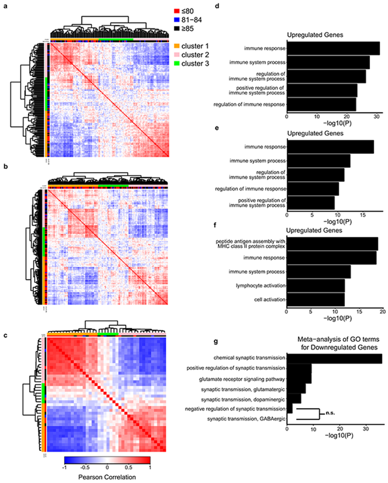
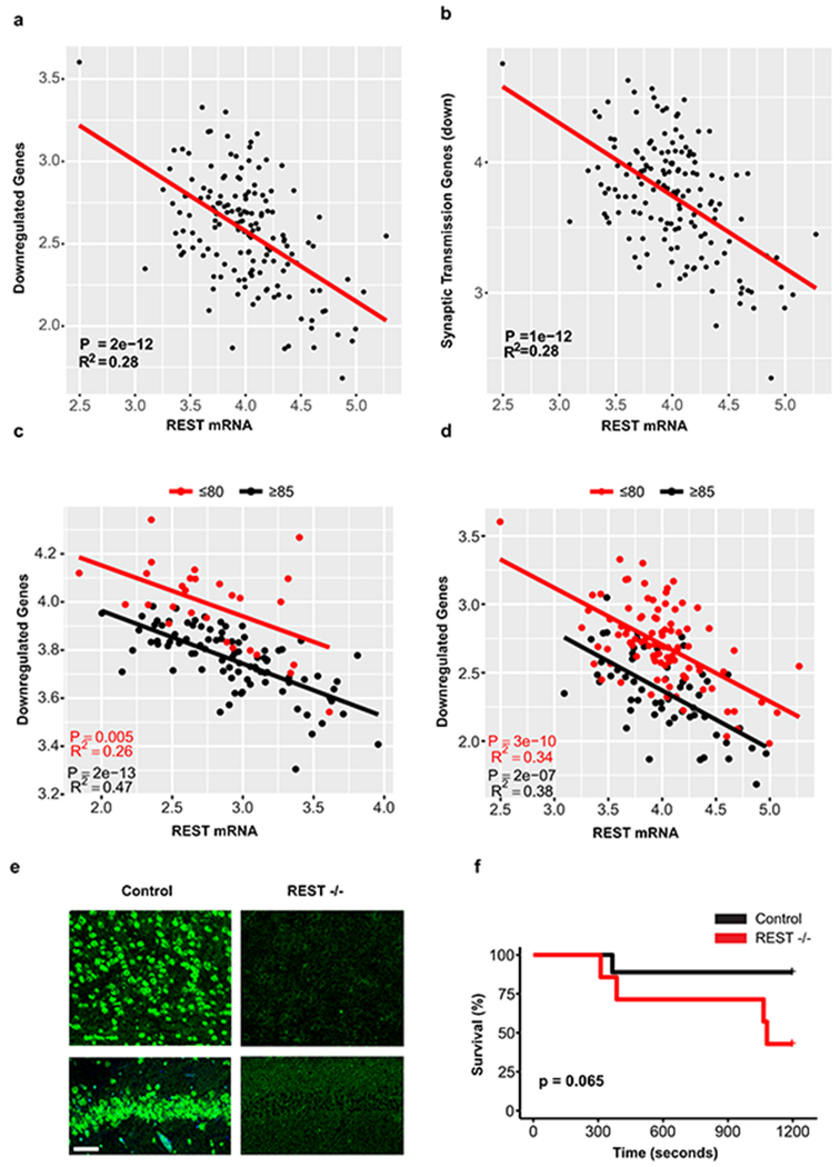
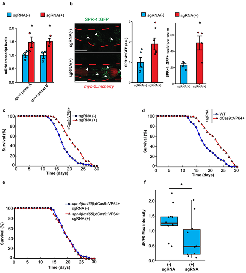
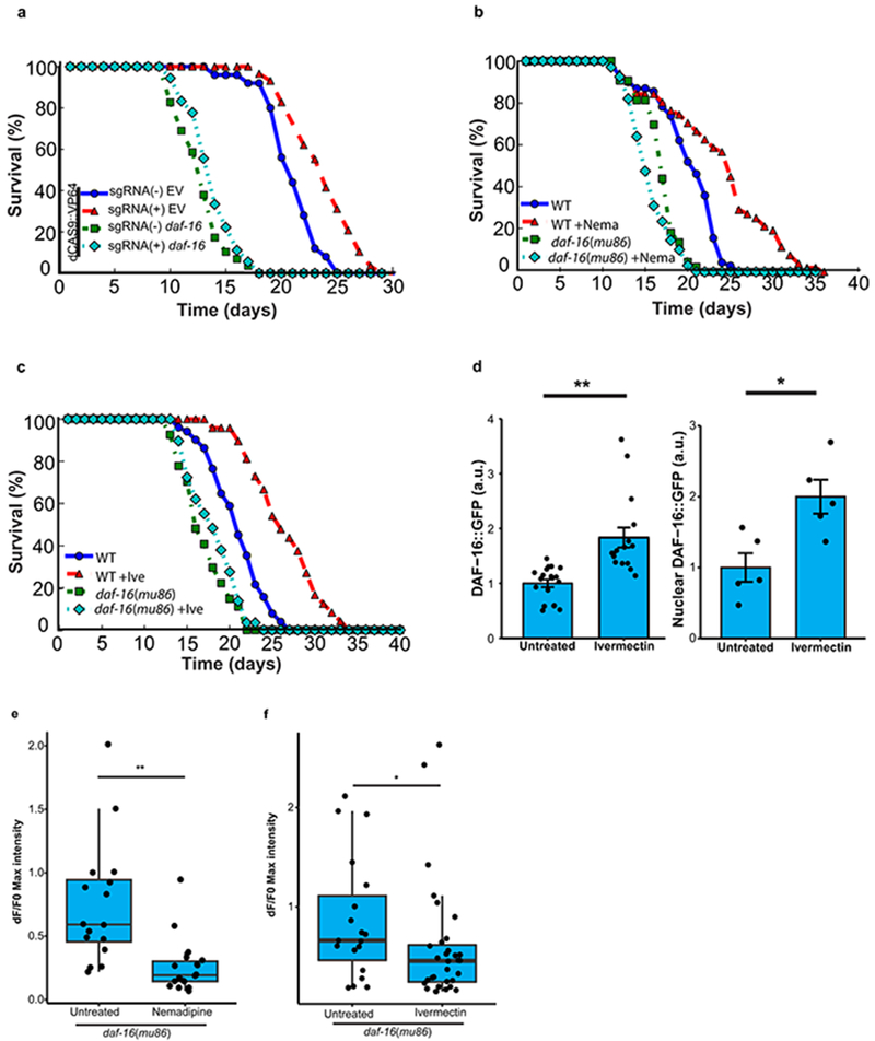
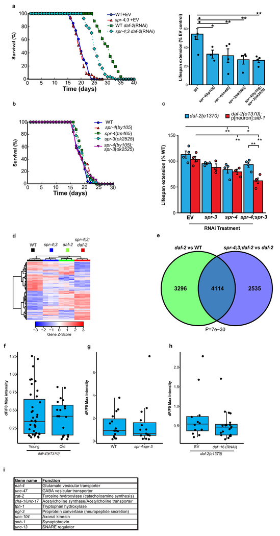
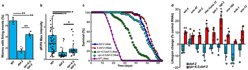
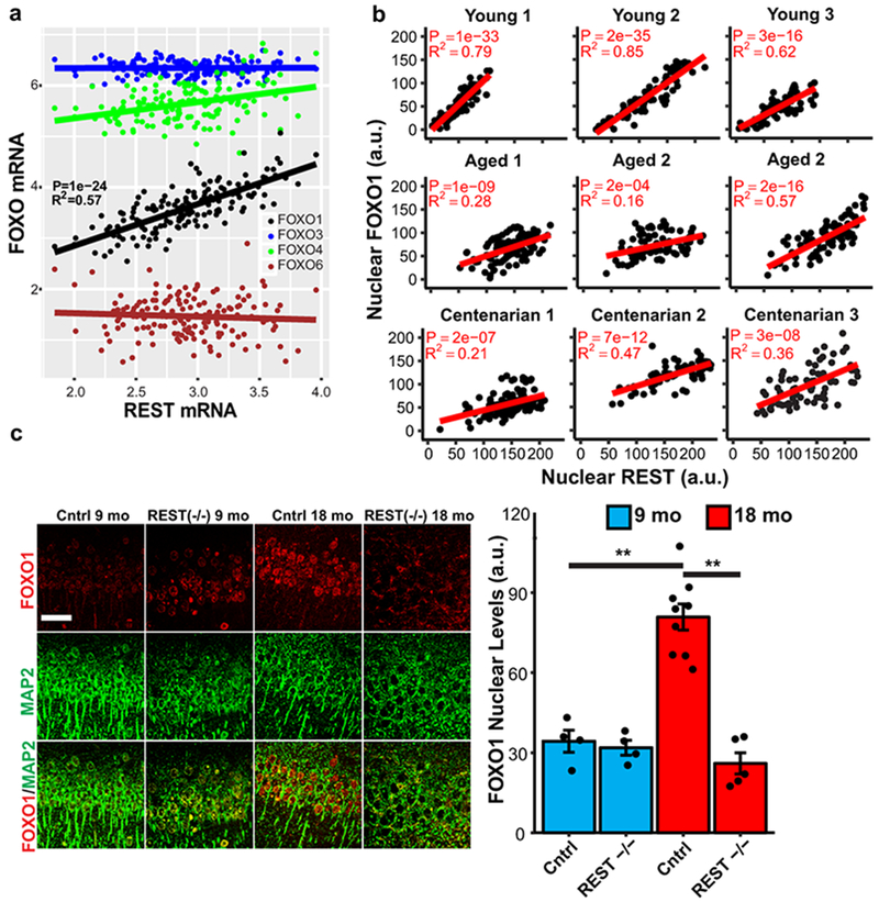
The mechanisms that extend lifespan in humans are poorly understood. Here we show that extended longevity in humans is associated with a distinct transcriptome signature in the cerebral cortex that is characterized by downregulation of genes related to neural excitation and synaptic function. In Caenorhabditis elegans, neural excitation increases with age and inhibition of excitation globally, or in glutamatergic or cholinergic neurons, increases longevity. Furthermore, longevity is dynamically regulated by the excitatory-inhibitory balance of neural circuits. The transcription factor REST is upregulated in humans with extended longevity and represses excitation-related genes. Notably, REST-deficient mice exhibit increased cortical activity and neuronal excitability during ageing. Similarly, loss-of-function mutations in the C. elegans REST orthologue genes spr-3 and spr-4 elevate neural excitation and reduce the lifespan of long-lived daf-2 mutants. In wild-type worms, overexpression of spr-4 suppresses excitation and extends lifespan. REST, SPR-3, SPR-4 and reduced excitation activate the longevity-associated transcription factors FOXO1 and DAF-16 in mammals and worms, respectively. These findings reveal a conserved mechanism of ageing that is mediated by neural circuit activity and regulated by REST.


Figures

Extended Data Figure 1.
Extended Data Figure 1.. Partitioning of the aging human population for analysis of gene expression in the brain.
a-c, Adjusted gene expression profiles for age-associated genes were compared between cognitively normal aged individuals to derive a matrix of Pearson correlation coefficients that indicate the degree of similarity between any two cases in the ROSMAP (a, dorsolateral prefrontal cortex, n=150 individuals) CommonMind Consortium (b, dorsolateral prefrontal cortex, n=174 individuals) and Gibbs (c, frontal cortex, n=40 individuals) cohorts. d-f, Most significantly enriched GO terms for upregulated genes in the cortex of cognitively normal individuals who lived to be ≥85 years old relative to individuals who lived to be ≤80 years old in the ROSMAP (d, n=117 individuals), CommonMind Consortium (e, n=155 individuals), and Gibbs (f, n=37 individuals) cohorts. P-values were calculated using Fisher’s exact test (see Methods). g, Meta-analysis of GO term enrichment for downregulated genes. Shown are selected GO terms related to excitatory and inhibitory synaptic transmission. The individual cohort enrichment p-values were combined using Stouffer’s method (see Methods). NS, FDR>0.1.
Extended Data Figure 2.
Extended Data Figure 2.. Ivermectin and nemadipine extend lifespan without interfering with worm motility.
a, Worms were transferred at day 8 to either standard NGM plates or plates containing ivermectin (1 pg/ml) or nemadipine (2 μM). Shown is a representative curve of an experiment repeated twice. Nemadipine versus WT: P=3.2 e-4; Ivermectin versus WT: P=2.2e-7 by log-rank test. Nemadipine, n=81; Ivermectin, n=82; WT, n=76. b. Day 2 worms treated with nemadipine or ivermectin for 24 hours were transferred to liquid culture and thrashing rate was assessed using the Nemametrix wMicrotracker (see Methods). Shown are mean motility scores for the first 60 minutes, ± S.E.M.. Untreated, n=17 wells; Ivermectin, n=17 wells; Nemadipine, n=16 wells. Each well contained ~10 worms. **P=1.7e-4 vs untreated, Mann–Whitney U test with multiple testing correction by Holm’s method. Results are representative of an experiment replicated twice.
Extended Data Figure 3.
Extended Data Figure 3.. Repression of multiple neurotransmitter systems extends lifespan in C. elegans.
a-h. C. elegans lines expressing the transgenic HisCl1 channel in the indicated neuronal populations were treated with 10 mM histamine (His+) starting at adult day 1 (a, c, e, g) or day 8 (b, d, f, h) and compared to untreated controls (His−). P-values are by log-rank. See Supplementary Table 22 for individual n values and statistics. i, Shown is the mean lifespan extension ± S.E.M. for worms treated with histamine at days 1 or 8 relative to untreated controls for at least three independent replicates, *P<0.05, **P<0.01 by Student’s t-test. HisCl1was driven using the GAL4SK:VP64 system for the GABAergic (GABA), glutamatergic (GLUT) and cholinergic systems, using unc-47, eat-4, and unc-17 drivers, respectively (see Supplementary Table 19 for details). j, Reduced ASH neuron excitation following inhibition of GABA activity at day 1 but not day 8. Shown is normalized maximum GCaMP fluorescence in day 1 and 8 unc-47:HisCl1 worms that were treated with 10 mM histamine [His (+)] on the indicated day, or untreated controls [His (−)]. Day 1 His(−): n=18 worms; Day 1 His(+), n=19 worms; Day 8 His(−): n=23 worms; Day 8 His(+): n=20 worms. *P=1.1e-3 by the Mann–Whitney U test.
Extended Data Figure 4.
Extended Data Figure 4.. Neural excitation, neuropeptide signaling and lifespan in C. elegans.
a, Increased excitation of ASH neurons following RNAi against the GABA vesicular transporter unc-47. GCaMP imaging was performed on worms with enhanced neuronal RNAi (See Figure 3 legend and Methods for details) for unc-47 (n=37) or controls (n=43) at day 2. **P= 6.8e-3 by the Mann–Whitney U test. b, RNAi for unc-47 reduces lifespan. Worms with enhanced neuronal RNAi were treated with unc-47 (n=31) or control RNAi (n=84). Shown is a representative lifespan analysis replicated 3 times. P= 1.3e-6 by log-rank test. c, Reduction of synaptic neurotransmission or neuropeptide signaling extend lifespan in C. elegans. Mutations in genes affecting glutamatergic neurotransmission (eat-4), presynaptic function (unc-13) and neuropeptide signaling (egl-3) exhibit comparable lifespan extension. WT, n=57; eat-4(nj2), n=54. P≤2.2e-16; unc-13(e51), n=92, P=3.6e-14; egl-3(gk238), n=35, P=8.3e-11 by log-rank test. Shown are curves representative of two independent replicates. d, Extension of lifespan by egl-3 RNAi in worms with enhanced neuronal RNAi. Shown are lifespan curves representative of two independent replicates. egl-3 RNAi (n=47 worms); Empty Vector (n=84 worms). P=3.5e-11 by the log-rank test.
Extended Data Figure 5.
Extended Data Figure 5.. Gene regulation and neural activity associated with REST and extended longevity.
a-b, Expression of genes downregulated in individuals ≥85 years versus ≤80 years old is inversely related to REST mRNA levels. Shown is linear regression analysis of normalized and adjusted REST mRNA levels and mean expression of a, all downregulated genes and b, downregulated genes associated with the synaptic transmission GO term. Data is from the CommonMind cohort. Each point represents an individual case, n=155 individuals. P-values were derived by a t-test for the slope of the regression line. Note similarity to the data for the ROSMAP cohort in Fig. 2a, b. c-d, Stratification by age group. Analysis of the ROSMAP cohort (c, n=117 individuals) and the CommonMind cohort (d, n=155 individuals) as in Fig 2a, but stratified by age group. P-values were derived by t-test for the slope of the regression line. e, Loss of REST expression in conditional REST knockout mice. Representative images of the cortex (top panel) and hippocampus (bottom panel) from RESTlx/lx (Control), and Nestin-Cre;RESTlx/lx (REST−/−) mice. Immunolabeling was performed with the anti-mouse REST-14 antibody directed against the REST C-terminal domain. Scale bar, 40 μm. Image shown is representative of an experiment replicated 4 times. f, Survival of REST −/− and control mice following administration of the seizure-inducing agent pentylentetrazole (PTZ, 40 mg/kg). P=0.065 for REST−/− versus control by the log-rank test. Control, n=9; REST−/−, n=7.
Extended Data Figure 6.
Extended Data Figure 6.. Induction of spr-4 extends lifespan and suppresses neural excitation in C. elegans.
a, spr-4 mRNA levels in worms expressing a stably integrated dCas9::VP64 transgene in the presence, sgRNA(+), or absence sgRNA(−), of 4 different sgRNAs targeting the spr-4 promoter. Transcript levels were determined by qRT-PCR and normalized to sgRNA(−) controls. Values are the mean ± S.E.M., n=3. A: *P= 0.041; B: P=0.020 by one-sided Student’s t-test. b, dCas9::VP64-mediated elevation of SPR-4 protein levels. Left panel: Representative images of the head region of heterozygous F1 progeny of the strains bearing a pspr-4::spr-4::gfp::spr-4utr transgene. Arrowheads indicate SPR-4::GFP positive nuclei. Dashed red lines indicate the outline of the worm body. Scale bar, 40 μm. Middle panel: Values represent the mean ± S.E.M. sgRNA(−), n=5 worms; sgRNA(+), n=5 worms with 7-38 measurements per worm; *P=0.022, one-sided Student’s t-test. Right panel: Values represent the mean ± S.E.M. sgRNA(−), n=4 worms; sgRNA(+), n=4 worms. P=0.011, one-sided Student’s t-test. Shown is a representative experiment replicated three times. c, Extended lifespan in worms expressing an integrated dCas9::VP64 transgene and sgRNAs targeting the spr-4 promoter [sgRNA(+)] (n=79 worms) relative to dCas9::VP64-expressing worms in the absence of sgRNAs [sgRNA (−)] (n=57 worms) P=5.5e-9, log-rank test. Representative of an experiment replicated 6 times. d, Lifespans of worms expressing sgRNA targeting the spr-4 promoter in the presence (n=87 worms) or absence (n=58 worms) of dCas9::VP64. P=3.7e-7, log-rank test. Representative of an experiment replicated twice. e, Lifespans of dCas9::VP64 expressing worms in the presence (n=51 worms) or absence (n=58 worms) of sgRNAs on the spr-4(tm465) loss-of-function mutant background. P=0.49, log-rank test. Representative of three independent replicates. f, Overexpression of spr-4 reduces neural excitation. GCaMP imaging was performed in ASH neurons in SPR-4 overexpressing (sgRNA+) and control (sgRNA-) worms in the lines described in c. Shown are maximum GCaMP fluorescence changes. sgRNA minus, n=12 worms; sgRNA plus, n=10 worms. *P=0.025, Mann–Whitney U test.
Extended Data Figure 7.
Extended Data Figure 7.. Lifespan extension by spr-4 overexpression and inhibition of neural excitation are daf-16-dependent.
a, Lifespan extension by overexpression of spr-4 is daf-16 dependent. Lifespans of worms overexpressing spr-4 (sgRNA+;dCAS9::VP64) or not overexpressing spr-4 (sgRNA-;dCAS9::VP64) following treatment with daf-16 RNAi or an empty vector control. sgRNA(+)EV (n=29 worms) versus sgRNA(−)EV (n=25 worms): P=2.7e-4; sgRNA(+)daf-16 (n=18 worms) versus sgRNA(−)daf-16 (n=29 worms) P=0.20 by log-rank test. Representative of 4 independent replicates. b, c, Lifespan extension by the neural excitation inhibitors ivermectin and nemadipine is daf-16-dependent. Shown are lifespan determinations for WT control and daf-16(mu86) mutant worms in the presence or absence of nemadipine (2μM) (b) or ivermectin (1pg/ml) (c). b,WT, n=69 worms; WT+Nema, n=51; daf-16, n=43; daf-16+Nema, n=67. WT+Nema vs WT, P=9.9e-8; daf-16+Nema vs daf-16, P=0.014 by log-rank test c, WT, n=78 worms; WT+Ive, n=77; daf-16, n=27; daf-16+Ive, n=29. WT+Ive vs WT, P=7.3e-8; daf-16+Ive vs daf-16, P=0.22; log-rank test. Curves are representative of an experiment replicated 2 (nemadipine) or 3 (ivermectin) times. d, Inhibition of neural excitation with ivermectin elevates DAF-16 levels. Worms expressing a DAF-16::GFP transgene were treated for 10 days with 1 pg/mL ivermectin and assessed by confocal microscopy. Left panel: Shown is total DAF-16::GFP. Values represent the mean ± S.E.M (untreated, n=19 worms, Ivermectin, n=16 worms). **P=2.5e-7, Mann-Whitney U test. Right panel: Shown is nuclear DAF-16::GFP (n=5 worms per group, 50-61 nuclei per worm). *P= 0.013 by Student’s t-test. Results are representative of an experiment replicated twice. e, DAF-16 is not required for inhibition of neural excitation by nemadipine. Shown are maximum ASH GCaMP intensity changes for day 2 daf-16(mu86) mutant worms treated for 24 hours with 2 μM nemadipine (untreated, n=16 worms; nemadipine, n=18 worms). P=9.4e-5, Mann-Whitney U test). f, DAF-16 is not required for inhibition of neural excitation by ivermectin. Shown are day 2 worms treated for 24 hours with 1 pg/mL ivermectin (control, n=19 worms; ivermectin, n=32 worms). P=0.030, Mann-Whitney U test).
Extended Data Figure 8.
Extended Data Figure 8.. SPR-3 and SPR-4 contribute to lifespan extension and gene regulation associated with reduced DAF-2 insulin/IGF-like signaling.
a, Loss of function of SPR-3 and SPR-4 reduces the lifespan extension of daf-2 RNAi. Left panel: Representative lifespan analysis of spr-4(by105);spr-3(ok2525) double mutant and wild-type (WT) worms following daf-2 or empty vector (EV) control RNAi. WT+EV, n=54 worms; spr-4;3+EV, n=58 worms; WT+daf-2, n=26 worms; spr-4;3+daf-2, n=54 worms. Right panel: Values represent mean percent lifespan extension ± S.E.M. (daf-2 RNAi versus EV control) in the indicated genotypes. WT, n=6 independent experiments; spr-4(by105), n=3, *P=0.017 versus WT; spr-4(tm465), n=4, **P=0.0062 versus WT; spr-3(ok2525), n=4, **P=0.0018 versus WT; spr-4(by105);spr-3(ok2525), n=4, **P=0.0016 versus WT by Students t-test. See Supplementary Table 22 for individual lifespan data and statistics. b, Lifespan is unaffected by spr-4 and spr-3 mutations in a wild-type background. WT, n=50 worms; spr-3(ok2525), n=31; spr-4(by105);spr-3(ok2525), n=32; spr-4(by105), n=34; spr-4(tm465), n=33.There were no reproducibly significant changes by the log-rank test in 3-6 independent experiments per genotype (see Supplementary Table 22). c, Quantification of lifespan extension in daf-2 mutant worms shown in Fig. 3b attributable to neuronal expression of spr-3 and spr-4. RNAi was targeted to neurons by neuronal expression of a sid-1 transgene in otherwise sid-1 null daf-2(1370) mutants (daf-2;p[neuron]:sid-1), and compared with untargeted RNAi in sid-1 wild-type daf-2(1370) mutants (daf-2). Values represent mean lifespan extension relative to the control sid-1(pk3321);p[neuron]:sid-1 worms treated with empty vector (EV) ± S.E.M (n= 3 independent experiments). Significant lifespan effects were not observed for RNAi in the absence of the daf-2 mutation. *P<0.05; **P<0.01 by Student’s t-test. d, Gene expression determined by RNA sequencing in day 2 adult worms. Differentially expressed genes (rows) and the indicated worm genotypes (columns) were clustered, and gene expression, transformed to a z-score per gene, is represented in a heat map. N=3 independent replicates per genotype. e, Venn diagram illustrating the overlap of differentially expressed genes in daf-2 single mutant vs WT and spr-4;3;daf-2 triple mutant vs daf-2 single mutant comparisons. P=7e-30, Fisher’s exact test with a one-sided alternative hypothesis. . f, Long-lived daf-2 mutants do not show an age-related increase in neural excitation. Shown is maximum ASH GCaMP intensity changes in day1-2 (n=39) and day 14-16 (n=20) daf-2(e1370) mutant worms. Note the absence of the age-related increase in excitation observed in wild-type aging worms (Fig. 1e). P=0.93, Mann-Whitney U test. g, The spr-4;spr-3 double mutant in a wild-type background does not significantly affect neural excitation in ASH neurons. WT, n=15 worms; spr-4;spr-3, n=15 worms. P= 0.62, Mann-Whitney U test. h, DAF-16 does not mediate suppression of neural excitation in the daf-2 mutant. RNAi for daf-16 was performed in daf-2(e1370) mutant worms on a sid-1(pk3321);p[neuron]:sid-1 background to augment RNAi in neurons (daf-16 RNAi, n=20 worms, empty vector (EV) control, n=12 worms). P=0.33, Mann–Whitney U test. i, Description of the genes targeted by RNAi in Figure 4d.
Extended Data Figure 9.
Extended Data Figure 9.. Regulation of DAF-16 by SPR-3 and SPR-4.
a, Reduced DAF-16 activation in spr-4;3 mutants following daf-2 RNAi. Left confocal panel: Shown are day 10 worms of the indicated genotypes expressing an integrated DAF-16::GFP transgene and treated with daf-2 RNAi or empty vector (EV) control since day 1 of adulthood. Images are maximum intensity z-projections. Scale bar, 40μm. Left bar graph: Values represent mean GFP intensity ± S.E.M. in the peri-pharyngeal regions of spr-4;3 double mutants relative to wild-type controls for a representative experiment replicated 4 times (see methods for details of analysis). (n=8-12 worms per replicate). **P=5.2e-5 by Welch’s t-test. Right confocal panel: Higher magnification views of DAF-16::GFP and DAPI-labeled nuclei. Images are magnified confocal z-planes. Scale bar, 10μm. Right bar graph: Values represent mean nuclear GFP intensity ± S.E.M. relative to the WT-EV control, n=5 worms per genotype and 51-89 nuclei per worm. *P=0.016, **P=5.5e-3 by ANOVA with post-hoc Tukey test. Values and images are representative of an experiment replicated 3 times. b, Gene expression determined by RNA sequencing in adult day 10 worms. Differentially expressed genes (rows) and replicates of the indicated worm genotypes (columns) were clustered, and gene expression, transformed to a z-score per gene, is represented in a heat map. n=3 independent replicates per genotype. c, Venn diagram illustrating the overlap of differentially expressed genes in day 10 daf-2 vs WT and spr-4;3;daf-2 vs daf-2 comparisons. P=4e-123, Fisher’s exact test with a one-sided alternative hypothesis. d, Overlap of class I daf-16 target genes (described in Methods) with genes downregulated in day 10 spr-4;3;daf-2 triple mutants relative to daf-2 single mutants. P-values were calculated using a hypergeometric distribution (see Methods). n.s, p=0.99 e, Ivermectin increases DAF-16::GFP levels in spr-4;3 worms following daf-2 RNAi. Left panel: Confocal imaging of GFP fluorescence in ivermectin-treated (10 pg/ml) and untreated worms. The red dashed lines indicate the worm body. Right panel: Quantification of DAF-16::GFP. Values represent mean GFP intensity ± S.E.M., WT/Untreated, n=12; WT/Ivermectin, n=10; spr-4;3/Untreated, n=10; spr-4;3/Ivermectin, n=10. **P=4.6e-4 (spr-4;3 vs WT untreated), P=2.6e-4 (spr-4;3 +Ivermectin vs spr-4;3 untreated) by Mann–Whitney U test with multiple testing correction by Holm’s method. Shown is a representative experiment replicated 3 times.
Extended Data Figure 10.
Extended Data Figure 10.. Coregulation of FOXO1 and REST in the aging brain and modulation by glutamatergic signaling.
a, Linear regression analysis of REST and FOXO mRNA levels in the prefrontal cortex of 174 cognitively-intact individuals (age≥60 years) from the CommonMind Consortium determined by RNA sequencing. P-values are derived from a t-test for the slope of the regression line and Bonferroni-corrected across all expressed genes. b, Colocalization of REST and FOXO1 in neurons of the aged human prefrontal cortex. Confocal immunofluorescence microscopy was performed in human prefrontal cortex using antibodies against REST (green, rabbit polyclonal; Bethyl), FOXO1 (red, goat polyclonal; LS-Bio) and the neuronal marker MAP2 (grey, chicken polyclonal; Abcam). Scale bar, 40 μm. The image shown is representative of immunofluorescence labeling performed in 30 individuals. See Supplementary Table 20 for additional information on antibodies. c, Inhibition of glutamatergic signaling in mouse cortical neuronal cultures elevates FOXO1 levels. Left panel: Primary mouse cortical neuronal cultures treated with kynurenic acid (KYNA, 5 μM), APV (50 μM), NBQX (2 μM) or vehicle (Control) were analyzed by confocal immunofluorescence for FOXO1 or MAP2 and labeled with DAPI. Boxed areas were magnified in the lower three rows. Note that most FOXO1 in cultured neurons is cytoplasmic, but a detectable nuclear component overlaps with DAPI. Scale bar, 40 μm. Right panel: Quantification of total and nuclear FOXO1 levels in MAP2-positive neurons. Values represent the mean ± S.E.M. Control, n=200; KYNA, n=326; APV, n=148; NBQX, n=197. FOXO1 Total/KYNA: **P=2.1e-8; FOXO1 Nuclear/KYNA **P=1.1e-4; FOXO1 Total/NBQX **P=8.8e-13; FOXO1 nuclear/NBQX **P=5.2e-6 by the Mann–Whitney U test with multiple testing correction by Holm’s method. Shown is a representative experiment replicated 3 times.
Figure 1.
Figure 1.
Neural excitation and longevity in humans and C. elegans. a, Analysis of the cortical transcriptome profile in cognitively intact aged individuals from the ROSMAP cohort. Unsupervised hierarchical clustering shows a transcriptional signature of down- and up-regulated genes associated with extended longevity. b-d, Most significantly enriched gene ontology (GO) terms for downregulated genes associated with extended longevity (≥85 versus ≤80 years of age) in the ROSMAP (dorsolateral prefrontal cortex, n=117) (b), CommonMind Consortium (dorsolateral prefrontal cortex, n=155) (c), and Gibbs (frontal cortex, n=37) (d) cohorts. P-values were calculated by Fisher’s exact test (see Methods). e, Aging C. elegans exhibit increased neuronal excitation. Shown are the maximum GCaMP fluorescence intensity changes in ASH neurons of young adult (day 1-2) and older (day 12-16) worms. Young, n=82 worms; Old, n=30 worms. *P=3.6e-4 by Mann–Whitney U test. f, The L-type calcium channel blocker nemadipine (2μM) represses neural excitation. Control, n=14; Nemadipine, n=13. *P=0.029, Mann–Whitney U test. g, Nemadipine extends lifespan. Worms were continuously treated with 2 μM nemadipine beginning at adult day 1, P=7.7e-11, log-rank test. Control, n=59; Nemadipine, n=50, replicated 3 times. h, The chloride channel agonist ivermectin (1pg/ml) reduces neural excitation. Control, n=18; Ivermectin, n=23. *P=0.038, Mann–Whitney U test. i, Extension of lifespan by continuous treatment with ivermectin beginning at adult day 1 (Control, n=35; 0.01 pg/ml: n=34, P= 0.62 ; 0.1 pg/ml: n=33, P= 1.5e-3; 1pg/ml: n=42, P= 1.9e-3, log-rank test), replicated 3 times. Summary statistics for all individual lifespan experiments are in Supplementary Table 22.
Figure 2.
Figure 2.
REST regulates neural excitation in the aging brain and is associated with extended longevity. a-b, Expression of genes downregulated in individuals with extended longevity (≥85 versus ≤80 years old) is inversely related to REST mRNA levels. Shown is linear regression analysis of the mean expression of a, all downregulated genes and b, downregulated genes associated with the synaptic transmission GO term. Data is from the ROSMAP cohort. Each point represents an individual case (n=117). P-values were derived by t-tests of the regression line slopes. c, Increased nuclear REST levels in the prefrontal cortex of centenarians. Left panel: Immunofluorescence labeling for REST (green, rabbit polyclonal; Bethyl laboratories) and DAPI (blue) in human prefrontal cortex. Scale bar, 40 μm. Right panel: Nuclear REST levels in cognitively intact individuals 70-80 years (n=9) and >100 years (n=7) of age. Values represent the mean ± S.E.M, **P=1.5e-4, Student’s t-test. d, REST represses neural excitation in the mouse cerebral cortex. Shown are images from PET-CT scanning of fluorodeoxyglucose (18F-FDG) uptake in 18-month-old Nestin-Cre;RESTlx/lx (REST−/−) and age-matched RESTlx/lx (Control) mice. e, Average standardized uptake value (SUV) at increasing time intervals after injection of 18F-FDG. Values represent the mean ± S.E.M., n=7 mice per group. *P<0.05, **P<0.01, Mann-Whitney U test. f, Increased epileptiform discharges in aged REST-deficient mice. Upper Panel: EEG recording from REST(−/−) and age-matched control mice. Lower Panel: Number of mice with at least one epileptiform discharge (≥ 3 secs) in a 48 hour recording. Control, n=9; REST−/−, n=7. *P=0.035, Fisher’s exact test. g, Seizure duration after administration of PTZ (40 mg/kg). Control, n=6; REST −/−, n=6 mice. *P=0.016, Mann–Whitney U test.
Figure 3.
Figure 3.
C. elegans REST orthologs mediate longevity in daf-2 loss-of-function mutants. a, The REST orthologs spr-4 and spr-3 are required for maximal longevity in daf-2 mutant worms. Lifespan analysis was performed on wild-type and daf-2(1370) loss-of-function mutant worms, and the indicated combinations of daf-2 and spr-4/spr-3 mutations. The spr-4/spr-3 mutations significantly reduced the lifespan of daf-2 mutant worms. n=29-59 worms per genotype, replicated at least three times per genotype. P<0.001 for all curves relative to daf-2, by log-rank test. b, Neuronal expression of spr-3 and spr-4 mediate lifespan extension in daf-2 mutant worms. Shown are lifespans of worms with neuronal targeting of RNAi by neuronal expression of a sid-1 transgene in otherwise sid-1 null daf-2(1370) mutants, or untargeted RNAi in sid-1 wild-type daf-2(1370) mutants. Lifespan effect of neuronal targeting of spr-4;3 RNAi versus EV control RNAi is significant by log rank test (P=2.5e-6), n=22-56 worms per curve replicated at least 4 times. c, SPR-3 and SPR-4 repress genes that mediate neural excitation. Shown are significantly enriched GO terms for upregulated genes related to neural excitation in RNA-seq analysis of day 2 spr-4;3;daf-2 triple mutants versus daf-2 single mutant worms. P-values were calculated using Fisher’s exact test (see Methods), n=3 biological replicates per genotype.
Figure 4.
Figure 4.
SPR-3 and SPR-4 suppress multiple neurotransmitter and neuropeptide systems to extend lifespan in daf-2 mutant worms. a, Neural excitation is suppressed in daf-2 mutants and partially restored by spr-4;3 mutations. GCaMP imaging was performed in ASH neurons. Shown is the fraction of worms with at least 1 firing event in a 2 minute recording. Values represent the mean ± S.E.M., n= 4-5 independent experiments. **P=7.9e-07 (daf-2 vs WT), P=1.5e-4 (spr-4;3;daf-2 vs daf-2); P= 0.0011(spr-4;3;daf-2 vs WT) by ANOVA with post-hoc Tukey test. b, Quantification of GCaMP fluorescence changes in day 2 worms: WT, n=53; daf-2, n=25; spr-4;3;daf-2, n=26. **P=3.1e-6 (daf-2 vs WT), P=1.5e-3 (spr-4;3;daf-2 vs daf-2); *P=0.018 (spr-4;3;daf-2 vs WT) , Mann–Whitney U test with multiple testing correction by Holm’s method. c, Inhibition of neural excitation by ivermectin (+Ive, 10 pg/ml) reverses lifespan reduction by spr-4;3 mutations in daf-2 mutant worms (P=1.1e-16 spr-4;3;daf-2 +Ive versus -Ive) daf-2-Ive, n=53 worms; daf-2+Ive, n=55; spr-4;3;daf-2-Ive, n=95; spr-4;3;daf-2+Ive, n=69; WT-Ive, n=64. d, Multiple neurotransmitter and neuropeptide signaling systems contribute to the effects of spr-4;3 mutants on longevity. Change in lifespan of spr-4;3;daf-2 triple mutant and daf-2 single mutant worms following neuronal RNAi for the indicated genes relative to empty vector control RNA. RNAi was targeted to neurons as described in Fig. 3b. *P<0.05, **P<0.01, Student’s t-test. n=3 independent experiments per group. Individual statistics are in Supplementary Table 22.
Figure 5.
Figure 5.
REST regulates FOXO1 expression in the mammalian brain. a, Linear regression analysis of REST and FOXO1 mRNA expression in the prefrontal cortex of cognitively intact individuals (ROSMAP cohort age 71-101 years, n=150) determined by RNA sequencing. P-values were derived by linear regression t-tests for the slope with Bonferroni correction for all expressed genes. b, Coordinate regulation of REST and FOXO1 in human prefrontal cortex. Nuclear REST and FOXO1 protein levels were determined by immunofluorescence microscopy in pyramidal neurons of the prefrontal cortex in individual young adult (20-38 yrs), aged (70-80 yrs), and centenarian (>100 yrs) cases. Each point represents a neuron double-labeled for REST and FOXO1. n=71-114 neurons per individual, P-values were derived as in a. c, FOXO1 induction in the aging mouse cortex is REST-dependent. Left panel: Immunocytochemical labeling for FOXO1 and the neuronal marker MAP2 in cortical neurons of RESTlx/lx (Control) and Nestin-Cre;RESTlx/lx (REST−/−) mice at 9 and 18 months of age. Scale bar is 40 μm. Right panel: Quantitation of FOXO1 nuclear levels. Values represent the mean ± S.E.M. Control 9 mo: n = 4 mice; Control 18 mo: n = 9 mice; REST−/− 9 mo: n = 4 mice; REST−/− 18 mo: n = 5 mice. **P=1.2e-5 cntrl 9 mo vs cntrl 18mo; **P=4.4e-7 cntrl 18 mo vs REST−/− 18 mo by ANOVA with post-hoc Tukey test.

Comment in

References

    1. Satoh A, Imai S-i, Guarente L. The brain, sirtuins, and ageing. Nat Rev Neurosci. 2017;18(6):362–74. - PubMed
    1. Bishop NA, Lu T, Yankner BA. Neural mechanisms of ageing and cognitive decline. Nature. 2010;464(7288):529–35. - PMC - PubMed
    1. Ailion M, Inoue T, Weaver CI, Holdcraft RW, Thomas JH. Neurosecretory control of aging in Caenorhabditis elegans. Proc Natl Acad Sci U S A. 1999;96(13):7394–7. - PMC - PubMed
    1. Apfeld J, Kenyon C. Regulation of lifespan by sensory perception in Caenorhabditis elegans. Nature. 1999;402(6763):804–9. - PubMed
    1. Alcedo J, Kenyon C. Regulation of C. elegans longevity by specific gustatory and olfactory neurons. Neuron. 2004;41(1):45–55. - PubMed

Publication types

MeSH terms