Review
. 2019 Sep 18:13:413.
doi: 10.3389/fncel.2019.00413. eCollection 2019.

Axonal Computations

Affiliations
Review

Axonal Computations

Pepe Alcami et al. Front Cell Neurosci. .

Abstract

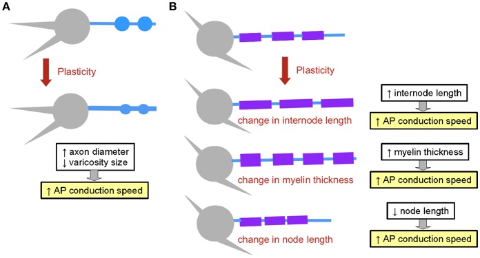
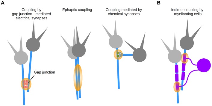
Axons functionally link the somato-dendritic compartment to synaptic terminals. Structurally and functionally diverse, they accomplish a central role in determining the delays and reliability with which neuronal ensembles communicate. By combining their active and passive biophysical properties, they ensure a plethora of physiological computations. In this review, we revisit the biophysics of generation and propagation of electrical signals in the axon and their dynamics. We further place the computational abilities of axons in the context of intracellular and intercellular coupling. We discuss how, by means of sophisticated biophysical mechanisms, axons expand the repertoire of axonal computation, and thereby, of neural computation.

Keywords: action potential generation; analog-digital signaling; axo-axonal coupling; capacitance; myelin; propagation; resistance.


Figures

Figure 1
Figure 1
Diversity of axons. (A) The axon of the BAC cell in the honeybee projects onto the left and right hemispheres, targeting a large number of regions. Modified from Zwaka et al. (2016) (CC-BY). (B) A cerebellar rat stellate cell axon illustrates a small local axon. Modified from Alcami and Marty (2013). (C) The extensive arborization of a cortical chandelier cell targets the axon initial segment of many postsynaptic cells (example shown in gray). Modified from Wang et al. (2016) (CC-BY).
Figure 2
Figure 2
Overview of axonal electrical signaling. Axonal signaling is formed by a mixture of analog and digital signaling. Insets: membrane potential traces show both the analog and digital components, the former being attenuated with distance. Electrical signals can propagate orthodromically or antidromically. Specialized regions of the axon are labeled.
Figure 3
Figure 3
Action potential generation. (A) Ultrastructure of the axon initial segment. A highly-structured spatial organization characterizes proteins at the AIS. (B) Action potential of a stellate cell from the cochlear nucleus with two components: a fast rising phase of the action potential contributed by the AIS and a second phase contributed by the somatodendritic (SD) compartment. Right, phase plane plot of this cell. Modified from Yang et al. (2016) (CC-BY).
Figure 4
Figure 4
Equivalent circuits of axons and structural biophysical specializations that improve conduction. (A) An axon can be reduced to an equivalent electrical circuit involving an axial resistor (parallel to the membrane) conveyed by the intracellular medium of the axon (axoplasm), and a parallel “RC circuit” formed by the membrane resistance and the membrane capacitance. (B) Conduction can be improved by increasing axon diameter, thereby decreasing longitudinal resistance to current flow. This allows for a larger current flow in the axon relative to the membrane resistor, and thereby a larger space constant and faster conduction. (C) An alternative circuit modification occurs with myelination: additional capacitances conveyed by myelin in series with axonal capacitance reduce the effective capacitance and additional resistance increase the effective resistance, increasing propagation speed and space constant, respectively. Note that the battery associated to the membrane has been omitted for simplification purposes. Orange arrows represent the current flow, and their thickness is indicative of the relative current flow in the axoplasm and in the radial direction out of the axon.
Figure 5
Figure 5
Structural plasticity mechanisms affecting the propagation of electrical signals. (A) Axon structural plasticity. Axons can change their diameter and bouton size. (B) Myelin structural plasticity can involve changes in internode length, in myelin thickness or in node length. The impact of each mechanism on AP propagation speed is outlined.
Figure 6
Figure 6
Coupling modalities between axons. (A) Three types of synapse-mediated coupling: through axo-axonal gap junctions (left), ephaptic coupling (middle), and chemical axonic synapses (right). (B) Indirect coupling via myelinating cells.

References

    1. Abbott B., Howarth J., Ritchie J. (1965). The initial heat production associated with the nerve impulse in crustacean and mammalian non-myelinated nerve fibres. J. Physiol. 178, 368–383. 10.1113/jphysiol.1965.sp007633 - DOI - PMC - PubMed
    1. Agudelo-Toro A., Neef A. (2013). Computationally efficient simulation of electrical activity at cell membranes interacting with self-generated and externally imposed electric fields. J. Neural Eng. 10:026019. 10.1088/1741-2560/10/2/026019 - DOI - PubMed
    1. Akkin T., Joo C., De Boer J. F. (2007). Depth-resolved measurement of transient structural changes during action potential propagation. Biophys. J. 93, 1347–1353. 10.1529/biophysj.106.091298 - DOI - PMC - PubMed
    1. Alcami P. (2018). Electrical synapses enhance and accelerate interneuron recruitment in response to coincident and sequential excitation. Front. Cell. Neurosci. 12:156. 10.3389/fncel.2018.00156 - DOI - PMC - PubMed
    1. Alcami P., Marty A. (2013). Estimating functional connectivity in an electrically coupled interneuron network. Proc. Natl. Acad. Sci. U.S.A. 110, E4798–E4807. 10.1073/pnas.1310983110 - DOI - PMC - PubMed

LinkOut - more resources