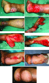
Reverse Flow Superficial Sural Artery Fasciocutaneous Flap: A Comparison of Outcome between Interpolated Flap Design versus Islanded Flap Design
- PMID: 31620333
- PMCID: PMC6790259
- DOI: 10.29252/wjps.8.3.316
Reverse Flow Superficial Sural Artery Fasciocutaneous Flap: A Comparison of Outcome between Interpolated Flap Design versus Islanded Flap Design
Abstract
Background: Complex soft-tissue defects of the distal third of the leg, proximal third of foot and similar wounds around the ankle represent formidable foes for plastic surgeons. This study compared the outcome of 2-staged interpolated flap design versus single stage islanded flap design of reverse flow superficial sural artery flap.
Methods: Thirty-four patients were enrolled, while half randomly underwent interpolated flap design (group A) and for half, islanded flap design (group B). The outcome measures were frequency of epidermolysis, flap-tip necrosis, partial flap loss, total flap loss and number of secondary procedures required for addressing these complications.
Results: Among patients, 79.41% were male and 20.58% were females. The age range was 12-51 years (mean: 28.82±10.76 years). The wound locations were hind foot (50%), ankles (17.64%), heel (14.70%), distal third of leg (11.76%) and dorsum of proximal third of foot (5.88%). In group B, epidermolysis was noted in 35.29% of flaps, and flap tip necrosis and partial flap necrosis in 17.64%. In group A, 5.88% were tip necrosis with no other problems. In group B, 76.47% of secondary procedures were done to address various flap related complications, whereas in group A, 5.88% additional procedures were required to address the flap tip necrosis.
Conclusion: The reverse flow superficial sural artery flap constituted a practical solution to address complex defects of the distal leg, ankle, heel and proximal foot. The 2-staged interpolated flap design considerably enhanced the flap reliability and reduced the frequency of venous congestion and resultant flap necrosis of variable proportions.
Keywords: Flap; Interpolated; Islanded; Necrosis; Reverse flow; Superficial sural artery.
Conflict of interest statement
The authors declare no conflict of interest.
Figures


References
-
- Lachica RD. Evidence-Based Medicine: Management of Acute Lower Extremity Trauma. Plast Reconstr Surg. 2017;139:287e–301e. - PubMed
-
- Follmar KE, Baccarani A, Baumeister SP, Levin LS, Erdmann D. The distally based sural flap. Plast Reconstr Surg. 2007;119:138e–48e. - PubMed
-
- Masquelet AC, Alain G. An atlas of flaps of the musculoskeletal system. London: CRC Press; 2001.
-
- Nakajima H, Imanishi N, Fukuzumi S, Minabe T, Fukui Y, Miyasaka T, Kodama T, Aiso S, Fujino T. Accompanying arteries of the lesser saphenous vein and sural nerve: anatomic study and its clinical applications. Plast Reconstr Surg. 1999;103:104–20. - PubMed
-
- Yang D, Morris SF. Reversed sural island flap supplied by the lower septocutaneous perforator of the peroneal artery. Ann Plast Surg. 2002;49:375–8. - PubMed
LinkOut - more resources
Full Text Sources
