. 2019 Oct;6(4):043502.
doi: 10.1117/1.JMI.6.4.043502. Epub 2019 Oct 15.

Resolution characterization of a silicon-based, photon-counting computed tomography prototype capable of patient scanning

Affiliations

Resolution characterization of a silicon-based, photon-counting computed tomography prototype capable of patient scanning

Joakim da Silva et al. J Med Imaging (Bellingham). 2019 Oct.

Abstract

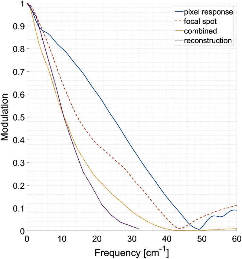
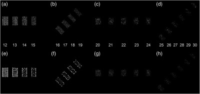
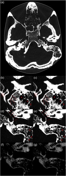
Photon-counting detectors are expected to bring a range of improvements to patient imaging with x-ray computed tomography (CT). One is higher spatial resolution. We demonstrate the resolution obtained using a commercial CT scanner where the original energy-integrating detector has been replaced by a single-slice, silicon-based, photon-counting detector. This prototype constitutes the first full-field-of-view silicon-based CT scanner capable of patient scanning. First, the pixel response function and focal spot profile are measured and, combining the two, the system modulation transfer function is calculated. Second, the prototype is used to scan a resolution phantom and a skull phantom. The resolution images are compared to images from a state-of-the-art CT scanner. The comparison shows that for the prototype 19 lp / cm are detectable with the same clarity as 14 lp / cm on the reference scanner at equal dose and reconstruction grid, with more line pairs visible with increasing dose and decreasing image pixel size. The high spatial resolution remains evident in the anatomy of the skull phantom and is comparable to that of other photon-counting CT prototypes present in the literature. We conclude that the deep silicon-based detector used in our study could provide improved spatial resolution in patient imaging without increasing the x-ray dose.

Keywords: computed tomography; photon-counting; resolution; silicon.


Figures

Fig. 1
Fig. 1
(a) In-plane pixel response and (b) focal spot profile along the center line measured using edge scan and shown at the isocenter.
Fig. 2
Fig. 2
(a) In-plane pixel response and (b) focal spot profile for different energy bins measured using an edge scan and shown at isocenter.
Fig. 3
Fig. 3
Focal spot profiles at (a) different distances perpendicular to the center line at the isocenter depth and (b) corresponding calculated focal spot FWHM values measured using tungsten wires.
Fig. 4
Fig. 4
MTFs from edge scans corresponding to pixel response, focal spot profile and their product projected at the isocenter. In addition, the MTF as measured from the PSF of the reconstructed image of a wire is shown.
Fig. 5
Fig. 5
Catphan 714 line pair images. (a)–(d) 12 to 30  lp/cm on the prototype system, reconstructed from two revolutions. (e) and (f) 12 to 19  lp/cm on the state-of-the-art system, dose matched to (a)–(d). (g) and (h) 20 to 30  lp/cm on the prototype scanner reconstructed at twice the dose of (a)–(d). (a), (b), (e), and (f) Reconstructed with 0.098-mm pixel size; (c), (d), (g), and (h) reconstructed with 0.059-mm pixel size.
Fig. 6
Fig. 6
Photon-counting CT images of the anthropomorphic skull phantom at clinical photon fluxes. (a) A reconstruction with 190-mm field-of-view and 0.19-mm pixel size whereas the remaining panels show detailed views with 0.16-mm pixel size. (b) and (d) Reconstructed from one revolution, corresponding to native slice thickness; (c) and (e) reconstructed from two revolutions, mimicking the number of photons per slice of a conventional CT detector. Arrow heads indicate septations in mastoid cells and lines indicate delineation of the hypoglossal canal, the sigmoidal sinus and the carotid canal. (f) and (g) The same detail as (d) and (e), reconstructed from two revolutions using only the low and high energy bins, respectively.

References

    1. Taguchi K., Iwanczyk J. S., “Vision 20/20: single photon counting x-ray detectors in medical imaging,” Med. Phys. 40(10), 100901 (2013).MPHYA610.1118/1.4820371 - DOI - PMC - PubMed
    1. Schmidt T. G., “Optimal ‘image-based’ weighting for energy-resolved CT,” Med. Phys. 36(7), 3018–3027 (2009).MPHYA610.1118/1.3148535 - DOI - PubMed
    1. Alvarez R. E., Macovski A., “Energy-selective reconstructions in x-ray computerized tomography,” Phys. Med. Biol. 21(5), 733–744 (1976).PHMBA710.1088/0031-9155/21/5/002 - DOI - PubMed
    1. Schlomka J. P., et al. , “Experimental feasibility of multi-energy photon-counting K-edge imaging in pre-clinical computed tomography,” Phys. Med. Biol. 53(15), 4031–4047 (2008).PHMBA710.1088/0031-9155/53/15/002 - DOI - PubMed
    1. Symons R., et al. , “Low-dose lung cancer screening with photon-counting CT: a feasibility study,” Phys. Med. Biol. 62(1), 202–213 (2017).PHMBA710.1088/1361-6560/62/1/202 - DOI - PMC - PubMed