. 2019 Sep 25;5(9):eaaw2853.
doi: 10.1126/sciadv.aaw2853. eCollection 2019 Sep.

Synthetic self-assembling ADDomer platform for highly efficient vaccination by genetically encoded multiepitope display

Affiliations

Synthetic self-assembling ADDomer platform for highly efficient vaccination by genetically encoded multiepitope display

Charles Vragniau et al. Sci Adv. .

Abstract

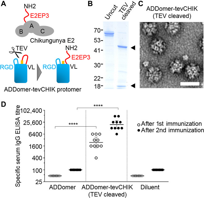
Self-assembling virus-like particles represent highly attractive tools for developing next-generation vaccines and protein therapeutics. We created ADDomer, an adenovirus-derived multimeric protein-based self-assembling nanoparticle scaffold engineered to facilitate plug-and-play display of multiple immunogenic epitopes from pathogens. We used cryo-electron microscopy at near-atomic resolution and implemented novel, cost-effective, high-performance cloud computing to reveal architectural features in unprecedented detail. We analyzed ADDomer interaction with components of the immune system and developed a promising first-in-kind ADDomer-based vaccine candidate to combat emerging Chikungunya infectious disease, exemplifying the potential of our approach.


Figures

Fig. 1
Fig. 1. Cryo-EM structure of ADDomer.
(A) ADDomer design. Three evolutionary nonconserved segments (boxed) in the protomer were engineered by BioBrick design into exchangeable cassettes. One cassette (yellow) is located within the VL, while two cassettes, RGD1 (light green) and RGD2 (dark green), are located within the loop containing a functional RGD tripeptide sequence. Numbers indicated amino acid boundaries of BioBrick cassettes as well as VL and RGD loop. (B) Left: A 3.5-Å cryo-EM map of the ADDomer particle formed by 60 protomers. The rigid core is colored blue, and more flexible regions comprising the loops are colored cyan and green. Right: The corresponding atomic model is shown in gray. One penton (center) is highlighted, with individual protomers colored red, orange, green, cyan, and blue, respectively. (C) Side view of a penton formed by five protomers. Flexible VL and RGD loop are drawn in dashed lines. (D) Closeup view of the RGD loop and VL in ADDomer. Residues 156 to 170 in VL and 314 to 366 in RGD loop in the BioBrick format maintain their flexibility, essential for functionalization by epitope insertion. (E) Superimposition of the fiber-binding region in the ADDomer cryo-EM structure (cyan) with apo Ad3 [Protein Data Bank (PDB) ID: 4AQQ; marine blue] and fiber peptide–bound Ad3 (PDB ID: 4AR2; magenta) crystal coordinates. The fiber peptide (PDB ID: 4AR2) is drawn in a ball-and-stick representation. (F) Individual ADDomer protomer is shown in a side view (left) with a putative metal-binding cluster boxed in between the crown (top) and jellyroll fold (bottom) domains. Zoomed-in views of the boxed region (right) depict the atomic model in the EM density contoured at two different levels, σ = 4 (blue) and σ = 10.5 (red). Four juxtaposed sulfurs (yellow spheres) likely coordinate a metal ion. (G) Left: Central channel of an ADDomer penton. Right: Ca2+ coordination as seen previously in the Ad3 penton base protein crystal structure (PDB ID: 4AR2) is not observed. The helices fencing the central channel are rearranged, resulting in a different conformation of E466. (H) Interface between two pentons (A1 and A2) in the ADDomer (cyan). The zoomed-in view depicts the boxed region with the corresponding Ad3 crystal structure (magenta) superimposed. Domain swapping is not observed in the ADDomer. Instead, this interface is stabilized by mutual hydrogen bonds between S61 side chains and E59 peptide backbones from neighboring protomers (right).
Fig. 2
Fig. 2. ADDomer internalizations.
(A) HeLa cells incubated with ADDomer. 4′,6-diamidino-2-phenylindole–stained nuclei are colored blue. ADDomer detected by punctuate-specific immunofluorescence is colored red. Scale bar, 20 μm. (B) Human monocytes (Mo; left) and MDDCs (right) were incubated with Alexa Fluor 488–labeled ADDomer (green). Efficient uptake of ADDomer was observed by in all cell types tested. Scale bars, 20 μm. (C) Lymph node biodistribution of Alexa Fluor 680–labeled ADDomer in mice. Ex vivo imaging of the main lymph nodes from one representative mouse 5 hours after subcutaneous injection (10 μg) in the right leg is depicted in a schematic drawing (left). Specific fluorescence signal was quantified (for 100 ms), and intensities (in arbitrary units) are plotted in spectral colors (bottom). (D) Ex vivo quantification (for 100 ms) of ADDomer in isolated lymph nodes from a cohort of mice (n = 8) is shown in a bar diagram. Fluorescence was measured at 1 and 5 hours, respectively, after intramuscular or subcutaneous injection of Alexa Fluor 680–labeled ADDomer (10 μg). Bar color coding is as follows: dark blue, intramuscular at 1 hour; brown, intramuscular at 5 hours; light blue, subcutaneous for 1 hour; red, subcutaneous for 5 hours; black, control (noninjected) mice (n = 5). SDs are shown as error bars. Axi, axillary; Bra, brachial; Sci, sciatic; Ing fpr, inguinal; Mes, mesenteric lymph nodes; RLU, relative light unit per pixel.
Fig. 3
Fig. 3. ADDomer immunizations.
(A) Top: Chikungunya envelope protein E2, composed of domains A, B, and C, is shown in a schematic representation. The major neutralizing epitope E2EP3 (red) comprises the N terminus of E2 with the amino acid sequence NH2-STDKDNFNVYKATRPYLAH. Bottom: This specific configuration of E2E3P was mimicked in ADDomer-tevCHIK as depicted by inserting the E2EP3 sequence (in red), preceded by a TEV protease cleavage site (orange) into the BioBrick cassette in VL of the ADDomer protomer. Cleavage by the protease exposes E2EP3 in a native-like configuration as shown schematically. (B) SDS–polyacrylamide gel electrophoresis section showing purified ADDomer-tevCHIK before and after cleavage by TEV protease. Molecular weight marker sizes are marked (left). Appearance of two bands (black triangles) indicates quantitative cleavage. (C) Representative negative-stain EM images of TEV-cleaved ADDomer-tevCHIK reveals stable dodecamers. Scale bar, 30 nm. (D) This TEV-cleaved sample was used in immunization experiments in mice (n = 10 per group). Scatter dot plot diagram depicts the outcome of the immunization experiments, revealing high individual IgG titers specific for Chikungunya E2E3P peptide in enzyme-linked immunosorbent assay (ELISA). Short horizontal line segments are geometric mean titers, and error bars represent the 95% confidence interval. Significant difference (****P < 0.0001) was determined between ADDomer-tevCHICK– and ADDomer-only immunized mice.

References

    1. Brune K. D., Howarth M., New routes and opportunities for modular construction of particulate vaccines: Stick, click, and glue. Front. Immunol. 9, 1432 (2018). - PMC - PubMed
    1. Frietze K. M., Peabody D. S., Chackerian B., Engineering virus-like particles as vaccine platforms. Curr. Opin. Virol. 18, 44–49 (2016). - PMC - PubMed
    1. Unzueta U., Céspedes M. V., Vázquez E., Miralles N. F., Mangues R., Villaverde A., Towards protein-based viral mimetics for cancer therapies. Trends Biotechnol. 33, 253–258 (2015). - PubMed
    1. Diaz D., Care A., Sunna A., Bioengineering strategies for protein-based nanoparticles. Genes 9, E370 (2018). - PMC - PubMed
    1. Bachmann M. F., Jennings G. T., Vaccine delivery: A matter of size, geometry, kinetics and molecular patterns. Nat. Rev. Immunol. 10, 787–796 (2010). - PubMed

Publication types

MeSH terms

LinkOut - more resources