. 2019 Dec 1;160(12):2929-2945.
doi: 10.1210/en.2019-00259.

Spermatogonial Type 3 Deiodinase Regulates Thyroid Hormone Target Genes in Developing Testicular Somatic Cells

Affiliations

Spermatogonial Type 3 Deiodinase Regulates Thyroid Hormone Target Genes in Developing Testicular Somatic Cells

M Elena Martinez et al. Endocrinology. .

Erratum in

Abstract

Premature overexposure to thyroid hormone causes profound effects on testis growth, spermatogenesis, and male fertility. We used genetic mouse models of type 3 deiodinase (DIO3) deficiency to determine the genetic programs affected by premature thyroid hormone action and to define the role of DIO3 in regulating thyroid hormone economy in testicular cells. Gene expression profiling in the neonatal testis of DIO3-deficient mice identified 5699 differentially expressed genes. Upregulated and downregulated genes were, respectively, involved according to DAVID analysis with cell differentiation and proliferation. They included anti-Müllerian hormone and genes involved in the formation of the blood-testis barrier, which are specific to Sertoli cells (SCs). They also included steroidogenic genes, which are specific to Leydig cells. Comparison with published data sets of genes enriched in SCs and spermatogonia, and responsive to retinoic acid (RA), identified a subset of genes that were regulated similarly by RA and thyroid hormone. This subset of genes showed an expression bias, as they were downregulated when enriched in spermatogonia and upregulated when enriched in SCs. Furthermore, using a genetic approach, we found that DIO3 is not expressed in SCs, but spermatogonia-specific inactivation of DIO3 led to impaired testis growth, reduced SC number, decreased cell proliferation and, especially during neonatal development, altered gene expression specific to somatic cells. These findings indicate that spermatogonial DIO3 protects testicular cells from untimely thyroid hormone signaling and demonstrate a mechanism of cross-talk between somatic and germ cells in the neonatal testis that involves the regulation of thyroid hormone availability and action.


Figures

Figure 1.
Figure 1.
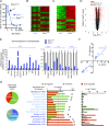
Thyroid hormone target genes in the developing testis. (A) Ontogeny of serum concentration of T3 in Dio3−/− mice relative to control values; P5 was chosen (arrow) to perform the RNA-sequencing experiment. (B and C) Heat map and sample clustering of differentially expressed genes in the testis due to T3 overexposure; selected genes included those with an average minimum expression of 1 read per kilobase per million mapped reads in at least one of the experimental groups and (B) a q value <0.05, and a fold change >1.5 or (C) a q value <0.01 and a fold change >3. (D) Volcano plot of the RNA-sequencing results highlighting differentially expressed genes. (E) Validation by qPCR in independent samples of 23 genes upregulated and downregulated by T3, as identified in the RNA-sequencing experiment. **P < 0.01, ***P < 0.001, as determined by a Student t test (n = 7 and n = 5 for the Dio3+/+ and Dio3−/− groups, respectively). (F) Correlation of the expression changes observed by RNA sequencing with those observed by qPCR in validated genes. (G) Functional analysis result highlights of differentially expressed genes. #P < 0.01, as determined by χ2 analysis of expected vs observed percentage distributions.
Figure 2.
Figure 2.
Ingenuity pathway analysis of the 1607 DEGs analyzed. Terms with top significance for upstream regulators, diseases and biofunctions, and canonical pathways are represented for all genes, upregulated genes, or downregulated genes. Represented P values were determined by Ingenuity.
Figure 3.
Figure 3.
Expression of markers of SC and GC differentiation in Dio3+/+ and Dio3−/− mouse testis. (A and B) Expression of (A) SC and (B) GC markers at two different ages. (C) Expression of markers of T3 action at the same ages. Data represent the mean ± SEM. *P < 0.05, **P < 0.01, ***P < 0.001, as determined by ANOVA and a Tukey post hoc test (n = 9 to 12 mice per experimental group).
Figure 4.
Figure 4.
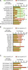
Cell-specific gene regulation in P5 Dio3−/− testis. (A–C) Subsets of 5699 DEGs in Dio3−/− testis that are specific to different (A) testicular cell types, (B) stages of spermatogenesis, and (C) spermatogonia subtypes, according to the single-cell RNA-sequencing data from Green et al. (47). Green and red dotted lines represent the percentage of upregulated and downregulated genes in all 5699 DEGs genes. #P < 0.001, as determined by χ2 test on observed vs predicted number of upregulated and downregulated genes. [From: Green CD, Ma Q, Manske GL, Shami AN, Zheng X, Marini S, Moritz L, Sultan C, Gurczynski SJ, Moore BB, Tallquist MD, Li JZ, Hammoud SS. A comprehensive roadmap of murine spermatogenesis defined by single-cell RNA-seq. Developmental Cell 2018;46:651–667.e610.]
Figure 5.
Figure 5.
DEGs in Dio3−/− testis that were also regulated in other testis gene profiling data sets. (A–C) Comparison with data reported by Evans et al. (48). (A) Percentage distribution of T3 upregulation and downregulation of genes according to cell type enrichment for genes regulated by T3 (left) or regulated by both T3 and RA (right). (B) T3-dependent vs RA-dependent fold change in gene expression for 155 genes regulated by T3 and RA, depicted according to cell type enrichment. The names of some of the genes are noted. (C) Percentage distribution of T3 upregulation and downregulation of genes according to cell type enrichment for 91 genes that are regulated in the same direction by both T3 and RA. Dotted lines indicate the percentage of upregulated and downregulated genes (green and red, respectively) in the full set of relevant genes. #P < 0.001, as determined by a χ2 test. (D) DEGs also regulated in a mouse model of androgen receptor deficiency in SCs (50). The expression changes in each model show a strong inverse correlation. (E and F) Data on DEGs also identified by transcriptome analyses (51) as markers of progenitor cells for fetal (E) LCs and (F) SCs. The vast majority of those markers were downregulated in the current study. ns, not significant. [From: Green CD, Ma Q, Manske GL, Shami AN, Zheng X, Marini S, Moritz L, Sultan C, Gurczynski SJ, Moore BB, Tallquist MD, Li JZ, Hammoud SS. A comprehensive roadmap of murine spermatogenesis defined by single-cell RNA-seq. Developmental Cell 2018;46:651–667.e610.]
Figure 6.
Figure 6.
Regulation of 166 DEGs in the Dio3−/− testis that were also differentially expressed in prepubertal monkey testis treated with LH (53). The direction of the changes in gene expression observed in both experiments did not correlate. The distribution of genes upregulated and downregulated in the two experiments (bar graph) suggests that the effects of T3 in the current experiment are predominantly opposite to those in the published experiment. P was determined by a χ2 test. FC, fold change. [From: Perrotta C, Buldorini M, Assi E, Cazzato D, De Palma C, Clementi E, Cervia D. The thyroid hormone triiodothyronine controls macrophage maturation and functions: protective role during inflammation. Am J Pathol 2014;184:230–247.]
Figure 7.
Figure 7.
Cell-specific inactivation of DIO3 in the testis. (A) Abundance of Dio3 recombined allele and levels of DIO3 enzymatic activity in the testis of neonatal Amh-cre/Dio3f/f mice. (B) In situ hybridization of Dio3 mRNA in the testis of wild-type mice and mice carrying a full deletion of the Dio3 gene (Del-Dio3−/−). Dio3 mRNA is shown in red, and tissue sections are counterstained with 4′,6-diamidino-2-phenylindole (DAPI; blue). Scale bars, ∼10 μm. Note that DNA at P21 exhibits a marked multinucleolar structure and only six to eight nuclei are shown per field. (C) Testicular immunofluorescence in P8 mice carrying a Stra8-cre transgene and a cre-dependent GFP reporter showing cell specificity of cre-recombination. Scale bars, 30 μm (left panel) and 10 μm (right panel). (D) DIO3 activity in the neonatal testis of mice carrying the Stra8-cre transgene that were heterozygous or homozygous for the floxed Dio3 allele. ***, # indicates ***P < 0.001, when compared with control; #P < 0.001, when compared with heterozygous, as determined by ANOVA and a Tukey post hoc test (n = 9, n = 8, and n = 7, respectively). (E) Body weight and testis weight at postnatal days 10, 21, and 60. **P < 0.01, Dio3f/f vs Stra8-cre/Dio3f/f as determined by the a Student t test (n = 6 to 8 for each experimental group).
Figure 8.
Figure 8.
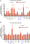
Gene expression of thyroid hormone–regulated genes in spermatogonia-specific DIO3-deficient neonatal testis. (A) P10 testis. (B) P21 testis. Genes written in green and blue are enriched in SCs and LCs, respectively. *P < 0.05, **P < 0.01, ***P < 0.001, Dio3f/f vs Stra8-cre/Dio3f/f as determined by the Student t test (n = 6 to 8 for each experimental group).
Figure 9.
Figure 9.
Testicular abnormalities in Dio3−/− and Stra8-cre/Dio3f/f mice. (A and D) Quantification of SCs per 100 μm of seminiferous tubule circumference (STC) in (A) Dio3−/− and (D) Stra8-cre/Dio3f/f mice by SOX9 immunofluorescence. (B and E) Quantification of spermatogonia per 103 μm2 of seminiferous tubule area (STA) in (B) Dio3−/− and (E) Stra8-cre/Dio3f/f mice by TRA98 immunofluorescence. (C and F) STA in (C) Dio3−/− and (F) Stra8-cre/Dio3f/f mice. Scale bars, ∼40 μm. *P < 0.05, **P < 0.01, ***P < 0.001, as determined by the Student t test (n = 4 mice per experimental group).
Figure 10.
Figure 10.
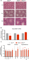
Testis histology and pituitary and hypothalamic gene expression in in Stra8-cre/Dio3f/f. (A) Hematoxylin and eosin (H&E) staining of Dio3f/f and Stra8-cre/Dio3f/f testis at different ages. (B) Ki67 mRNA expression in Dio3f/f and Stra8-cre/Dio3f/f testis at different ages. (C) Adult expression in Stra8-cre/Dio3f/f tissues of genes relevant to the reproductive axis that showed abnormal expression in global DIO3 deficiency (34). **P < 0.01, ***P < 0.001, as determined by the Student t test (n = 5 and n = 6 per group). [From: Martinez ME, Karaczyn A, Stohn JP, Donnelly WT, Croteau W, Peeters RP, Galton VA, Forrest D, St Germain D, Hernandez A. The type 3 deiodinase is a critical determinant of appropriate thyroid hormone action in the developing testis. Endocrinology 2016;157:1276–1288.]

Similar articles

Cited by

References

    1. Picut CA, Remick AK, de Rijk EP, Simons ML, Stump DG, Parker GA. Postnatal development of the testis in the rat: morphologic study and correlation of morphology to neuroendocrine parameters. Toxicol Pathol. 2015;43(3):326–342. - PubMed
    1. Gao Y, Lee WM, Cheng CY. Thyroid hormone function in the rat testis. Front Endocrinol (Lausanne). 2014;5:188. - PMC - PubMed
    1. Wagner MS, Wajner SM, Maia AL. The role of thyroid hormone in testicular development and function. J Endocrinol. 2008;199(3):351–365. - PMC - PubMed
    1. Hernandez A. Thyroid hormone role and economy in the developing testis. Vitam Horm. 2018;106:473–500. - PubMed
    1. Bunick D, Kirby J, Hess RA, Cooke PS. Developmental expression of testis messenger ribonucleic acids in the rat following propylthiouracil-induced neonatal hypothyroidism. Biol Reprod. 1994;51(4):706–713. - PubMed

Publication types