

Prefrontal theta modulates sensorimotor gamma networks during the reorienting of attention
- PMID: 31621977
- PMCID: PMC7268018
- DOI: 10.1002/hbm.24819
Prefrontal theta modulates sensorimotor gamma networks during the reorienting of attention
Abstract
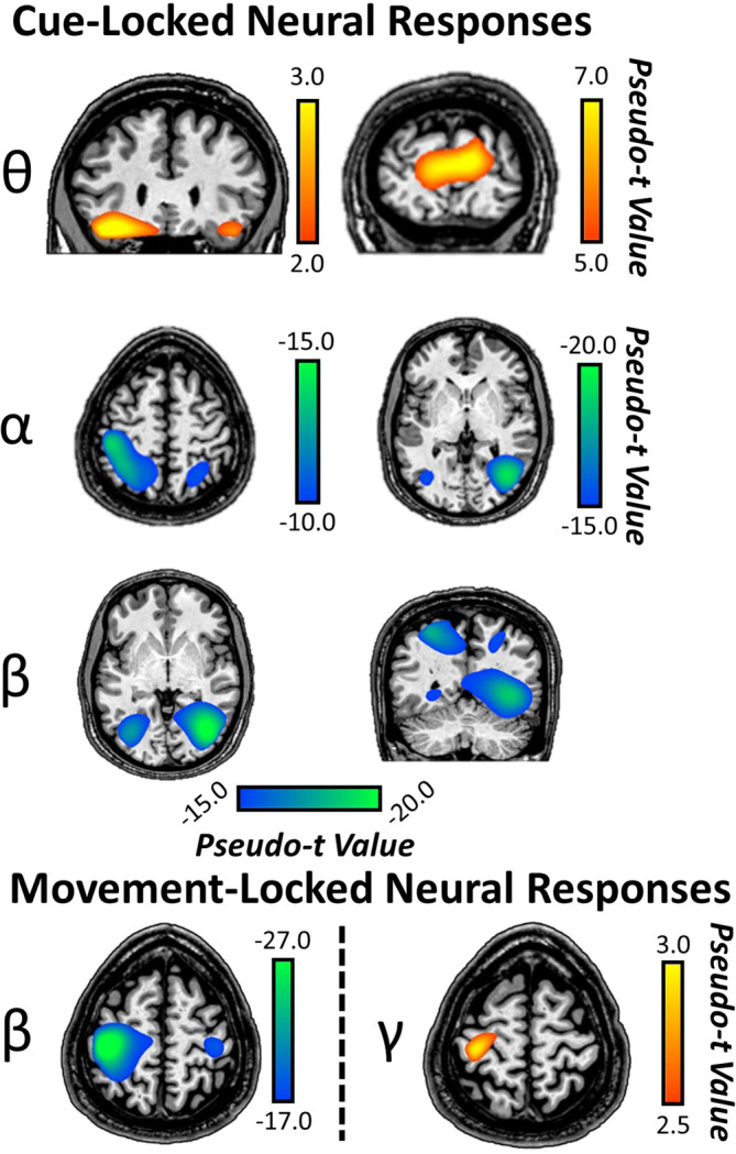
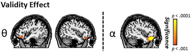
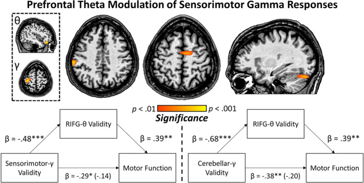
The ability to execute a motor plan involves spatiotemporally precise oscillatory activity in primary motor (M1) regions, in concert with recruitment of "higher order" attentional mechanisms for orienting toward current task goals. While current evidence implicates gamma oscillatory activity in M1 as central to the execution of a movement, far less is known about top-down attentional modulation of this response. Herein, we utilized magnetoencephalography (MEG) during a Posner attention-reorienting task to investigate top-down modulation of M1 gamma responses by frontal attention networks in 63 healthy adult participants. MEG data were evaluated in the time-frequency domain and significant oscillatory responses were imaged using a beamformer. Robust increases in theta activity were found in bilateral inferior frontal gyri (IFG), with significantly stronger responses evident in trials that required attentional reorienting relative to those that did not. Additionally, strong gamma oscillations (60-80 Hz) were detected in M1 during movement execution, with similar responses elicited irrespective of attentional reorienting. Whole-brain voxel-wise correlations between validity difference scores (i.e., attention reorienting trials-nonreorienting trials) in frontal theta activity and movement-locked gamma oscillations revealed a robust relationship in the contralateral sensorimotor cortex, supplementary motor area, and right cerebellum, suggesting modulation of these sensorimotor network gamma responses by attentional reorienting. Importantly, the validity difference effect in this distributed motor network was predictive of overall motor function measured outside the scanner and further, based on a mediation analysis this relationship was fully mediated by the reallocation response in the right IFG. These data are the first to characterize the top-down modulation of movement-related gamma responses during attentional reorienting and movement execution.
Keywords: Posner cueing task; magnetoencephalography; oscillations; top-down.
© 2019 The Authors. Human Brain Mapping published by Wiley Periodicals, Inc.
Conflict of interest statement
The authors report no financial, institutional, or commercial conflicts of interest.
Figures
Similar articles
-
Oscillatory dynamics in the dorsal and ventral attention networks during the reorienting of attention.Hum Brain Mapp. 2018 May;39(5):2177-2190. doi: 10.1002/hbm.23997. Epub 2018 Feb 6. Hum Brain Mapp. 2018. PMID: 29411471 Free PMC article.
-
Cortical Oscillatory Activity and Motor Control in Pediatric Stroke Patients With Hemidystonia.Hum Brain Mapp. 2025 Apr 1;46(5):e70204. doi: 10.1002/hbm.70204. Hum Brain Mapp. 2025. PMID: 40186512 Free PMC article.
-
The developmental trajectory of sensorimotor cortical oscillations.Neuroimage. 2019 Jan 1;184:455-461. doi: 10.1016/j.neuroimage.2018.09.018. Epub 2018 Sep 12. Neuroimage. 2019. PMID: 30217545 Free PMC article.
-
Revisiting Persistent Neuronal Activity During Covert Spatial Attention.Front Neural Circuits. 2021 Jun 30;15:679796. doi: 10.3389/fncir.2021.679796. eCollection 2021. Front Neural Circuits. 2021. PMID: 34276314 Free PMC article. Review.
-
The reorienting system of the human brain: from environment to theory of mind.Neuron. 2008 May 8;58(3):306-24. doi: 10.1016/j.neuron.2008.04.017. Neuron. 2008. PMID: 18466742 Free PMC article. Review.
Cited by
-
Oscillatory markers of neuroHIV-related cognitive impairment and Alzheimer's disease during attentional interference processing.Aging (Albany NY). 2023 Jan 19;15(2):524-541. doi: 10.18632/aging.204496. Epub 2023 Jan 19. Aging (Albany NY). 2023. PMID: 36656738 Free PMC article.
-
High-definition transcranial direct-current stimulation of left primary motor cortices modulates beta and gamma oscillations serving motor control.J Physiol. 2025 Mar;603(6):1627-1644. doi: 10.1113/JP287085. Epub 2025 Feb 26. J Physiol. 2025. PMID: 40009440
-
Chronic Cannabis users exhibit altered oscillatory dynamics and functional connectivity serving visuospatial processing.J Psychopharmacol. 2024 Aug;38(8):724-734. doi: 10.1177/02698811241265764. Epub 2024 Aug 1. J Psychopharmacol. 2024. PMID: 39087306 Free PMC article.
-
EMG-projected MEG high-resolution source imaging of human motor execution: Brain-muscle coupling above movement frequencies.Imaging Neurosci (Camb). 2024 Jan 9;2:1-20. doi: 10.1162/imag_a_00056. eCollection 2024 Jan 1. Imaging Neurosci (Camb). 2024. PMID: 39290632 Free PMC article.
-
Modeling the contribution of theta-gamma coupling to sequential memory, imagination, and dreaming.Front Neural Circuits. 2024 Jun 14;18:1326609. doi: 10.3389/fncir.2024.1326609. eCollection 2024. Front Neural Circuits. 2024. PMID: 38947492 Free PMC article.
References
-
- Baron, R. M. , & Kenny, D. A. (1986). The moderator‐mediator variable distinction in social psychological research: Conceptual, strategic, and statistical considerations. Journal of Personality and Social Psychology, 51(6), 1173–1182. - PubMed
Publication types
MeSH terms
Grants and funding
LinkOut - more resources
Full Text Sources
