. 2019 Nov 14;4(22):e126315.
doi: 10.1172/jci.insight.126315.

Prelamin A mediates myocardial inflammation in dilated and HIV-associated cardiomyopathies

Affiliations

Prelamin A mediates myocardial inflammation in dilated and HIV-associated cardiomyopathies

Daniel Brayson et al. JCI Insight. .

Abstract

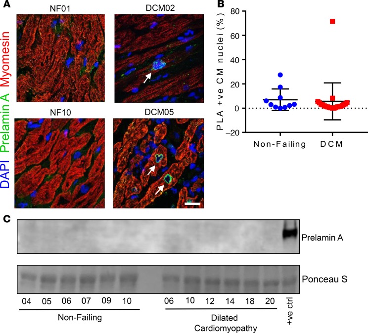
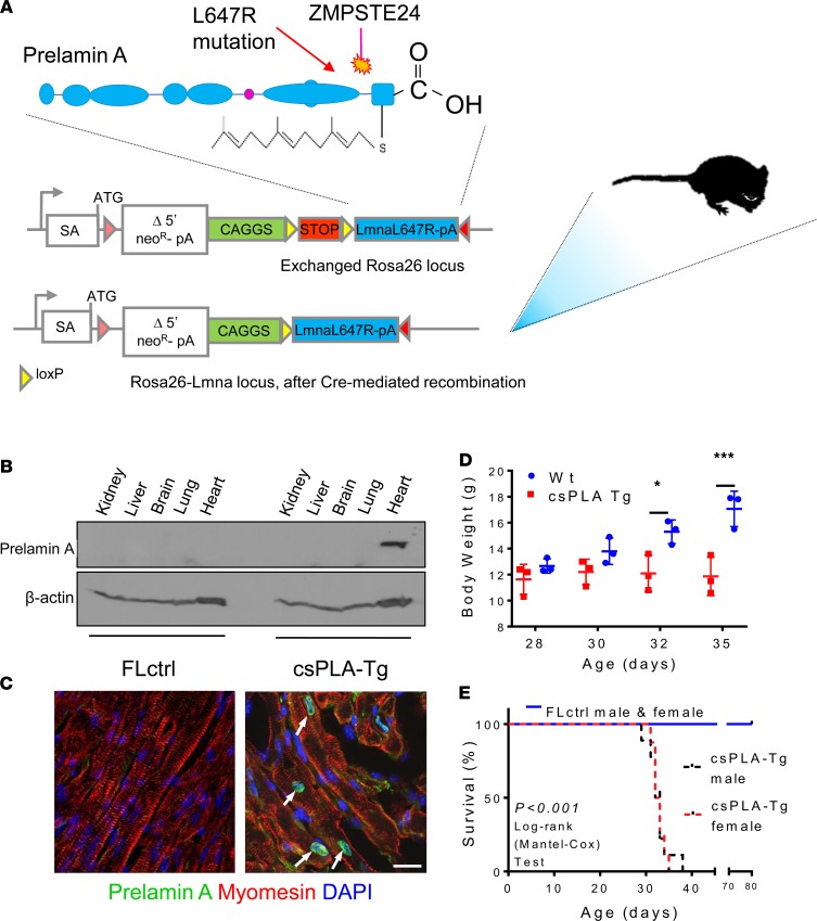
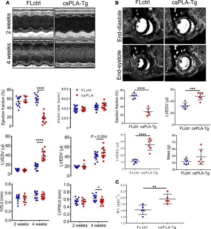
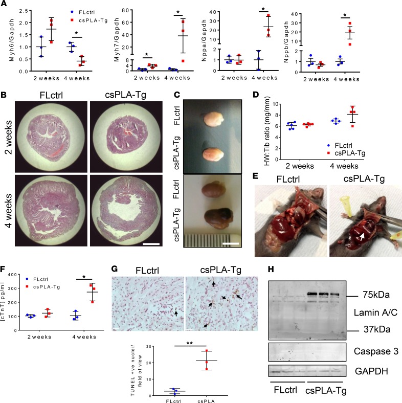
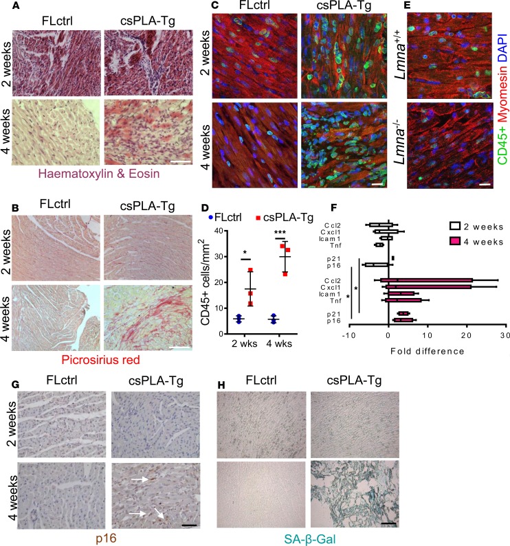
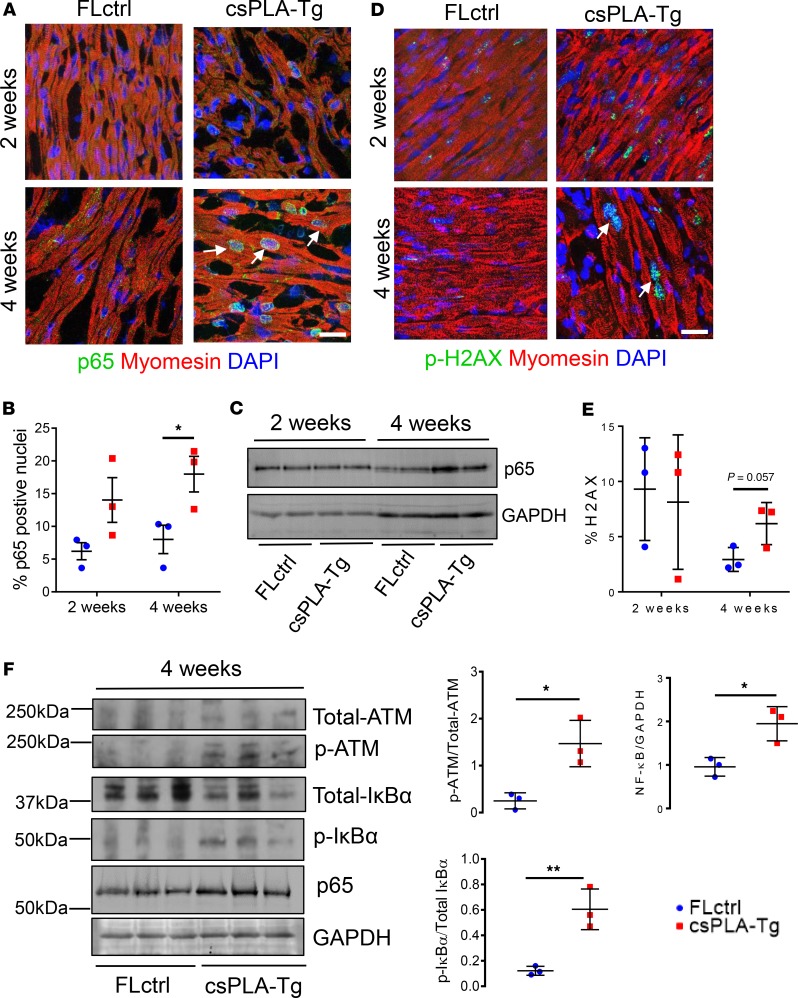
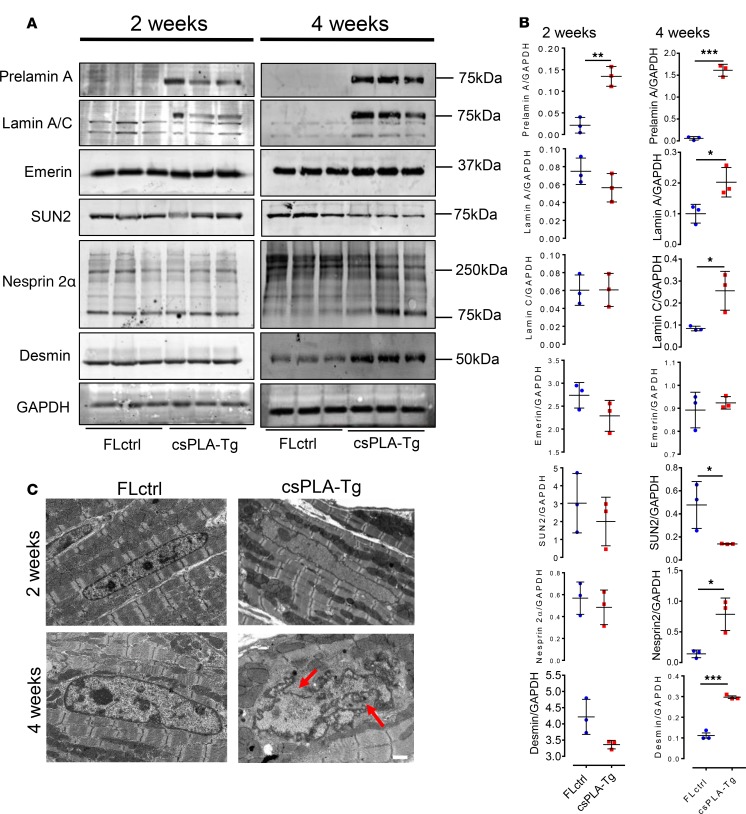
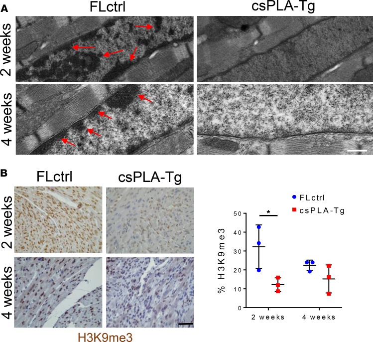
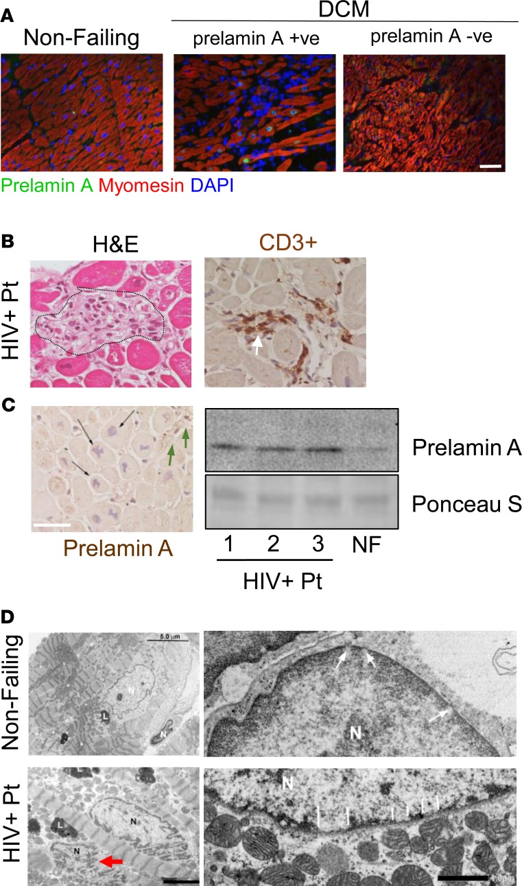
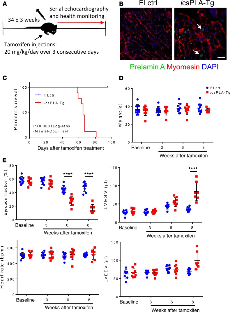
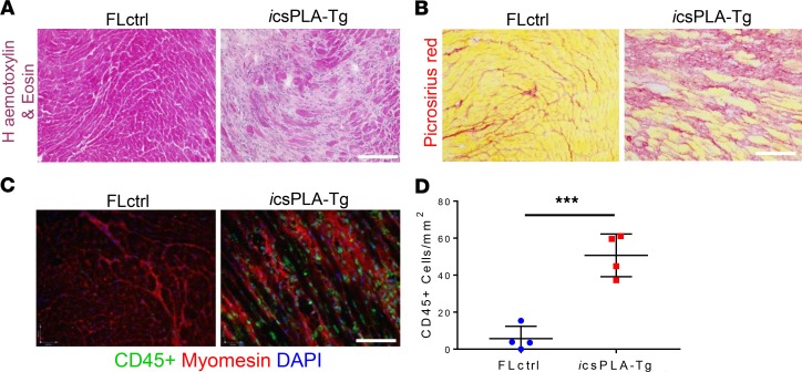
Cardiomyopathies are complex heart muscle diseases that can be inherited or acquired. Dilated cardiomyopathy can result from mutations in LMNA, encoding the nuclear intermediate filament proteins lamin A/C. Some LMNA mutations lead to accumulation of the lamin A precursor, prelamin A, which is disease causing in a number of tissues, yet its impact upon the heart is unknown. Here, we discovered myocardial prelamin A accumulation occurred in a case of dilated cardiomyopathy, and we show that a potentially novel mouse model of cardiac-specific prelamin A accumulation exhibited a phenotype consistent with inflammatory cardiomyopathy, which we observed to be similar to HIV-associated cardiomyopathy, an acquired disease state. Numerous HIV protease therapies are known to inhibit ZMPSTE24, the enzyme responsible for prelamin A processing, and we confirmed that accumulation of prelamin A occurred in HIV+ patient cardiac biopsies. These findings (a) confirm a unifying pathological role for prelamin A common to genetic and acquired cardiomyopathies; (b) have implications for the management of HIV patients with cardiac disease, suggesting protease inhibitors should be replaced with alternative therapies (i.e., nonnucleoside reverse transcriptase inhibitors); and (c) suggest that targeting inflammation may be a useful treatment strategy for certain forms of inherited cardiomyopathy.

Keywords: AIDS/HIV; Cardiology; Cardiovascular disease.


Conflict of interest statement

Conflict of interest: The authors have declared that no conflict of interest exists.

Figures

Figure 1
Figure 1. Prelamin A accumulated in a heart of a patient with dilated cardiomyopathy (DCM).
(A) Confocal micrographs of human heart sections from DCM patients and nonfailing (NF) controls subjected to immunofluorescence staining to detect prelamin A (green), myomesin (red), and DAPI (blue). Arrows point to prelamin A+ CM nuclei, many of which exhibit nuclear morphology defects. Scale bar: 10 μm. (B) The number of nuclei that stained positively for prelamin A were quantified as a percentage of CM nuclei for nonfailing (blue circles, n = 10 nonfailing donor samples) and DCM (red squares, n = 21 samples from explanted DCM hearts) myocardial sections (mean ± SD). (C) Western blotting did not detect prelamin A in a selection of DCM patient samples. There was not enough sample remaining from DCM05 to run on a Western blot.
Figure 2
Figure 2. Targeted transgenesis of prelamin A led to nuclear accumulation in CMs and resulted in premature death in mice.
(A) Schematic representation showing the site of prelamin A (LMNA-L647R) cDNA insertion and the modifications required for conditional expression. SA, splice acceptor site; neoR, neomycin resistance; pA, polyadenylation signal. (B) Western blotting for prelamin A showing expression was restricted to heart tissue. (C) Confocal micrographs of myocardium stained for prelamin A showing nuclear rim localization in csPLA-Tg hearts. Scale bar: 10 μm. Arrows indicate prelamin A expressing nuclei. (C) Growth curves showing that csPLA-Tg mice stop growing after 30 days. n = 3 males/group. Two-way ANOVA with repeated measures with Sidak’s post hoc test for multiple comparisons was performed. *P < 0.05, ***P < 0.001. (D) Kaplan-Meier survival analysis showing that csPLA-Tg male and female mice exhibited attenuated survival compared with FLctrl counterparts. n = 7 FLctrl males, 8 FLctrl females, 9 csPLA-Tg males, 8 csPLA-Tg females. Log-rank Mantel-Cox test was performed. P < 0.0001.
Figure 3
Figure 3. Cardiac function was attenuated in 4-week-old csPLA-Tg mice.
(A) Representative images of echocardiographs and corresponding graphs of analysis performed on movies acquired in B-mode showing severely compromised cardiac function in 4-week-old mice (n = 12/group [6 females, 6 males], except csPLA-Tg 2 weeks, which was n = 9 [7 females, 2 males]). Values are mean ± SD. Two-way ANOVA, no repeated measures, was performed with Sidak’s post hoc test for multiple comparisons. *P < 0.05, ****P < 0.0001. (B) Representative cardiac MRI images of myocardium in end-systole and end-diastole and corresponding graphs displaying a decrease in ejection fraction alongside increases in left ventricle end-diastolic (LVEDV) and LV end-systolic volume (LVESV). Mass was statistically unchanged, but with increased variation, and concurs with postmortem heart weight measurements. (C) Increased relaxation time (R1) of gadolinium contrast in 4-week-old csPLA-Tg myocardium, indicative of fibrosis remodeling. n = 6 males/group. Values are mean ± SD. Student’s 2-tailed t test was performed. **P < 0.01, ***P < 0.001, ****P < 0.0001. IVS;d, intraventricular septal thickness in diastole; LVPW;d, left ventricle posterior wall thickness in diastole.
Figure 4
Figure 4. csPLA-Tg mice displayed signs of heart failure and necrotic cell death.
(A) Fetal gene expression is dysregulated in csPLA-Tg hearts at 4 weeks, indicating heart failure; n = 3 females/group. Values are mean ± SD. Two-Way ANVOA, no repeated measures, was performed with Sidak’s post hoc test for multiple comparisons. *P < 0.05. (B) Low-magnification micrographs showing cross-sectional view of the heart stained with H&E, indicating 4-week-old csPLA-Tg mice possessed dilated cardiac chambers. Scale bar: 2 mm. (C) Photographs showing csPLA-Tg appeared enlarged at 4 weeks. Scale bar: 5 mm. (D) No significant difference in heart weight to tibia length ratio was observed. Values are mean ± SD. n = 3/group. (E) Dissection of chest cavities showing transudative pleural effusions in csPLA-Tg mice. (F) Blood plasma subjected to ELISA showed elevated levels of circulating cardiac Troponin T (TnT), indicative of cardiac damage in 4-week-old mice. n = 3 females/group. Values are mean ± SD. Two-Way ANOVA, no repeated measures, with Sidak’s post hoc test for multiple comparisons was performed. *P < 0.05. (G) TUNEL staining was performed on 4-week-old csPLA-Tg heart sections, and image quantification showed a significant increase in TUNEL+ nuclei in csPLA-Tg heart sections at 4 weeks. n = 3 females/group. Values are mean ± SD. Student’s 2-tailed t test was performed. **P < 0.01. Arrows indicate TUNEL positive nuclei. Scale = 30 µm. (H) Western blotting showed that cleavage of caspase 3 and lamins A/C, which occurs at ~37 kDa and occurs during apoptosis, did not occur in 4-week-old csPLA-Tg myocardium.
Figure 5
Figure 5. Fibrotic remodeling of csPLA-Tg myocardium occurred in tandem with inflammation and senescence.
(A) Light micrographs showing myocardial disarray in 4-week-old csPLA-Tg myocardium stained with H&E. Scale bar: 30 μm. (B) Light micrographs showing Picrosirius red–stained myocardium to indicate fibrosis in 4-week-old csPLA-Tg myocardium shown by excessive red staining. Scale bar: 30 μm. (C and D) Quantitative fluorescence immunostaining for CD45 shows presence of CD45+ cells in 2- and 4-week-old csPLA-Tg myocardium. Scale bar: 10 μm. Values are mean ± SD. n = 3 females/group. Two-way ANOVA with Sidak’s post hoc test for multiple comparisons was performed. *P < 0.05, ***P < 0.001. (E) CD45+ immunostaining of Lmna–/–myocardium showed no evidence of infiltration by CD45+ leukocyte populations. (F) qPCR showing the cytokine profile of csPLA-Tg myocardial mRNA. n = 4 females/group. (G) Immunohistochemical staining showing expression of p16 in 4-week-old csPLA-Tg myocardium. (H) Senescence-associated β-galactosidase was expressed in 4 week csPLA-Tg myocardium. Scale = 30 µm (G and H).
Figure 6
Figure 6. NF-κB signaling was activated in 4-week-old csPLA-Tg CMs and mediated by persistent DNA damage.
(A) Subcellular localization of p65 subunit of NF-κB in csPLA-Tg myocardium, highlighted by white arrows. (B) Quantification of p65 micrographs showing increases in the number of nuclei expressing p65 for 2- and 4-week-old hearts counted as a percentage of total nuclei. n = 3 females/group. Values are mean ± SD. *P < 0.05. Two-way ANOVA, no repeated measures, with Sidak’s test for multiple comparisons was performed. (C) Western blot showing increase of NF-κB subunit p65 in 4-week-old csPLA-Tg myocardium. (D) Confocal micrographs of fluorescence immunostaining showing DNA damage marker γ-H2AX (white arrows). Scale bar: 10 μm. (E) Quantification of γ-H2AX micrographs. n = 3 females/group. Values are mean ± SD. Two-way ANOVA, no repeated measures, with Sidak’s post hoc test for multiple comparisons was performed. (F) Western blots of 4-week-old myocardial lysates showing phosphorylation status of ATM and IκBα and graphs showing corresponding densitometry analyses. Values are mean ± SD. n = 3 females/group. Unpaired 2-tailed t test was performed. *P < 0.05, **P < 0.001.
Figure 7
Figure 7. Prelamin A accumulation led to disorganization of molecular structure at 4 weeks and loss of chromatin and histone marks at 2 weeks.
(A and B) Western blot analysis showing the protein expression changes occurring at 4 weeks in structural proteins of the nuclear envelope-lamin A/C, emerin, SUN2, nesprin 2α, and also the cytoskeleton-desmin, with corresponding semiquantitative densitometry analysis. n = 3 females/group. Values are mean ± SD. Unpaired 2 tailed t test was performed on age-matched groups. Welch’s correction was applied to data for lamin A, lamin C, emerin, SUN2, and nesprin 2α at 4 weeks. *P < 0.05, **P < 0.01, ***P < 0.001. (C) Electron micrographs showing nuclear shape and size changes in csPLA-Tg myocardium; red arrows point to regions of nuclear in-folding characteristic of nuclear morphology defects. Scale bar: 1 μm.
Figure 8
Figure 8. Prelamin A accumulation resulted in loss of heterochromatin and H3K9me3 repressive histone marks in the myocardium of 2-week-old mice.
(A) Representative electron micrographs show heterochromatin displacement and loss of chromocentres. Scale bar: 500 nm. Arrows indicate regions of condensed chromatin. (B) Quantitative IHC showed a profound loss of H3K9me3 staining as a percentage of total Hematoxylin stain in 2-week-old csPLA myocardium. Scale bar: 30 μm. Values are mean ± SD. n = 3 females/group. Two-way ANOVA, no repeated measures, with Sidak’s test for multiple comparisons was performed.*P < 0.05.
Figure 9
Figure 9. Prelamin A accumulated in hearts of patients with HIV-associated cardiomyopathy under retroviral therapy.
(A) The DCM patient sample in which prelamin A accumulated showed profound mononuclear infiltration unlike other DCM samples. Scale bar: 40 μm. (B) H&E and CD3+ IHC showing inflammation in HIV+ myocardium consistent with the csPLA-Tg model. Scale bar: 30 μm. (C) IHC showing focal prelamin A accumulation in CM nuclei (black arrows) and non-CM populations (green arrows) of HIV+ myocardium supported by Western blotting, which detected accumulation of prelamin A in hearts of HIV patients. (D) Electron micrographs showing nuclear morphology defects in HIV+ myocardium (red arrow; scale bar: 3 μm) and nuclear pore complexes surrounded by evenly spread heterochromatin in nondiseased myocardium (large white arrows) and heterochromatin displacement in HIV+ myocardium (small white arrows). Arrows are located at the inner nuclear membrane of lower right panel of D. Scale bar: 1 μm. N, nucleus; L, lipid bodies.
Figure 10
Figure 10. Induction of prelamin A expression in adulthood leads to a progressive and fatal decline in cardiac function in inducible csPLA-Tg (icsPLA-Tg) mice.
(A) Schematic showing the protocol for assessing the effect of prelamin A acquired in adult mouse myocardium via utilization of a tamoxifen inducible MerCreMer promoter. (B) Immunofluorescence staining of heart sections showing the expression of prelamin A in cardiomyocytes of icsPLA-Tg mice. Scale bar: 20 μm. (C) Kaplan-Meier survival analysis showing that csPLA-Tg male and female mice died early compared with FLctrl counterparts. Log-rank Mantel-Cox. P < 0.0001. n = 9/group. (D) Body weights remained constant during the course of the protocol. (E) Cardiac function declined progressively over the course of the protocol. Values are mean ± SD. n = 9–8 males and 1 female/group. Two-way ANOVA with repeated measures and Sidak’s test for multiple comparisons was performed. ****P < 0.0001.
Figure 11
Figure 11. Induction of prelamin A in mouse hearts induces cardiac remodeling and inflammation.
(A) H&E and (B) Picrosirius red staining showed myocardial disarray and fibrosis, respectively. (C) Immunoflourescence micrographs showing CD45+ cells were evident in icsPLA-Tg myocardium. Scale bars: 30 μm. (D) Quantification of CD45+ cells showing an increase in leukocyte population in icsPLA-Tg myocardium. Values are mean ± SD. n = 4 males/group. Unpaired student’s t test was performed. ***P < 0.001.

References

    1. van Berlo JH, et al. Meta-analysis of clinical characteristics of 299 carriers of LMNA gene mutations: do lamin A/C mutations portend a high risk of sudden death? J Mol Med. 2005;83(1):79–83. doi: 10.1007/s00109-004-0589-1. - DOI - PubMed
    1. Fatkin D, et al. Missense mutations in the rod domain of the lamin A/C gene as causes of dilated cardiomyopathy and conduction-system disease. N Engl J Med. 1999;341(23):1715–1724. doi: 10.1056/NEJM199912023412302. - DOI - PubMed
    1. Brayson D, Shanahan CM. Current insights into LMNA cardiomyopathies: Existing models and missing LINCs. Nucleus. 2017;8(1):17–33. doi: 10.1080/19491034.2016.1260798. - DOI - PMC - PubMed
    1. Andre P, Schneebeli S, Vigouroux C, Lascols O, Schaaf M, Chevalier P. Metabolic and cardiac phenotype characterization in 37 atypical Dunnigan patients with nonfarnesylated mutated prelamin A. Am Heart J. 2015;169(4):587–593. doi: 10.1016/j.ahj.2014.12.021. - DOI - PubMed
    1. Brodsky GL, Bowersox JA, Fitzgerald-Miller L, Miller LA, Maclean KN. The prelamin A pre-peptide induces cardiac and skeletal myoblast differentiation. Biochem Biophys Res Commun. 2007;356(4):872–879. doi: 10.1016/j.bbrc.2007.03.062. - DOI - PubMed

Publication types