Review
. 2019 Nov-Dec:110:49-63.
doi: 10.1016/j.diff.2019.07.004. Epub 2019 Sep 6.

Reproductive tract biology: Of mice and men

Affiliations
Review

Reproductive tract biology: Of mice and men

Gerald R Cunha et al. Differentiation. 2019 Nov-Dec.

Abstract

The study of male and female reproductive tract development requires expertise in two separate disciplines, developmental biology and endocrinology. For ease of experimentation and economy, the mouse has been used extensively as a model for human development and pathogenesis, and for the most part similarities in developmental processes and hormone action provide ample justification for the relevance of mouse models for human reproductive tract development. Indeed, there are many examples describing the phenotype of human genetic disorders that have a reasonably comparable phenotype in mice, attesting to the congruence between mouse and human development. However, anatomic, developmental and endocrinologic differences exist between mice and humans that (1) must be appreciated and (2) considered with caution when extrapolating information between all animal models and humans. It is critical that the investigator be aware of both the similarities and differences in organogenesis and hormone action within male and female reproductive tracts so as to focus on those features of mouse models with clear relevance to human development/pathology. This review, written by a team with extensive expertise in the anatomy, developmental biology and endocrinology of both mouse and human urogenital tracts, focusses upon the significant human/mouse differences, and when appropriate voices a cautionary note regarding extrapolation of mouse models for understanding development of human male and female reproductive tracts.

Keywords: Adenosis; Alpha-fetoprotein; Benign prostatic hyperplasia; Clitoris; Hypospadias; Mullerian duct; Penis; Prepuce; Prostate; Uterus; Vagina.


Figures

Figure 1.
Figure 1.
The mouse penile glans (A) lies within an extensive preputial space defined by the hair-bearing external prepuce, which forms the prominent elevation in the perineum (C). Comparison of (A & B) suggests homology between the human prepuce and the mouse “internal prepuce” (both red) in so far as both are integral to the distal aspect of the penis and encircle the glans. (C & D) Side views of male (C) and female (D) mouse perineal appendages (labeled external genitalia). The MUMP (male urogenital mating protuberance) is a fibrocartilaginous process that extends ~1mm beyond the urethral meatus in mice (A).
Figure 2.
Figure 2.
Adult human and mouse penile anatomy. (A) Drawing of human penis in mid-sagittal view and (B) in transverse section. Note junction of the pendulous external portion and the internal portion of the human penis indicated by the dotted line in (A). (C) Photograph of a dissected adult mouse penis. The junction between the internal and external portions of the mouse penis occurs at the right-angle bend denoted by the dotted line and associated labels, “internal” and “external”. The forceps hold one of the bilateral crura. The internal portion contains the body of the mouse penis, while the external portion is called the glans. The glans in (C) cannot be seen because it lies within the preputial space (dotted lines); the position of the glans is indicated byformula image
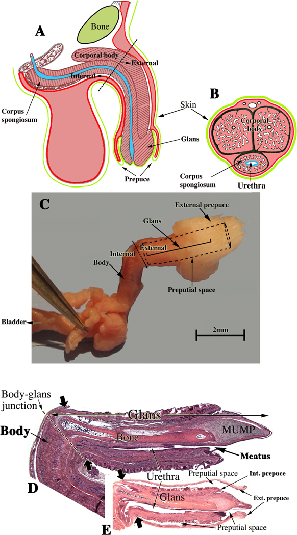
. (D) Mid- sagittal hematoxylin–eosin stained section of the adult mouse penis with the external prepuce removed. (E) The mouse glans penis within the proximal portion of the external prepuce. Note reflection of epithelium of the external prepuce onto the surface of the penis indicated by the large solid arrows in both (D & E). (Adapted from Rodriguez et al. 2011 and Cunha et al. 2015 with permission).
Figure 3.
Figure 3.
Diagrams of divergent development of mouse (A) and human (B) urethral development. (A) The proximal portion of the mouse penile urethra (within the glans) develops via direct canalization of the urethral plate, while the distal aspect of the mouse penile urethra and the urethral meatus forms via epithelial fusion. (B) The distal portion of the human penile urethra (within the glans) develops via direct canalization of the urethral plate, while the proximal portion of the human penile urethra within the shaft forms via fusion of the urethral folds.
Figure 4.
Figure 4.
Human fetal prostates at 15 (A) and 19 weeks (B) stained for smooth muscle actin. At 15 weeks (A) smooth muscle has differentiated ventrally and is just beginning to appear dorsally. By 19 weeks (B) smooth muscle has differentiated circumferentially around the developing prostate. Ductal branching has occurred in the smooth-muscle- poor central region as well as in the peripheral smooth-muscle-rich region.
Figure 5.
Figure 5.
Diagram of the reproductive tract of a neonatal mouse. The fused Müllerian ducts form the cervical canal (Cx) and the Müllerian vagina (M. vag.). The unfused Müllerian ducts form the uterine horns and oviducts. The Müllerian vagina contacts the solid sinus vagina whose epithelium is derived from the urogenital sinus.
Figure 6.
Figure 6.
Diagrams of adult human and mouse female reproductive tracts. In humans most of the Müllerian ducts fuse in the midline and form the uterus, cervix and contribute to vaginal development, with the unfused cranial portions forming the uterine tubes. In mice most of the Müllerian ducts remain unfused to form the oviducts and uterine horns, with the fused portion forming the cervix and contributing to the vagina.
Figure 7.
Figure 7.
Sagittal sections of a 21-week female reproductive tract. (A) depicts the upper portion of the vagina, cervix and uterus (H&E stain). The vaginal epithelium is many layers thick due to endogenous estrogenic stimulation. Note the abrupt transition in epithelial differentiation at the vaginal/cervical border. (B) PAX2 staining of the vaginal/cervical border shows prominent PAX2 immunostaining (indicative of Müllerian duct origin) of the pseudostratified stratified relatively thin cervical epithelium and PAX2-negative stratified squamous vaginal epithelium. (C) Epithelium of the uterine corpus is strongly PAX2-reactive as expected for a Müllerian epithelium. (D & E) FOXA1 nuclear immunostaining was seen uniformly throughout the entire vagina, and FOXA1 nuclear immunostaining abruptly stopped at the vaginal/cervical border (D-E) in mirror image to PAX2 immunostaining (B). (modified from Robboy et al., 2017 with permission). (B & D) are serial sections of the same specimen.
Figure 8.
Figure 8.
Light sheet microscopy of a human female reproductive tract at 9.5 weeks stained with an antibody to E-cadherin. The mesonephros and Wolffian ducts (WD) are present. Cranially, the unfused MDs are destined to form the uterine tubes. Midline fusion of the MDs has created the uterovaginal canal that terminates caudally by joining the urogenital sinus (UGS).

References

    1. Aboseif S, El-Sakka A, Young P and Cunha G (1999) Mesenchymal reprogramming of adult human epithelial differentiation. Differentiation 65:113–118. - PubMed
    1. Andrews GS (1951) The histology of the human foetal and prepubertal prostates. J Anat 85:44–54. - PMC - PubMed
    1. Atkins KA (2019) Normal Histology of the Uterus and Fallopian Tubes In: Mills S (ed) Histology for Pathologists. Lippincott, Philadelphia, pp 1059–1106.
    1. Ayala G, Tuxhorn JA, Wheeler TM, Frolov A, Scardino PT, Ohori M, Wheeler M, Spitler J and Rowley DR (2003) Reactive stroma as a predictor of biochemical-free recurrence in prostate cancer. Clin Cancer Res 9:4792–4801. - PubMed
    1. Baskin L, Shen J, Sinclair A, Cao M, Liu X, Liu G, Isaacson D, Overland M, Li Y and Cunha GR (2018) Development of the human penis and clitoris. Differentiation 103:74–85. - PMC - PubMed

Publication types