. 2019 Oct 3;11(10):914.
doi: 10.3390/v11100914.

Immunological Effects and Viral Gene Expression Determine the Efficacy of Oncolytic Measles Vaccines Encoding IL-12 or IL-15 Agonists

Affiliations

Immunological Effects and Viral Gene Expression Determine the Efficacy of Oncolytic Measles Vaccines Encoding IL-12 or IL-15 Agonists

Paul S Backhaus et al. Viruses. .

Abstract

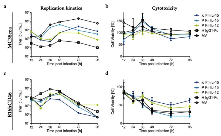
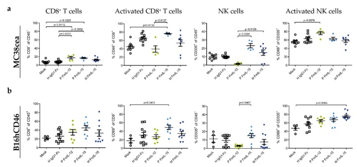
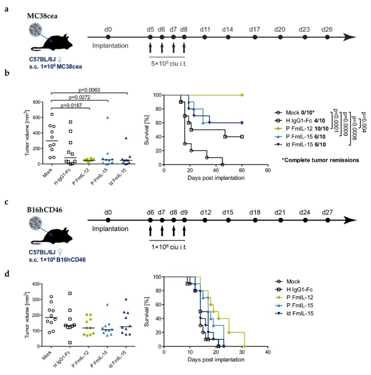
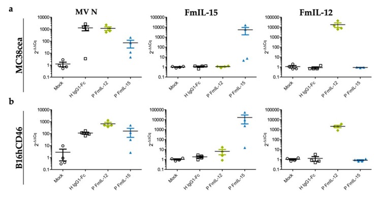
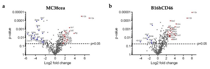
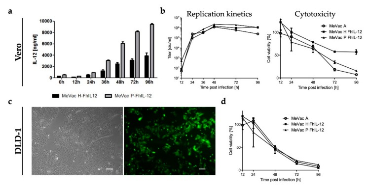
Tumor-targeted immunomodulation using oncolytic viral vectors is currently being investigated as a promising strategy in cancer therapy. In a previous study, we showed that a measles virus Schwarz vaccine strain (MeVac) vector encoding an interleukin-12 fusion protein (FmIL-12) is an effective immunotherapy in the MC38cea murine colon adenocarcinoma model. We hypothesized that MeVac encoding interleukin-15 may mediate enhanced T and NK cell responses and thus increase the therapeutic efficacy, especially in NK cell-controlled tumors. Therefore, we generated MeVac vectors encoding an interleukin-15 superagonist, FmIL-15. Replication and oncolytic capacity, transgene expression, and functionality of MeVac FmIL-15 vectors were validated in vitro. Effects on the tumor immune landscape and therapeutic efficacy of both FmIL-12 and FmIL-15 vectors were studied in the MC38cea and B16hCD46 tumor models. Treatment with MeVac FmIL-15 increased T and NK cell infiltration in both models. However, MeVac FmIL-12 showed more robust viral gene expression and immune activation, resulting in superior anti-tumor efficacy. Based on these results, MeVac encoding a human IL-12 fusion protein was developed for future clinical translation.

Keywords: cancer immunotherapy; interleukin-12; interleukin-15; measles virus; oncolytic virus.


Conflict of interest statement

R.V., C.E.E. and G.U. are listed as inventors on patents regarding RNA viruses for cancer immunotherapy owned by Heidelberg University. G.U. serves as CMO and CSO for CanVirex, which is developing immune-modulating oncolytic viruses. All other authors declare no conflict of interest.

Figures

Figure 1
Figure 1
Cloning of oncolytic measles vaccines encoding FmIL-12 or FmIL-15. (a) Design of the FmIL-12, FmIL-15, and IgG1-Fc expression cassettes. The FmIL-12 cassette consists of the mIL-12 p40 and p35 subunits separated by a (G4S)3 linker preceded by a Kozak sequence. The signal peptide sequence was deleted from the p35 subunit (Δp35). The FmIL-15 cassette consists of a Kozak sequence, sequences encoding the immunoglobulin kappa light chain leader (Igκ ld), the mIL-15Rα sushi domain, a GG(SGG)6 polypeptide linker, and mIL-15 lacking the signal peptide and propeptide (ΔmIL-15). Flanking MluI and PauI restriction sites were added for insertion into the MeVac genome. The IgG1-Fc cassette in control vectors includes Kozak and Igκ ld sequences, HA and myc tags, and the Fc region of human immunoglobulin G type 1 flanked by MluI and AscI restriction sites. (b) Genome schematic. The FmIL-12 expression cassette was inserted into an additional transcription unit downstream of the MeVac P open reading frame (position T2). The FmIL-15 expression cassette was inserted downstream of the MeVac ld or P open reading frame, respectively (positions T1 or T2). The IgG1-Fc cassette was inserted downstream of the H open reading frame (position T3). Retargeting to carcinoembryonic antigen (CEA) was achieved by replacing hemagglutinin (H) with an H variant harboring mutated receptor-binding sites and a CEA-specific scFv fused to its C-terminus (Hbl-αCEA). N, P, M, F, H, and L: Nucleocapsid, phosphoprotein, matrix, fusion, hemagglutinin, and large (polymerase) open reading frames. ld, tr: leader and trailer.
Figure 2
Figure 2
Secretion and bioactivity of MeVac-encoded FmIL-15. (a) MC38cea cells were inoculated with MeVac ld FmIL-15 Hbl-αCEA at a multiplicity of infection (MOI) of 3 in triplicates. FmIL-15 levels in the cell culture supernatant were determined using an ELISA for the murine IL-15/IL-15R complex between 0 and 96 h after inoculation. Mean values of triplicate infections with standard deviations are shown. (b) FmIL-15 was obtained from supernatants of Vero-αHis cells infected with MeVac ld FmIL-15 Hbl-αCEA. Cytokine-dependent CTLL-2 cells were starved from cytokines for 4 h and then stimulated with FmIL-15, rmIL-2 (positive control), or RPMI + 10% FCS (unstimulated). FmIL-15 and rmIL-2 were used in 2-fold serial dilutions from 125 ng/mL to 0.5 ng/mL. After 48 h, cell viability was determined with an XTT cell viability assay. Cell viability is depicted as fold change over cell viability of unstimulated CTLL-2 cells (mock, fold change = 1). Mean values of triplicate stimulations with standard deviations are shown. (c) Undiluted supernatants from Vero-αHis cells infected with MeVac ld FmIL-15 Hbl-αCEA (corresponding to 2500 ng/mL FmIL-15), MeVac ld EGFP (encoding enhanced green fluorescent protein) Hbl-αCEA, or MeVac Hbl-αCEA (without transgene) were used for the CTLL-2 stimulation assay, as described in (b). (d) Previously starved CTLL-2 cells were stimulated with diluted supernatants from Vero-αHis cells infected with MeVac ld FmIL-15 Hbl-αCEA (corresponding to a saturating concentration of 100 ng/mL FmIL-15) or mock stimulation (medium only). After 48 h of stimulation, phosphorylated STAT5 (pSTAT5), total STAT5, and β-actin were detected by Western blot. (e) Cells were treated as described in (d) and mRNA levels of the IL-15 target genes B cell lymphoma 2 (Bcl-2), Cytokine-inducible SH2-containing protein (Cish), Fas ligand (Fasl), and Granzyme B (Gzmb) were determined by quantitative reverse transcription PCR (RT-qPCR). Data are plotted as 2-ΔΔCp values with mL13a as reference gene. Data were analyzed by unpaired t-test. Dots representing individual samples with a line at the mean are shown.
Figure 3
Figure 3
In vitro replication and cytotoxicity of MeVac variants. Murine tumor cells (MC38cea and B16hCD46) were inoculated with the indicated MeVac variants at an MOI of 3 in triplicates. (a,c) To generate one-step growth curves, cells were harvested at indicated timepoints and progeny virus titers of pooled triplicates were determined by titration assay. (b,d) XTT cell viability assays were performed at designated timepoints after infection. Viability of mock-infected cells was defined as 100%. The mean and standard deviation of octuplicate titration wells (a,c) or triplicate infections (b,d) are shown. Note that MeVac variants retargeted to CEA (Hbl-αCEA) were used for inoculation of MC38cea cells, while MeVac variants with native tropism (CD46) were used in case of B16hCD46 cells.
Figure 4
Figure 4
Tumor-infiltrating lymphocytes after MeVac treatment. MC38cea (a) or B16hCD46 (b) tumor cells were implanted subcutaneously in the right flank of C57BL/6J mice. When tumors had reached a size of 50–100 mm³, mice received intratumoral injections of indicated MeVac variants on four consecutive days. Flow cytometry of tumor-infiltrating lymphocytes was performed one day after the fourth treatment. Symbols representing individual animals and the group mean with standard deviation are shown.
Figure 5
Figure 5
Therapeutic efficacy. C57BL/6J mice bearing s.c. MC38cea (a,b) or B16hCD46 (c,d) tumors were treated by intratumoral (i.t.) injections of 1 × 10⁶ ciu of indicated MeVac variants on four consecutive days. (a,c) Treatment schedules. (b,d) Left panels: Tumor volume distribution on day 13 (b) and 10 (d), which was the last day when all mice were alive. Values for individual animals and group median are shown. Right panels: Kaplan–Meier survival analysis.
Figure 6
Figure 6
Viral gene expression in vivo. Samples (n = 4 per group) from MC38cea tumors (a) and B16hCD46 tumors (b) were collected 24 h after the last treatment and total RNA was extracted. RT-qPCR was performed with primers specific for MV N, FmIL-15, and FmIL-12 with mL13A as reference.
Figure 7
Figure 7
Immune expression profiling. RNA was extracted from n = 4 tumors from each treatment group and expression profiling was carried out using the NanoString Mouse Immunology Panel. Volcano plots show differential gene expression between the FmIL-15 and FmIL-12 treatment groups in the (a) MC38cea and (b) B16hCD46 tumor models. Each dot represents one gene. The top 20 significantly upregulated genes in the FmIL-12 versus the FmIL-15 group (Log2 Fold change >1 and p-value < 0.05) are shown in red. The top 20 significantly downregulated genes in the FmIL-12 versus the FmIL-15 group (Log2 Fold change <−1 and p-value < 0.05), i.e., upregulated in the MeVac FmIL-15 group, are shown in blue.
Figure 8
Figure 8
Oncolytic measles vaccine vectors encoding a human IL-12 fusion protein. (a) Vero cells were infected at an MOI of 3 and FhIL-12 in cell culture supernatants was quantified by ELISA. (b) Left panel: To generate one-step growth curves, Vero cells were infected at an MOI of 3 and viral progeny at designated timepoints were quantified by titration assay. Right panel: XTT cell viability assay was performed after infection of Vero cells at an MOI of 1. (c) DLD-1 human colon cancer cells were subjected to mock treatment (left panel) or inoculated with MeVac ld EGFP at an MOI of 1. Images were acquired 48 h after inoculation. Scale bars: 100 µm. (d) DLD-1 cells were inoculated with indicated MeVac variants at an MOI of 1 and cell viability was determined by XTT assay at designated timepoints. Cell viability data is presented as a percentage of mock (100%). Mean and standard deviation of triplicate samples are shown.

References

    1. Harrington K., Freeman D.J., Kelly B., Harper J., Soria J.C. Optimizing oncolytic virotherapy in cancer treatment. Nat. Rev. Drug Discov. 2019;18:689–706. doi: 10.1038/s41573-019-0029-0. - DOI - PubMed
    1. Twumasi-Boateng K., Pettigrew J.L., Kwok Y.Y.E., Bell J.C., Nelson B.H. Oncolytic viruses as engineering platforms for combination immunotherapy. Nat. Rev. Cancer. 2018;18:419–432. doi: 10.1038/s41568-018-0009-4. - DOI - PubMed
    1. Pol J., Kroemer G., Galluzzi L. First oncolytic virus approved for melanoma immunotherapy. Oncoimmunology. 2016;5:e1115641. doi: 10.1080/2162402X.2015.1115641. - DOI - PMC - PubMed
    1. Robinson S., Galanis E. Potential and clinical translation of oncolytic measles viruses. Expert Opin. Biol. Ther. 2017;17:353–363. doi: 10.1080/14712598.2017.1288713. - DOI - PMC - PubMed
    1. Aref S., Bailey K., Fielding A. Measles to the rescue: A review of oncolytic measles virus. Viruses. 2016;8:294. doi: 10.3390/v8100294. - DOI - PMC - PubMed

Publication types

MeSH terms

LinkOut - more resources