. 2019 Oct 25;366(6464):454-460.
doi: 10.1126/science.aax7526. Epub 2019 Oct 17.

Spatiotemporal expansion of primary progenitor zones in the developing human cerebellum

Affiliations

Spatiotemporal expansion of primary progenitor zones in the developing human cerebellum

Parthiv Haldipur et al. Science. .

Abstract

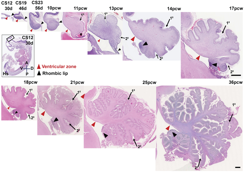
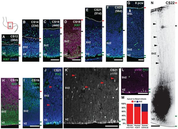
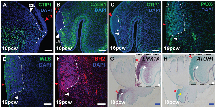
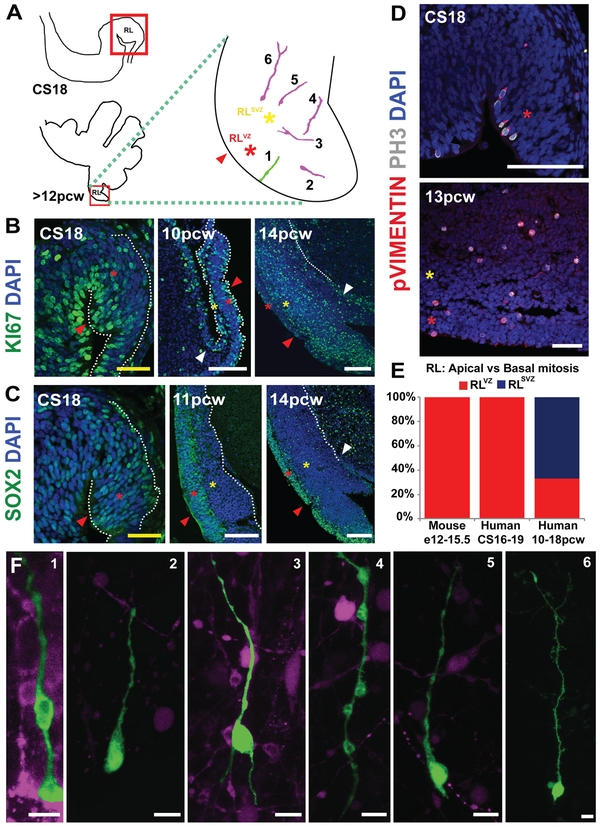
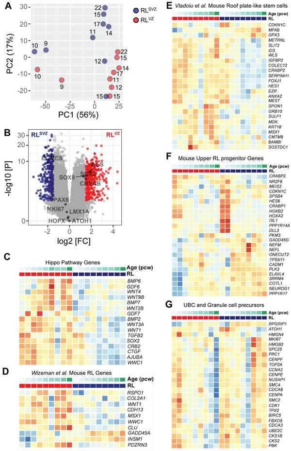
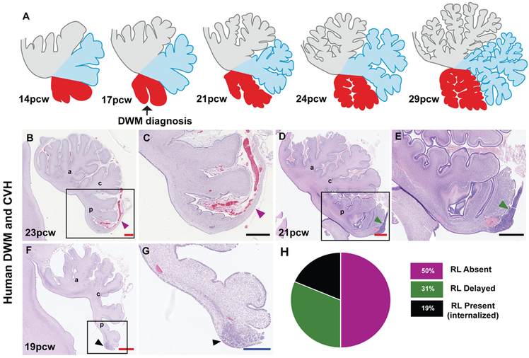
We present histological and molecular analyses of the developing human cerebellum from 30 days after conception to 9 months after birth. Differences in developmental patterns between humans and mice include spatiotemporal expansion of both ventricular and rhombic lip primary progenitor zones to include subventricular zones containing basal progenitors. The human rhombic lip persists longer through cerebellar development than in the mouse and undergoes morphological changes to form a progenitor pool in the posterior lobule, which is not seen in other organisms, not even in the nonhuman primate the macaque. Disruptions in human rhombic lip development are associated with posterior cerebellar vermis hypoplasia and Dandy-Walker malformation. The presence of these species-specific neural progenitor populations refines our insight into human cerebellar developmental disorders.


Figures

Fig. 1:
Fig. 1:. An outline of human cerebellar development.
H&E-stained midsagittal sections of the developing human cerebellum. The two main zones of neurogenesis, the ventricular zone (red arrowhead) and rhombic lip (black arrow head) are marked. Primary and secondary fissures are noted (arrows). Scale bar = 100 μm
Fig. 2:
Fig. 2:. The human cerebellar VZ is expanded into a SVZ.
Midsagittal sections of the human embryonic cerebellum stained with KI67 (A-G) and SOX2 (D), reveal VZ expansion. (H) β-III Tubulin (TUJ1) and CTIP1 (I) expression suggest neuronal differentiation beginning around CS19 takes place in the SVZ. (G) External granule layer (EGL) first appears at 8pcw. (J-L) Mitosis and mitotic radial glia are observed in both the VZ and SVZ as evidenced by phospho-histone H3 (PH3) (J) and phospho-vimentin (pVIM) (K, L) expression. (M) A significant increase in the proportion of cerebellar basal progenitors (J-K, red arrowheads) is seen between the human and mouse cerebellum, and between CS18-23 (Chi-square, df: 132.5, 2; P value <0.0001). (N) DiO labeling of VZ/SVZ progenitors at CS22 show radial glial fibers traversing the thickness of the cerebellum. Sections were counterstained using DAPI. Green and black dashed lines and the dotted line mark the boundaries of the VZ, SVZ and pia respectively. Scale bar = 100 μm.
Fig. 3:
Fig. 3:. The human cerebellar rhombic lip expresses classic markers.
(A) VZ born Purkinje cells expressing CTIP1 (A,C) and Calbindin (B) migrate around the RL (red arrowhead). The RL expresses classic markers like PAX6 (D), WLS (E), TBR2 (F) and LMX1A (G). (H) ATOH1 is expressed by cells exiting the RL into the external granule layer (white arrowhead). Sections were counterstained with DAPI (A-F) and Fast green (G,H). Scale bar = 100 μm (white), 500 μm (blue).
Fig. 4:
Fig. 4:. The human cerebellar RL is compartmentalized into ventricular and subventricular zones.
(A) Illustration of the cerebellar regions studied in this figure. The RL (red arrowhead) is expanded into ventricular (RLVZ, red asterisk) and subventricular zones (RLSVZ, yellow asterisk). (B) KI67 expression reveals extensive proliferation in the RL. (C) The RL is compartmentalized into a SOX2-rich RLVZ and SOX2-sparse RLSVZ. (D) Phosphohistone-H3 and phospho-vimentin expression indicate the presence of mitotic ventricular and subventricular-basal progenitors. (E) The proportion of basal progenitors increases significantly following the splitting of the RL (Chi-square, df: 137.8, 2; P value <0.0001). (F) DiI and lentiviral labeling of an organotypic slice of the human cerebellum reveal diverse morphologies of RL ventricular and basal progenitors. Scale bar = 100 μm (white), 50 μm (yellow).
Fig. 5:
Fig. 5:. RNA-seq of human RL compartments.
(A) Principal component (PC) analysis indicates the largest source of variation among the RNA-seq samples was age accounting for 56% of the variance in the data. Samples microdissected from RLSVZ are blue and RLVZ are red. The numbers beside each circle represent sample age (pcw). (B) Volcano plot illustrating differential expression of genes in RLVZ versus RLSVZ. Red and blue dots represent genes expressed significantly higher in RLVZ or RLSVZ, respectively. (C-G) Heatmaps of gene expression for each human sample. Samples are grouped by RLVZ (red) and RLSVZ (blue), then ascending age (9-22pcw). (C) Expression of Hippo pathway genes and (D-G) top genes for RL-related cell clusters identified by single-cell RNA-sequencing of the mouse cerebellum from E10-P14 (23,24).
Fig. 6:
Fig. 6:. Internalized RL may be a feature specific to human cerebellar development.
(A) Model for human cerebellar development indicates growth of the posterior vermis correlates with spatiotemporal RL expansion. (B-G) Analysis of H&E-stained sagittal sections of the human cerebellum from cases diagnosed with Dandy-Walker malformation (DWM) and cerebellar vermis hypoplasia (CVH) indicates the RL is absent in 50% of cases (B,C) while being severely diminished remaining cases (D-G). The anterior (a), central (c) and posterior lobes (p) are indicated. (H) Pie chart representing the absence of RL in 50% of tested samples. Scale bar = 100 μm (black), 0.5 mm (blue) and 1 mm (red).

Comment in

References

    1. Lange W, Cell number and cell density in the cerebellar cortex of man and some other mammals. Cell Tissue Res 157, 115 (1975). - PubMed
    1. Van Essen DC, Surface-based atlases of cerebellar cortex in the human, macaque, and mouse. Ann N Y Acad Sci 978, 468 (December, 2002). - PubMed
    1. Larsell O. a. J. J., The Comparative Anatomy and Histology of the Cerebellum The Human Cerebellum, Cerebellar Connections and Cerebellar Cortex. University of Minnesota Press; Minneapolis, (1972).
    1. Haldipur P et al., Preterm delivery disrupts the developmental program of the cerebellum. PLoS One 6, e23449 (2011). - PMC - PubMed
    1. Rakic P, Sidman RL, Histogenesis of cortical layers in human cerebellum, particularly the lamina dissecans. J Comp Neurol 139, 473 (August, 1970). - PubMed

Publication types