. 2019 Nov 29;294(48):18077-18091.
doi: 10.1074/jbc.RA119.010716. Epub 2019 Oct 17.

The molecular basis of endolytic activity of a multidomain alginate lyase from Defluviitalea phaphyphila, a representative of a new lyase family, PL39

Affiliations

The molecular basis of endolytic activity of a multidomain alginate lyase from Defluviitalea phaphyphila, a representative of a new lyase family, PL39

Shiqi Ji et al. J Biol Chem. .

Abstract

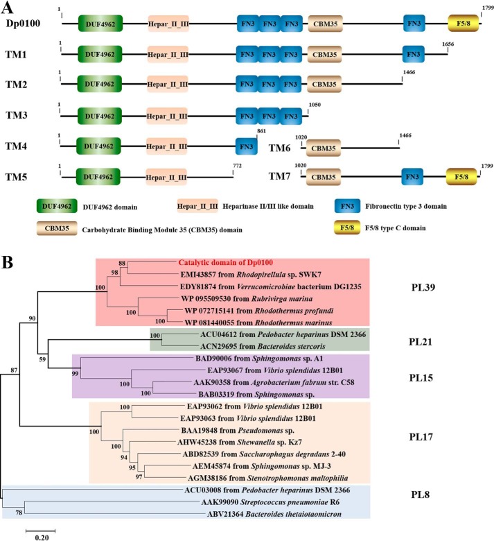
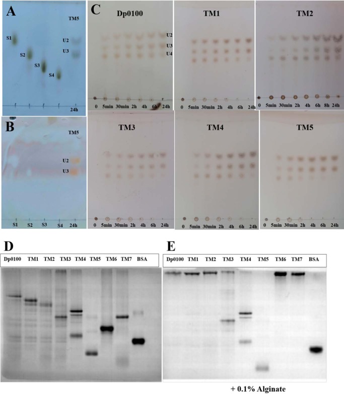
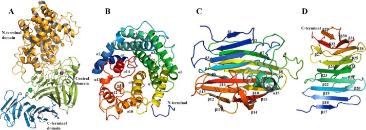
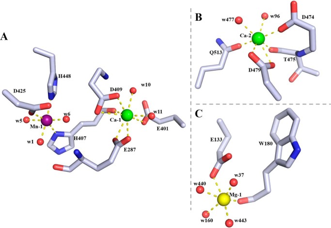
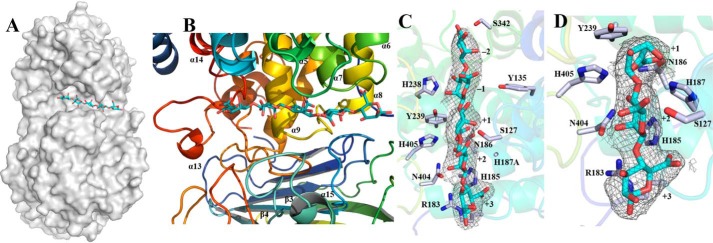
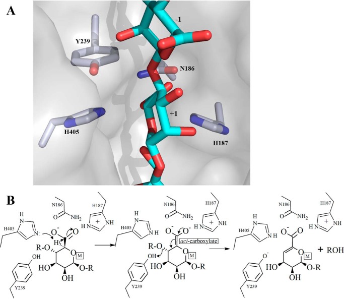
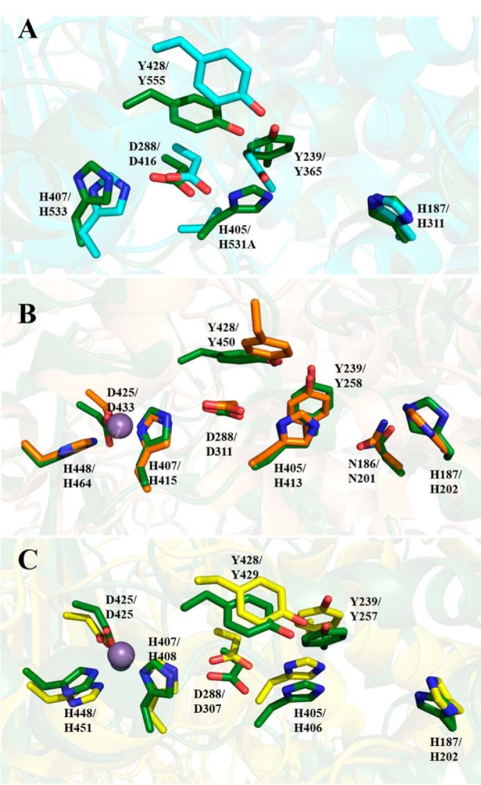
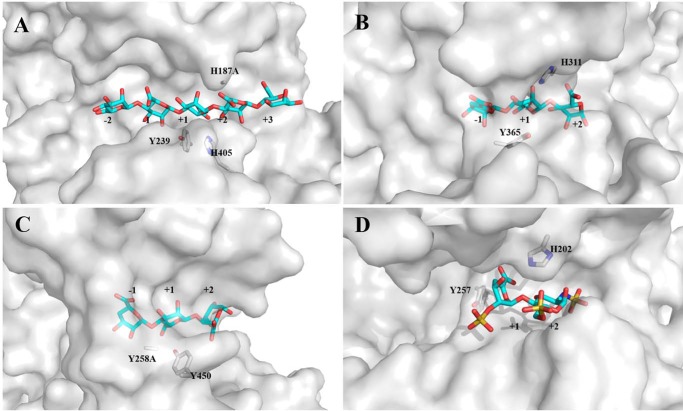
Alginate is a polymer containing two uronic acid epimers, β-d-mannuronate (M) and α-l-guluronate (G), and is a major component of brown seaweed that is depolymerized by alginate lyases. These enzymes have diverse specificity, cleaving the chain with endo- or exotype activity and with differential selectivity for the sequence of M or G at the cleavage site. Dp0100 is a 201-kDa multimodular, broad-specificity endotype alginate lyase from the marine thermophile Defluviitalea phaphyphila, which uses brown algae as a carbon source, converting it to ethanol, and bioinformatics analysis suggested that its catalytic domain represents a new polysaccharide lyase family, PL39. The structure of the Dp0100 catalytic domain, determined at 2.07 Å resolution, revealed that it comprises three regions strongly resembling those of the exotype lyase families PL15 and PL17. The conservation of key catalytic histidine and tyrosine residues belonging to the latter suggests these enzymes share mechanistic similarities. A complex of Dp0100 with a pentasaccharide, M5, showed that the oligosaccharide is located in subsites -2, -1, +1, +2, and +3 in a long, deep canyon open at both ends, explaining the endotype activity of this lyase. This contrasted with the hindered binding sites of the exotype enzymes, which are blocked such that only one sugar moiety can be accommodated at the -1 position in the catalytic site. The biochemical and structural analyses of Dp0100, the first for this new class of endotype alginate lyases, have furthered our understanding of the structure-function and evolutionary relationships within this important class of enzymes.

Keywords: alginate lyase; carbohydrate-binding protein; crystal structure; metalloenzyme; oligosaccharide; structure–function; substrate specificity.


Conflict of interest statement

The authors declare that they have no conflicts of interest with the contents of this article

Figures

Figure 1.
Figure 1.
A, schematic diagram of the modular structure of Dp0100 and its truncation mutants. B, phylogenetic analysis of the catalytic domain of Dp0100 (TM5) and its close relatives from PL8, PL15, PL17, and PL21. The percentage of replicate trees in which the associated taxa clustered together in the bootstrap test (1000 replicates) is shown next to the branches (57). The tree is drawn to scale, with branch lengths in the same units as those of the evolutionary distances used to infer the phylogenetic tree.
Figure 2.
Figure 2.
TLC analysis of the products released from alginate and affinity of Dp0100 and its TM1–TM7 constructs for soluble alginate by native affinity gel electrophoresis. A and B, marker sugars (1st lane, sodium d-mannuronate (S1); 2nd lane, sodium d-dimannuronate (S2); 3rd lane, sodium d-trimannuronate (S3); and 4th lane, sodium d-tetramannuronate (S4)) and the products of TM5 (unsaturated U2 and U3, 5th lane) after 24 h of incubation with alginate stained by the sulfuric acid/ethanol and TBA methods, respectively. Note that unsaturated sugars on the plate run faster than those of the equivalent saturated oligosaccharides (S1–S4) as reported previously (58, 59), and the saturated sugars are not stained by the TBA method (35, 59). C, unsaturated oligosaccharide products (U2, U3, and U4) of Dp0100 and TM1–TM5. 1st to 7th lanes represent incubation for 0, 5, and 30 min and 2, 4, 6, and 24 h, respectively. Plates in C were visualized by spraying with sulfuric acid in ethanol. D, 10% native-PAGE without alginate. E, 10% native-PAGE supplemented with 0.1% (w/v) alginate. 1st to 8th lanes represent Dp0100 and TM1–TM7 respectively; 9th lane contains BSA (0.2 μg) as a control.
Figure 3.
Figure 3.
Structure of the catalytic domain of Dp0100 (TM5). A, schematic diagram of the fold of TM5 showing its N-terminal (orange), central (green), and C-terminal domains (sky blue). The positions of the Mn2+, Ca2+, and Mg2+ ions are shown as purple, green, and yellow spheres, respectively. B–D, schematic diagrams of the N-terminal, central, and C-terminal domains drawn in a rainbow format (blue to red from the N to C termini) to show their secondary structure elements, which are labeled. Figure was prepared using PyMOL (56).
Figure 4.
Figure 4.
Metal-binding sites in the catalytic domain of Dp0100. A, binding site of the Mn2+ ion (Mn-1, purple sphere) and its neighboring Ca2+ ion (Ca-1, green sphere). Amino acid residues (drawn in atom colors (oxygen, red; carbon, gray; and nitrogen, blue)) and water molecules (red spheres) in the coordination spheres of the metal ions are shown. B, second Ca2+ ion–binding site (Ca-2) colored as in A. C, Mg2+ ion–binding site (Mg-1, yellow sphere) drawn as in A. Dotted lines indicate interactions between metal ions and ligands in the coordinate sphere. Figure was prepared using PyMOL (56).
Figure 5.
Figure 5.
Substrate-binding groove of TM5. A, surface representation to show the binding site of M5 (shown as sticks and drawn in atom colors (oxygen, red; carbon, cyan)) in the catalytic domain of Dp0100. B, schematic diagrams to show the binding site of M5 and the surrounding elements of secondary structure (colored as in Fig. 3). C and D, electron density surrounding the substrates (shown as in A) in the (2FoFc) omit map (contoured at 1.0σ) in complexes of the H187A mutant with M5 and the WT enzyme with ΔMM, respectively. The sugar-binding subsites are numbered in C and D. Key residues interacting with the substrates are highlighted (drawn in atom colors (oxygen, red; carbon, gray; and nitrogen, blue)) with the remainder of the enzyme shown transparently in cartoon format. Figure was prepared using PyMOL (56).
Figure 6.
Figure 6.
Schematic diagram of the active site of Dp0100 and the proposed mechanism. A, relative orientation of key functional groups on the enzyme with respect to the substrate is shown. The coordinates and the associated molecular surface displayed are those of the WT enzyme combined with M5 oligosaccharide. The position of the latter is modeled based on the superposition of the WT enzyme onto the structure of the complex of the inactive mutant, H187A, with M5. The sugar-binding subsites are numbered. The proximity of the residues proposed to be involved in the acid/base catalysis (His-405 and Tyr-239) can be seen. Figure was prepared using PyMOL (56). The color scheme is the same as that for Fig. 4. B, proposal for the catalytic mechanism of depolymerization of polyM by Dp0100.
Figure 7.
Figure 7.
Superposition of Dp0100 and related PL family enzymes. A–C, conserved residues around the catalytic center between TM5 (6JP4), PL15 alginate lyase (3AFL), PL17 (4NEI), and PL21 (2FUT), respectively. The conserved residues of these structures are drawn in oxygen, red; nitrogen, blue; and carbon, as green, teal, orange, and yellow, respectively. Residue numbers of Dp0100 are shown first alongside those of their counterparts in the other PL family enzymes. Figure was prepared using PyMOL (56).
Figure 8.
Figure 8.
Surface representations of the substrate-binding site in Dp0100 and related enzymes. A, H187A mutant in Dp0100 in complex with M5. B, H531A mutant of the PL15 alginate lyase from A. tumefaciens in complex with ΔGGG. C, Y258A mutant of the PL17 alginate lyase from S. degradans in complex with ΔMMG. D, heparinase II of PL21 from P. heparinus in complex with a disaccharide product from heparin. Selected residues proposed to be important in catalysis and the oligosaccharides are shown as sticks and drawn in atom colors (oxygen, red; carbon, cyan/gray for the oligosaccharide/protein, respectively, sulfur, yellow; and nitrogen, blue). Figure was prepared using PyMOL (56).

References

    1. Chung I. K., Beardall J., Mehta S., Sahoo D., and Stojkovic S. (2011) Using marine macroalgae for carbon sequestration: a critical appraisal. J. Appl. Phycol. 23, 877–886 10.1007/s10811-010-9604-9 - DOI
    1. Smith S. V. (1981) Marine macrophytes as a global carbon sink. Science 211, 838–840 10.1126/science.211.4484.838 - DOI - PubMed
    1. Enquist-Newman M., Faust A. M., Bravo D. D., Santos C. N., Raisner R. M., Hanel A., Sarvabhowman P., Le C., Regitsky D. D., Cooper S. R., Peereboom L., Clark A., Martinez Y., Goldsmith J., Cho M. Y., et al. (2014) Efficient ethanol production from brown macroalgae sugars by a synthetic yeast platform. Nature 505, 239–243 10.1038/nature12771 - DOI - PubMed
    1. Wargacki A. J., Leonard E., Win M. N., Regitsky D. D., Santos C. N., Kim P. B., Cooper S. R., Raisner R. M., Herman A., Sivitz A. B., Lakshmanaswamy A., Kashiyama Y., Baker D., and Yoshikuni Y. (2012) An engineered microbial platform for direct biofuel production from brown macroalgae. Science 335, 308–313 10.1126/science.1214547 - DOI - PubMed
    1. Gacesa P. (1988) Alginates. Carbohyd. Polym. 8, 161–182 10.1016/0144-8617(88)90001-X - DOI

Publication types

MeSH terms

Substances

Supplementary concepts