. 2020 May 8;29(7):1068-1082.
doi: 10.1093/hmg/ddz231.

Functional characterization of a novel PBX1 de novo missense variant identified in a patient with syndromic congenital heart disease

Affiliations

Functional characterization of a novel PBX1 de novo missense variant identified in a patient with syndromic congenital heart disease

Dimuthu Alankarage et al. Hum Mol Genet. .

Abstract

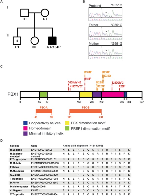
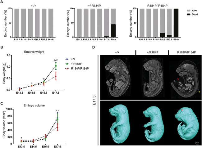
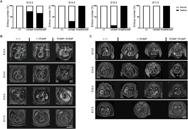
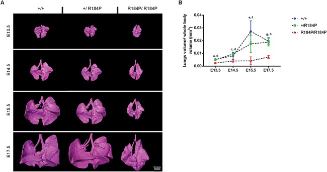
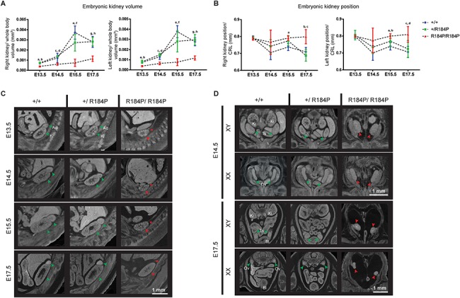
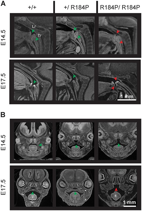
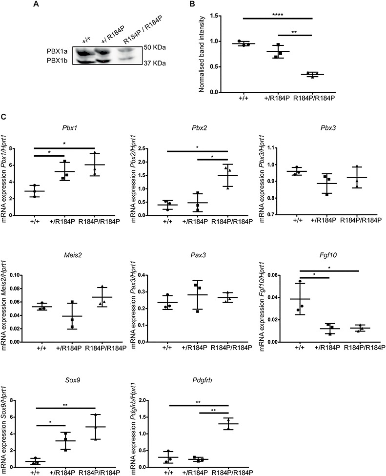
Pre-B cell leukemia factor 1 (PBX1) is an essential developmental transcription factor, mutations in which have recently been associated with CAKUTHED syndrome, characterized by multiple congenital defects including congenital heart disease (CHD). During analysis of a whole-exome-sequenced cohort of heterogeneous CHD patients, we identified a de novo missense variant, PBX1:c.551G>C p.R184P, in a patient with tetralogy of Fallot with absent pulmonary valve and extra-cardiac phenotypes. Functional analysis of this variant by creating a CRISPR-Cas9 gene-edited mouse model revealed multiple congenital anomalies. Congenital heart defects (persistent truncus arteriosus and ventricular septal defect), hypoplastic lungs, hypoplastic/ectopic kidneys, aplastic adrenal glands and spleen, as well as atretic trachea and palate defects were observed in the homozygous mutant embryos at multiple stages of development. We also observed developmental anomalies in a proportion of heterozygous embryos, suggestive of a dominant mode of inheritance. Analysis of gene expression and protein levels revealed that although Pbx1 transcripts are higher in homozygotes, amounts of PBX1 protein are significantly decreased. Here, we have presented the first functional model of a missense PBX1 variant and provided strong evidence that p.R184P is disease-causal. Our findings also expand the phenotypic spectrum associated with pathogenic PBX1 variants in both humans and mice.


Figures

Figure 1
Figure 1
A novel de novo variant in PBX1 was identified in a patient with syndromic CHD by WES. (A) Pedigree of the family with the affected individual (black) carrying the de novo PBX1 variant PBX1:c.551G>C p.R184P. The patient presented with TOF with absent pulmonary valve and later also diagnosed with bronchiolitis, autism, hyperechogenic kidneys, skeletal (clinodactyly) and craniofacial defects (broad nasal tip, thick lips, prominent philtrum and anteverted ears) (B) De novo presence of the variant was confirmed by Sanger sequencing. The older sister’s DNA was not available for testing (NT). Asterisk represents the position of the nucleotide variation in the proband (c.551G>C) versus the reference allele in the parents (c.551G). (C) A schematic representation of the PBX1 protein indicating the functional domains; PREP1 dimerization domain (green), PBX dimerization domain (yellow), inhibitory helix (purple), homeodomain (pink) and cooperativity helices (Blue). The positions of the pathogenic loss-of-function (red) and missense (orange) variants that are currently reported in the literature are presented. (D) Amino acid sequence alignment of reference and mutated human PBX1 protein (residues 181–195) with selected eukaryotes shows that R184 is within a highly conserved region of the protein and that the arginine residue is highly conserved between species.
Figure 2
Figure 2
Functional assessment of the R184P knock-in mouse model. (A). Embryo survival was analysed at six stages during embryonic development, embryonic day (E) 11.5, E13.5, E14.5, E15.5, E17.5 and birth. Embryonic death was observed in both homozygous and heterozygous embryos. Death during development was not observed in wild-type littermates (n > 40 per stage). (B) Measurement of body weights of wild-type, heterozygous and homozygous embryos shows that homozygous embryos displayed lower body weight compared to wild-type and heterozygous embryos during development (n ≥ 12 per stage; a, P < 0.01; b, P < 0.05; c, P < 0.01; d, P < 0.001). (C) Whole body volume quantification of embryos during development shows that homozygous embryos have lower body volumes during late development (n ≥ 12 per stage; a, P < 0.05; b, P < 0.05; c, P < 0.05). (D) Whole embryo analysis by microCT shows the dysmorphic homozygous embryos at E17.5. Top panel: red arrow, umbilical hernia; yellow arrow, subcutaneous oedema. Bottom panel: red arrow, open eyes. Results are presented as mean ± SD.
Figure 3
Figure 3
Analysis of heart defects observed in the R184P knock-in mouse model. (A) Heart defects were observed in homozygous embryos throughout development and in a proportion of heterozygous embryos until E15.5 (n ≥ 14 per stage). (B) Homozygous embryos exhibited PTA at all stages examined (E13.5–E17.5). (C) VSDs were observed in homozygous embryos examined at all stages. VSDs were also observed in a proportion of heterozygous embryos at E13.5, E14.5 and E15.5 (see A of this figure). AO, aorta; MPA, main pulmonary trunk; RVOT, right ventricular outflow tract; PTA, persistent truncus arteriosus; RV, right ventricle; LV, left ventricle; VSD, ventricular septal defect. Green arrows, typical heart morphology; red arrows, variation from the norm or presence of a defect.
Figure 4
Figure 4
Analysis of lung development in R184P knock-in mouse model. (A) Lungs in the homozygous embryos were smaller and dysmorphic compared to heterozygous and wild-type lungs at all analysed stages. (B) Quantification of the 3D reconstructions of the lungs showed a significant decrease in lung volume of the homozygous embryos at all stages (n ≥ 12 per stage; a and b, P < 0.0001; c, P < 0.05; d, P < 0.0001; e and f, P < 0.01; g and h, P < 0.0001). Results are presented as mean ± SD.
Figure 5
Figure 5
Analysis of urogenital phenotypes of the R184P knock-in mouse model. (A) Quantification of 3D reconstructions of the embryonic kidneys showed that both kidneys (right kidney and left kidney) of the homozygous embryos remained consistently smaller than the kidneys of the heterozygous and wild-type embryos at all stages examined. (Right kidney: n ≥ 12; a, P < 0.001; b, P < 0.0001; c, P < 0.05; d, P < 0.0001; e, P < 0.001; f, P < 0.01; g, P < 0.001; h, P < 0.0001. Left kidney: n ≥ 12; a and b, P < 0.001; c, P < 0.01; d, P < 0.0001; e, P < 0.0001; f, P < 0.01; g and h, P < 0.0001). (B) Measurement of the position of the kidneys within the body cavity relative to crown-to-rump length (CRL). In homozygous embryos, the position of the kidneys remains unchanging between E13.5 and E17.5. In both heterozygous and wild-type embryos, the kidneys ascend within the body cavity. (Right kidney: n ≥ 13; a, P < 0.01; b, P < 0.01; c, P < 0.001. Left kidney: n ≥ 13; a, P < 0.001; b, P < 0.05; c, P < 0.01; d, P < 0.001). (C) MicroCT analysis of the hypoplastic, pelvic kidneys of the homozygous embryos. Adrenal glands are absent in the homozygous embryos from the earliest stage examined at E13.5. (D) Gonads of homozygous R184P XY and XX embryos are dysmorphic and situated ectopically, presented at E14.5 and E17.5. Ad, adrenal glands; Bl, bladder; Ki, kidneys; Li, liver; Ov, ovary; Ts, testis. Green arrows, typical organ development. Red arrows, deviation from the norm or presence of defect. Red asterisk, absence of adrenal gland. Results are presented as mean ± SD.
Figure 6
Figure 6
Defects in trachea, larynx and palate were observed in R184P homozygous mice. (A) Larynges of the homozygous embryos fail to open during development. In wild-type and heterozygous embryos, the larynx is open from E13.5. The trachea of the homozygous embryos remains atretic at all examined stages. The trachea of wild-type and heterozygous embryos are patent by E17.5 (n ≥ 14 per stage). (B) The secondary palate of the homozygous embryos does not fuse. Palatal defects are not observed in wild-type and heterozygous embryos (n ≥ 14 per stage). Data are presented at E14.5 and E17.5. Lr, larynx; Tr, trachea. Green arrows, typical organ development. Red arrows, deviation from the norm or presence of defect.
Figure 7
Figure 7
Assessment of PBX1 protein and mRNA expression in the R184P mouse model. (A) Western blot analysis of PBX1 protein expression in E11.5 embryos revealed that the protein is present at lower levels in the homozygous mutant embryos. (B) Quantification of PBX1 protein normalized to total protein loaded per lane (n = 3 embryos per genotype, **P < 0.01, ****P < 0.0001). (C) Analysis of the Pbx1, Pbx2, Pbx3, Meis2, Pax3, Fgf10, Sox9 and Pdgfrb transcripts in E11.5 embryos relative to Hprt1 transcript levels. The expression of Pbx1 and Sox9 are up-regulated in heterozygous and homozygous embryos. Expression of Pbx2 and Pdgfrb are up-regulated in homozygous embryos. The expression of Fgf10 is down-regulated in heterozygous and homozygous embryos. The expression of Pbx3, Meis2 and Pax3 are not altered (n = 3 embryos per genotype,*P < 0.05, **P < 0.01). Results are presented as mean ± SD.

References

    1. van der Linde D., Konings E.E.M., Slager M.A., Witsenburg M., Helbing W.A., Takkenberg J.J.M. and Roos-Hesselink J.W. (2011) Birth prevalence of congenital heart disease worldwide. J Am. Coll. Cardiol., 58, 2241–2247. - PubMed
    1. Blue G.M., Kirk E.P., Giannoulatou E., Sholler G.F., Dunwoodie S.L., Harvey R.P. and Winlaw D.S. (2017) Advances in the genetics of congenital heart disease a Clinician's guide. J. Am. Coll. Cardiol., 69, 860–870. - PubMed
    1. Alankarage D., Ip E., Szot J.O., Munro J., Blue G.M., Harrison K., Cuny H., Enriquez A., Troup M., Humphreys D.T. et al. (2019) Identification of clinically actionable variants from genome sequencing of families with congenital heart disease. Genetics In Medicine., 21, 1111–1120. - PubMed
    1. Simmons M.A. and Brueckner M. (2017) The genetics of congenital heart disease...Understanding and improving long-term outcomes in congenital heart disease: a review for the general cardiologist and primary care physician. Curr. Opin. Pediatr., 29, 520–528. - PMC - PubMed
    1. Szot J.O., Cuny H., Blue G.M., Humphreys D.T., Ip E., Harrison K., Sholler G.F., Giannoulatou E., Leo P., Duncan E.L. et al. (2018) A screening approach to identify clinically actionable variants causing congenital heart disease in exome data. Circ. Genom. Precis. Med., 11:e001978. - PubMed

Publication types

MeSH terms

Substances