. 2019 Dec 1;28(23):3954-3969.
doi: 10.1093/hmg/ddz213.

Model system identification of novel congenital heart disease gene candidates: focus on RPL13

Affiliations

Model system identification of novel congenital heart disease gene candidates: focus on RPL13

Analyne M Schroeder et al. Hum Mol Genet. .

Abstract

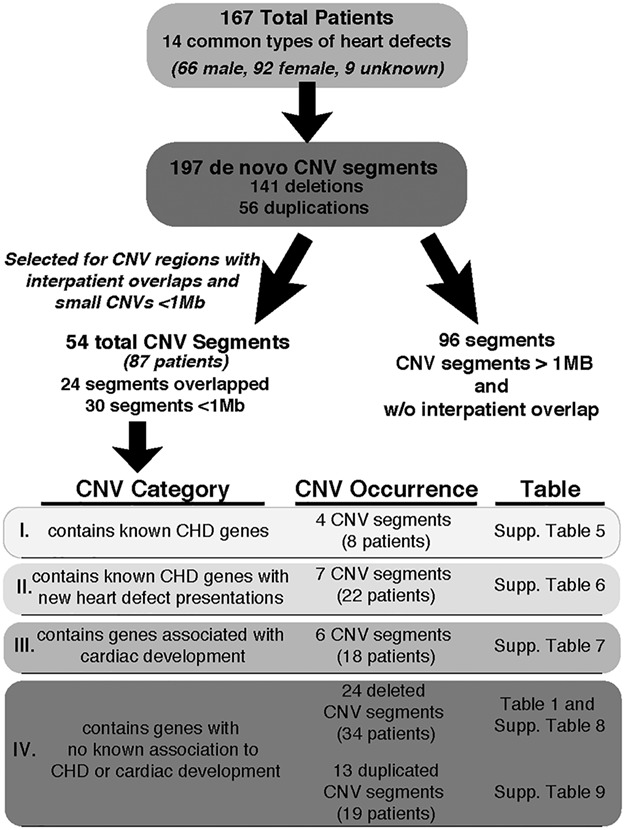
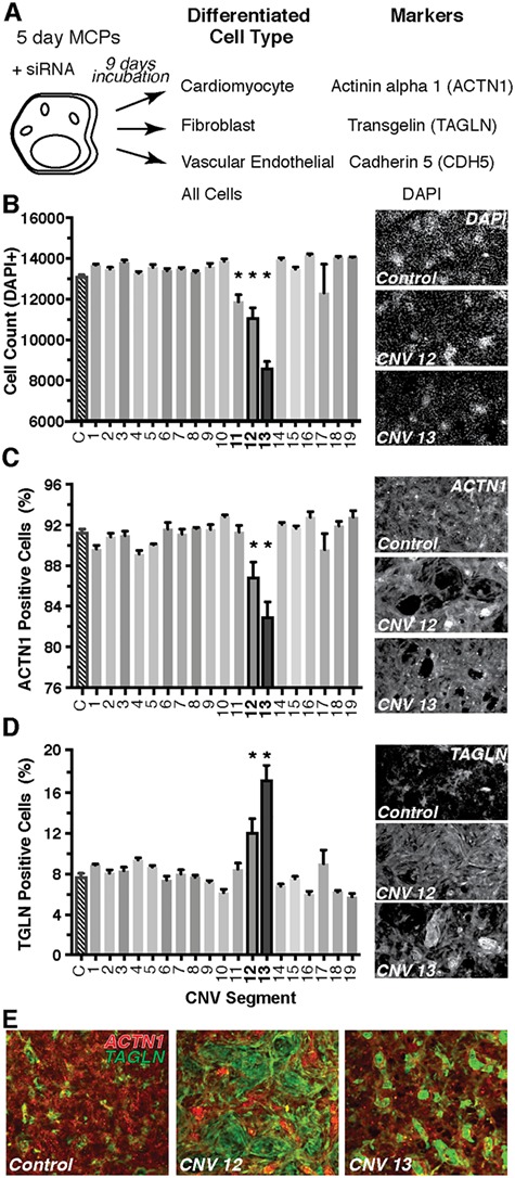
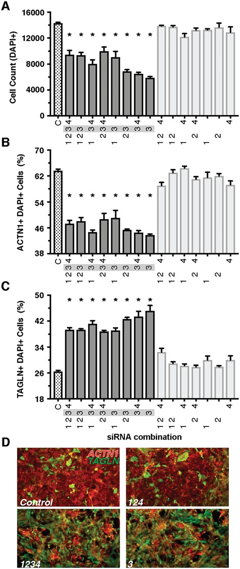
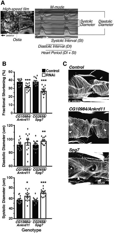
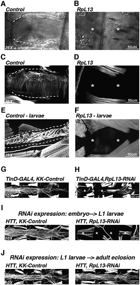
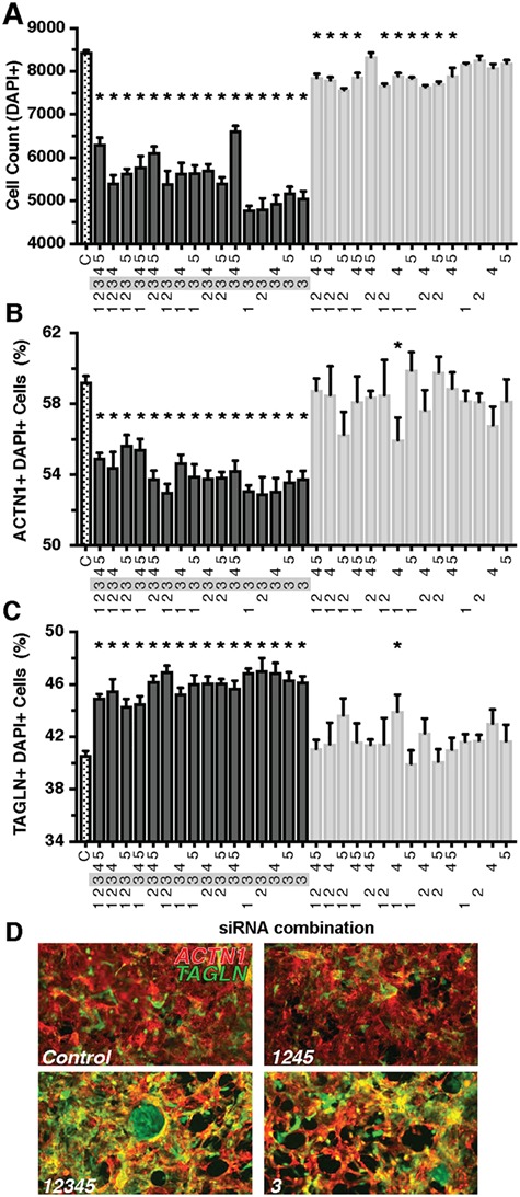
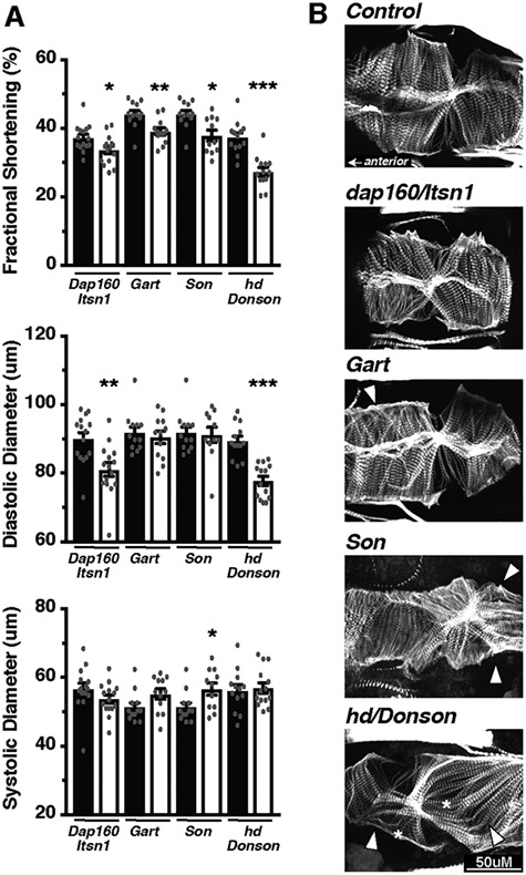
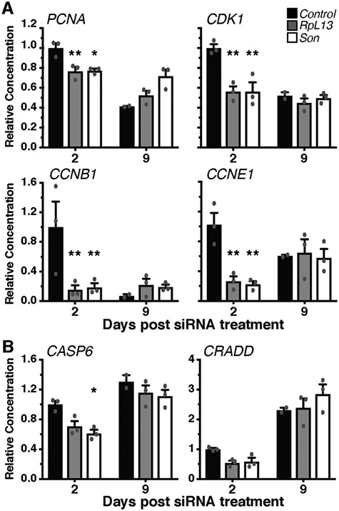
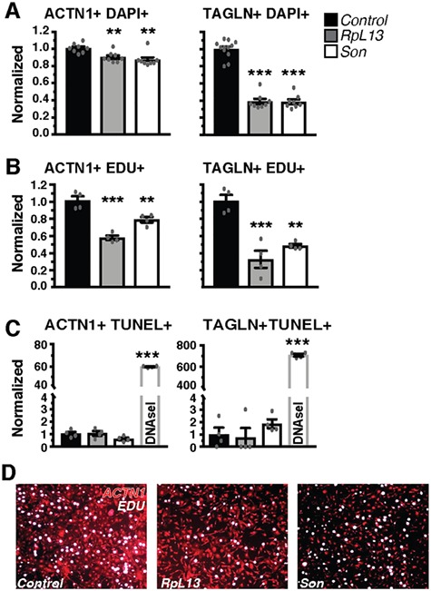
Genetics is a significant factor contributing to congenital heart disease (CHD), but our understanding of the genetic players and networks involved in CHD pathogenesis is limited. Here, we searched for de novo copy number variations (CNVs) in a cohort of 167 CHD patients to identify DNA segments containing potential pathogenic genes. Our search focused on new candidate disease genes within 19 deleted de novo CNVs, which did not cover known CHD genes. For this study, we developed an integrated high-throughput phenotypical platform to probe for defects in cardiogenesis and cardiac output in human induced pluripotent stem cell (iPSC)-derived multipotent cardiac progenitor (MCPs) cells and, in parallel, in the Drosophila in vivo heart model. Notably, knockdown (KD) in MCPs of RPL13, a ribosomal gene and SON, an RNA splicing cofactor, reduced proliferation and differentiation of cardiomyocytes, while increasing fibroblasts. In the fly, heart-specific RpL13 KD, predominantly at embryonic stages, resulted in a striking 'no heart' phenotype. KD of Son and Pdss2, among others, caused structural and functional defects, including reduced or abolished contractility, respectively. In summary, using a combination of human genetics and cardiac model systems, we identified new genes as candidates for causing human CHD, with particular emphasis on ribosomal genes, such as RPL13. This powerful, novel approach of combining cardiac phenotyping in human MCPs and in the in vivo Drosophila heart at high throughput will allow for testing large numbers of CHD candidates, based on patient genomic data, and for building upon existing genetic networks involved in heart development and disease.


Figures

Figure 1
Figure 1
Flow chart highlighting the criteria and categorization used to identify de novo CNVs from CHD patients. List of 14 heart defects: Ventricular septal defect, atrial septal defect, AVCD, tetralogy of fallot, coarctation of aorta, aortic valve stenosis, pulmonic stenosis, truncus arteriosus, hypoplastic left heart, anomalous pulmonary venous return, Ebstein anomaly, transposition of the great arteries, bicuspid aortic valve and mitral valve prolapse.
Figure 2
Figure 2
Screening of CNV segments for deviations in human MCP proliferation and differentiation. A differentiation assay of MCPs was used to screen through the deleted gene candidates within each CNV (A). Briefly, MCPs spontaneously differentiate into cells of mesodermal lineage including cardiomyocytes, fibroblasts or vascular endothelial. Any changes in the proportion of these various cell types following siRNA treatment would indicate a role in differentiation. All available siRNAs for the genes within each CNV segment were combined and tested. CNV segments 11–13 produced a decrease in total cell count as marked by nuclear DAPI staining (B). Only CNVs 12 and 13 produced a decrease in the proportion of cardiomyocytes (ACTN1, C) and an increase in fibroblasts (TAGLN, D). Representative images of each staining are to the right of each panel. Composite images depicting the shift in staining from cardiomyocytes (red) to fibroblast (green) in CNVs 12 and 13 cultures (E) *P < 0.05.
Figure 3
Figure 3
Combinatorial analysis of CNV Segment 12 implicates RpL13 as a candidate gene. Combinations that included the siRNA for RpL13 (gene 3) resulted in lower total cell count (A), decreased proportion of cardiomyocytes (B) and increased proportion of fibroblasts (C). Representative images of control (D, top) and treatment combinations that include RpL13 siRNA (D, bottom). A decrease in red ACTN1 staining and an increase in green TAGLN staining are observed in RpL13 treated conditions. Gene 1- Ankrd11; gene 2: Spg7; gene 3: RpL13; gene 4: Cpne7; Snord68 siRNA not available*P < 0.05.
Figure 4
Figure 4
Within CNV segment 12, Ankrd11 and Spg7 produce moderate alterations in heart function and structure. The Drosophila heart was used as an in vivo model system to assess the effects of candidate gene KD specifically in the heart using Hand4.2-GAL4 on function and structure (A). Hearts were filmed with a high-speed camera and analyzed using SOHA analysis, following which hearts were fixed and stained for phalloidin to demarcate filamentous actin. Within CNV segment 12, a moderate decrease in FS (top) caused largely by increased SD (bottom) was detected in Ankrd11 and Spg7 KD hearts (B). Phalloidin staining of the hearts visualized the cytoskeletal structure, whereby KD of Spg7 produced actin filament disorganization, whereas Ankrd11 KD produced minimal changes (C). Arrow heads indicate myofibrillar disorganization, whereas asterisks indicate gaps/holes in the actin filament structure. *P < 0.05, **P < 0.01 and ***P < 0.001.
Figure 5
Figure 5
KD of RpL13 in Drosophila heart leads to absence of the heart. KD of the ribosomal subunit RpL13 in the fly heart leads to complete loss of the heart in adults as visualized by brightfield microscopy (A, B) as well as actin staining using fluorescently tagged phalloidin (C, D). Absence of the heart was also observed in 3rd instar larvae (E, F). Embryonic dorsal mesoderm expression of RpL13 RNAi using the TinD-GAL4; midline-GFP driver ( led to a mostly absent heart with posterior remnants that were constricted (G,H). Using a temperature sensitive driver (HTT; Hand4.2-GAL4, Tubulin-GAL80ts, Tubulin-GAL80ts) ( to regulate Hand4.2-GAL4 KD of RpL13 allowed us to temporally regulate RpL13 expression during development. RpL13-RNAi expression during embryonic and early L1-larval stages resulted in a no-heart phenotype (I), while RpL13-RNAi expression starting at L1 larval stages through adult eclosion did not lead to any loss of heart structure (J), suggesting a developmental role for RpL13 specifically during embryonic stages. Asterisk denotes absent heart structures.
Figure 6
Figure 6
Combinatorial Analysis of CNV Segment 13 implicates Son in impeding cardiomyocyte differentiation in MCP cells. Combinations that included the siRNA for Son (Gene 3) resulted in lower total cell count (A). Certain combinations of siRNAs against genes other than Son also produced small decreases in total cell count. Only Son produced a significant decrease in the proportion of cardiomyocytes (B) and increased proportion of fibroblasts (C). Representative images of control (D, top) and treatment combinations that include Son siRNA (bottom). A decrease in red ACTN1 staining and an increase in green TAGLN staining are observed in Son-treated conditions. Gene 1- Itsn1; Gene 2: Gart; Gene 3: Son; Gene 4: Donson; Gene 5: Cryzl1.*P < 0.05.
Figure 7
Figure 7
All genes within CNV segment 13 caused some degree of cardiac dysfunction in the fly. Within CNV segment 13 (A), the KD of genes individually in fly hearts produced a decrease in FS (top). This FS reduction was caused by a decrease in DD when Dap160/Itsn1 or hd/Donson was knocked down (middle), while Son caused an increase in SD (bottom). KD of Dap160/Itsn1 and Gart produced minimal changes to the actin cytoskeletal organization (B), while KD of Son and hd/Donson produced more disorganized actin fibers (B). Arrow heads indicate myofibrillar disorganization, whereas asterisks indicate gaps/holes in the actin filament structure. *P < 0.05, **P < 0.01 and ***P < 0.001.
Figure 8
Figure 8
RpL13 and Son KD in MCPs transiently reduce mRNA expression of cell-cycle genes but not cell-death related genes. mRNA levels of cell cycle (A) and cell death (B) relevant genes following either RpL13 or Son KD. A decrease in all cell-cycle genes tested was detected two days post-transfection that returned to control levels at day 9. No changes in the expression of cell-death related genes were detected except for a decrease in Casp6 2 days after Son siRNA transfection. *P < 0.05 and **P < 0.01.
Figure 9
Figure 9
RpL13 and Son KD impedes proliferation but does not increase cell death. Effects on total cell count (A), cell proliferation (EdU incorporation, (B) and cell death (TUNEL, C) were assessed in more pure and mature cultures of cardiomyocytes (ACTN1+) and fibroblasts (TAGLN+). A decrease in total cell count (DAPI+, A) was caused primarily by a decrease in proliferation (EdU+, B) and not an induction of cell death (TUNEL+, C). As a positive control, DNase-I treatment of cells, caused a significant increase in TUNEL positive cells in both cardiomyocytes and fibroblasts. Representative images of EdU staining of cardiomyocyte cultures treated with RpL13 and Son siRNA (D). *P < 0.05, **P < 0.01 and ***P < 0.001.

References

    1. Dolk H., Loane M., Garne E. and European Surveillance of Congenital Anomalies Working, G (2011) Congenital heart defects in Europe: prevalence and perinatal mortality, 2000 to 2005. Circulation, 123, 841–849. - PubMed
    1. Acuna-Hidalgo R., Veltman J.A. and Hoischen A. (2016) New insights into the generation and role of de novo mutations in health and disease. Genome Biol., 17, 241. - PMC - PubMed
    1. Khoshnood B., de Vigan C., Vodovar V., Goujard J., Lhomme A., Bonnet D. and Goffinet F. (2006) Trends in antenatal diagnosis, pregnancy termination and perinatal mortality in infants with congenital heart disease: evaluation in the general population of Paris 1983-2000. J. Gynecol. Obstet. Biol. Reprod. (Paris), 35, 455–464. - PubMed
    1. Gilboa S.M., Salemi J.L., Nembhard W.N., Fixler D.E. and Correa A. (2010) Mortality resulting from congenital heart disease among children and adults in the United States, 1999 to 2006. Circulation, 122, 2254–2263. - PMC - PubMed
    1. Khairy P., Ionescu-Ittu R., Mackie A.S., Abrahamowicz M., Pilote L. and Marelli A.J. (2010) Changing mortality in congenital heart disease. J. Am. Coll. Cardiol., 56, 1149–1157. - PubMed

Publication types

MeSH terms