Review
. 2020 Feb;199(2):119-130.
doi: 10.1111/cei.13386. Epub 2019 Oct 31.

How to dissect the plasticity of antigen-specific immune response: a tissue perspective

Affiliations
Review

How to dissect the plasticity of antigen-specific immune response: a tissue perspective

D Amodio et al. Clin Exp Immunol. 2020 Feb.

Abstract

Generation of antigen-specific humoral responses following vaccination or infection requires the maturation and function of highly specialized immune cells in secondary lymphoid organs (SLO), such as lymph nodes or tonsils. Factors that orchestrate the dynamics of these cells are still poorly understood. Currently, experimental approaches that enable a detailed description of the function of the immune system in SLO have been mainly developed and optimized in animal models. Conversely, methodological approaches in humans are mainly based on the use of blood-associated material because of the challenging access to tissues. Indeed, only few studies in humans were able to provide a discrete description of the complex network of cytokines, chemokines and lymphocytes acting in tissues after antigenic challenge. Furthermore, even fewer data are currently available on the interaction occurring within the complex micro-architecture of the SLO. This information is crucial in order to design particular vaccination strategies, especially for patients affected by chronic and immune compromising medical conditions who are under-vaccinated or who respond poorly to immunizations. Analysis of immune cells in different human tissues by high-throughput technologies, able to obtain data ranging from gene signature to protein expression and cell phenotypes, is needed to dissect the peculiarity of each immune cell in a definite human tissue. The main aim of this review is to provide an in-depth description of the current available methodologies, proven evidence and future perspectives in the analysis of immune mechanisms following immunization or infections in SLO.

Keywords: OMICs sciences; computational immunology; cytometry; immune system; lymphocytes; microscopy.


Conflict of interest statement

The authors have no conflicts of interest to declare.

Figures

Figure 1
Figure 1
Tissue‐specific cell subsets at low numbers (as small as 1 cell), stained for surface molecules and analyzed by flow cytometry can be sorted from multiple subsets according to biological interest into tubes previously coated with specific polymerase chain reaction (PCR) buffer. After RNA extraction and cDNA reverse transcription, novel microfluidics‐based chip can be loaded with multiple assays and samples then mixed and analyzed for real time–PCR (RT–PCR). After data collection with specific r packages, the bioinformatics pipeline can be used to obtain multiple gene expression end‐points (e.g. outliers, differentially expressed genes (DEGs), differentially induced genes (DIGs) and pathway analysis by gene set enrichment analysis (GSEA).
Figure 2
Figure 2
Enzyme‐linked immunospot (ELISPOT) and FluoroSpot assays can be used for the quantification of antibody‐secreting cells (ASCs) present in the tissue‐derived cells. Antigen‐specific responses are examined by wells coated with specific antigens at a definite dilution. ELISPOT allows the quantification of ASCs at the single‐cell level through a chromogenic detection, while FluoroSpot employs fluorescence detection allowing the quantification of immunoglobulin (Ig)G and IgM or IgA simultaneously. These assays can also be applied to the identification of specific cytokine‐secreting T cells.
Figure 3
Figure 3
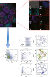
Specifically for lymph node (LN) immunology, multiplexed imaging allows for the evaluation of the heterogeneity within specific cell compartments [i.e. T follicular helper cells (Tfh) based on the expression of other markers‐CD57], the presence of potential suppressor CD4 T cells [forkhead box protein 3 (FoxP3+)] as well as the specific positioning of particular subset with respect to GC areas – dark zone versus light zone). The lower panels show the gating strategy that can be applied according to the markers used for the confocal image.
Figure 4
Figure 4
The illustration summarizes the most available technique at our disposal to study the immune system, highlighting the main advantages and limitations. These tools should be managed under the umbrella of the systems immunology. The lymph node at the center is to strengthen the necessity of performing studies on tissues. The lymph node image is taken and adapted from Servier Medical Art at: http://smart.servier.com/.

References

    1. Zhang Y, Garcia‐Ibanez L, Toellner KM. Regulation of germinal center B‐cell differentiation. Immunol Rev 2016; 270:8–19. - PMC - PubMed
    1. Davis MM. A prescription for human immunology. Immunity 2008; 29:835–8. - PMC - PubMed
    1. Mestas J, Hughes CC. Of mice and not men: differences between mouse and human immunology. J Immunol 2004; 172:2731–8. - PubMed
    1. Morita R, Schmitt N, Bentebibel SE et al Human blood CXCR5(+)CD4(+) T cells are counterparts of T follicular cells and contain specific subsets that differentially support antibody secretion. Immunity 2011; 34:108–21. - PMC - PubMed
    1. Roederer M. Multiparameter FACS analysis. Curr Protoc Immunol 2002; 49:5–8. - PubMed

Publication types