. 2020 Feb;594(4):717-727.
doi: 10.1002/1873-3468.13640. Epub 2019 Nov 7.

A missense mutation in the catalytic domain of O-GlcNAc transferase links perturbations in protein O-GlcNAcylation to X-linked intellectual disability

Affiliations

A missense mutation in the catalytic domain of O-GlcNAc transferase links perturbations in protein O-GlcNAcylation to X-linked intellectual disability

Veronica M Pravata et al. FEBS Lett. 2020 Feb.

Abstract

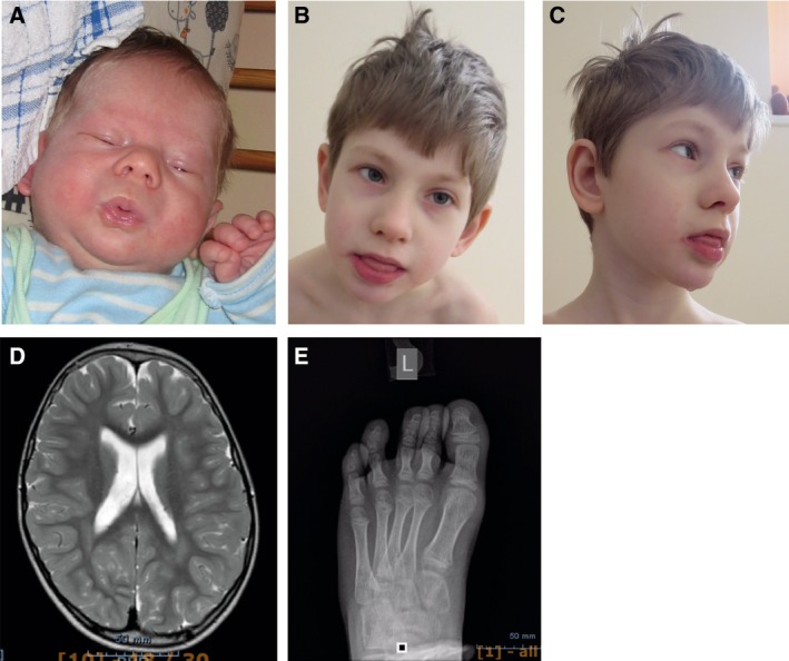
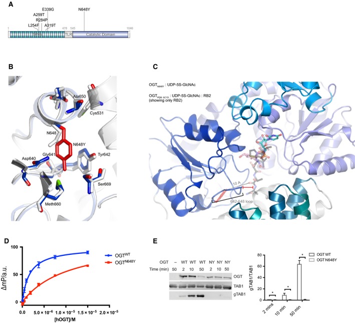
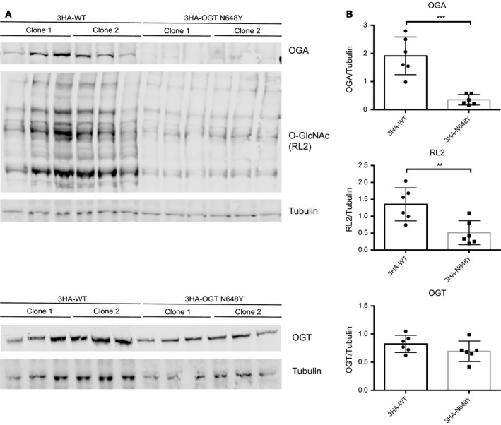
X-linked intellectual disabilities (XLID) are common developmental disorders. The enzyme O-GlcNAc transferase encoded by OGT, a recently discovered XLID gene, attaches O-GlcNAc to nuclear and cytoplasmic proteins. As few missense mutations have been described, it is unclear what the aetiology of the patient phenotypes is. Here, we report the discovery of a missense mutation in the catalytic domain of OGT in an XLID patient. X-ray crystallography reveals that this variant leads to structural rearrangements in the catalytic domain. The mutation reduces in vitro OGT activity on substrate peptides/protein. Mouse embryonic stem cells carrying the mutation reveal reduced O-GlcNAcase (OGA) and global O-GlcNAc levels. These data suggest a direct link between changes in the O-GlcNAcome and intellectual disability observed in patients carrying OGT mutations.

Keywords: O-GlcNAc; OGT; OGlcNAC transferase; XLID; intellectual disability; neurodevelopment.


Figures

Figure 1
Figure 1
Clinical pictures from patient carrying the N648Y mutation in the catalytic domain of OGT. (A) Facial asymmetry at birth. (B) Facial view at 7 years. (C) Profile view at 7 years. Note coarse facial features, drooling. (D) MRI image showing brain atrophy and mega cisterna magna. (E) X‐ray image of the left foot—cone‐shape epiphyses.
Figure 2
Figure 2
Effects of the N648Y mutation on OGT structure and activity. (A) Schematic diagram of OGT highlighting the TPRs, TPR‐like repeat, the Catalytic domain, the N648Y mutation (bold) and all the previous identified mutations in OGT. TPR, tetratricopeptide repeat domain; TLR, tetratricopeptide repeat‐like domain. (B) OGT superimposed complexes of OGTWT (in light grey; PDB: http://www.rcsb.org/pdb/search/structidSearch.do?structureId=5C1D; 43) and OGTN648Y (in blue; PDB: http://www.rcsb.org/pdb/search/structidSearch.do?structureId=6Q4M) showing the mutated site and the proximal residues. N648Y mutated residue is shown in red. (C) OGTN648Y in complex with superimposed RB2 peptide from OGTWT (PDB: http://www.rcsb.org/pdb/search/structidSearch.do?structureId=5C1D) showing the putative location of the peptide. The loop 642–648 of OGTN648Y is indicated including the distance from the superimposed peptide. (D) FP assay showing the binding of the UDP‐peptide bisubstrate conjugate to OGTWT and OGT N648Y. (E) Immunoblots showing OGT glycosyltransferase activity against TAB1 and gTAB1. Quantification of gTAB1 normalised to TAB1 signal. N = 3, mean ± SD. Multiple t‐test using the Holm–Sidak method. * corresponds to P = 0.021 (2 min), P = 0.017 (5 min) and P = 0.008 (50 min) TAB1, TAK1‐binding protein antibody; gTAB1, glycosylated TAB1 antibody.
Figure 3
Figure 3
The N648Y mutation leads to reduced protein O‐GlcNAcylation in 3HA‐N648Y mES cells. (A) Immunoblots showing OGA, protein O‐GlcNAcylation (RL2) and OGT levels in 3HA‐WT and 3HA‐N648Y undifferentiated mES cells. (B) Quantification of western blotting of OGA, protein O‐GlcNAcylation (RL2) and OGT levels normalised to tubulin signal. n = 3, mean ± SD. Unpaired t‐test. ** corresponds to P = 0.0068 (RL2); *** corresponds to P = 0.0003 (OGA).

References

    1. Maulik PK, Mascarenhas MN, Mathers CD, Dua T and Saxena S (2011) Prevalence of intellectual disability: a meta‐analysis of population‐based studies. Res Dev Disabil 32, 419–436. - PubMed
    1. American Psychiatric Association (2013) Diagnostic and Statistical Manual of Mental Disorders. American Psychiatric Association, Washington, D.C.
    1. Wieczorek D (2018) Autosomal dominant intellectual disability. Med Genet 30, 318–322. - PMC - PubMed
    1. Kvarnung M and Nordgren A (2017) Intellectual disability & rare disorders: a diagnostic challenge. Adv Exp Med Biol 1031, 39–54. - PubMed
    1. Neri G, Schwartz CE, Lubs HA and Stevenson RE (2018) X‐linked intellectual disability update 2017. Am J Med Genet 39, 1–14. - PMC - PubMed

Publication types

Substances