. 2019 Oct 17;9(10):279.
doi: 10.3390/brainsci9100279.

Pre- and Post-Treatment with Novel Antiepileptic Drug Oxcarbazepine Exerts Neuroprotective Effect in the Hippocampus in a Gerbil Model of Transient Global Cerebral Ischemia

Affiliations

Pre- and Post-Treatment with Novel Antiepileptic Drug Oxcarbazepine Exerts Neuroprotective Effect in the Hippocampus in a Gerbil Model of Transient Global Cerebral Ischemia

Ji Hyeon Ahn et al. Brain Sci. .

Abstract

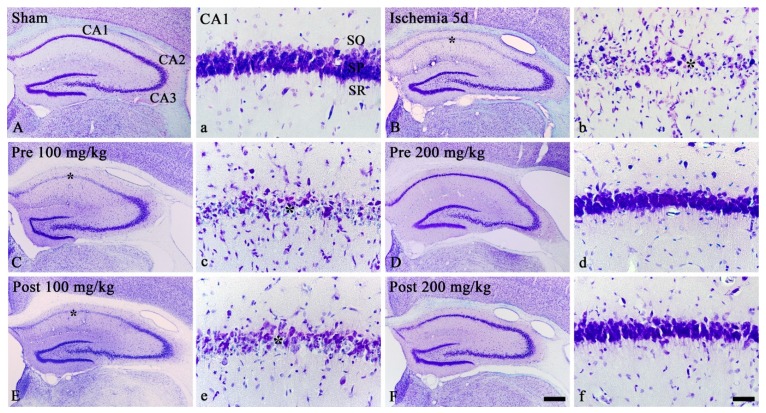
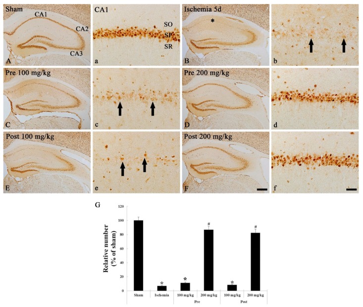
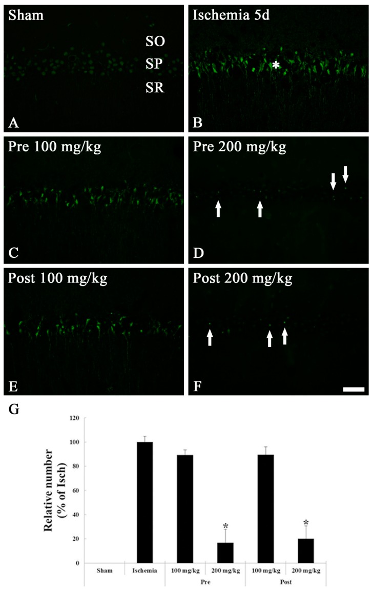
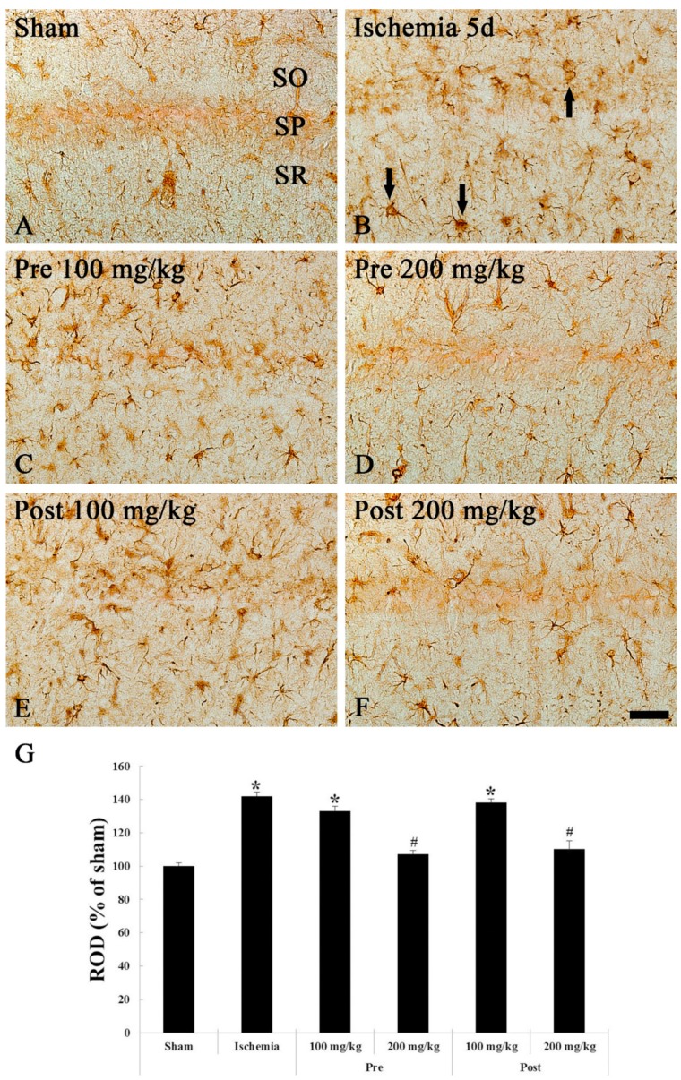
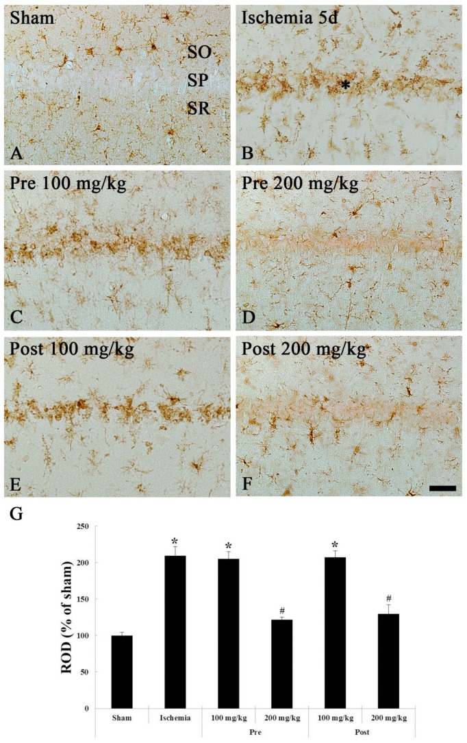
Oxcarbazepine, an antiepileptic drug, has been reported to modulate voltage-dependent sodium channels, and it is commonly used in epilepsy treatment. In this study, we investigated the neuroprotective effect of oxcarbazepine in the hippocampus after transient ischemia in gerbils. Gerbils randomly received oxcarbazepine 100 or 200 mg/kg before and after transient ischemia. We examined its neuroprotective effect in the cornu ammonis 1 subfield of the gerbil hippocampus at 5 days after transient ischemia by using cresyl violet staining, neuronal nuclei immunohistochemistry and Fluoro-Jade B histofluorescence staining for neuroprotection, and by using glial fibrillary protein and ionized calcium-binding adapter molecule 1 immunohistochemistry for reaction of astrocytes and microglia, respectively. Pre- and post-treatment with 200 mg/kg of oxcarbazepine, but not 100 mg/kg of oxcarbazepine, protected pyramidal neurons of the cornu ammonis 1 subfield from transient ischemic damage. In addition, pre- and post-treatment with oxcarbazepine (200 mg/kg) significantly ameliorated astrocytes and microglia activation in the ischemic cornu ammonis 1 subfield. In brief, our current results indicate that post-treatment as well as pre-treatment with 200 mg/kg of oxcarbazepine can protect neurons from ischemic insults via attenuation of the glia reaction.

Keywords: anti-epileptic drug; glial activation; neuroprotection; pyramidal neurons; transient cerebral ischemia.


Conflict of interest statement

The authors have no financial conflict of interest.

Figures

Figure 1
Figure 1
CV staining in the gerbil hippocampus of the sham (A,a), ischemia (B,b), OXC pretreated ischemia (C,c,D,d) and OXC posttreated ischemia (E,e,F,f) groups at 5 days after TGCI. In the ischemia and both 100 mg/kg OXC treated ischemia groups, most of CV-positive cells in the stratum pyramidale (SP) (asterisks) of the CA1 region are damaged or lost. However, in both 200 mg/kg OXC treated ischemia groups, CV-positive cells are not damaged. CA, cornu ammonis; CV, cresyl Violet; OXC, oxcarbazepine; SO, stratum oriens; SR, stratum radiatum; TGCI, transient global cerebral ischemia. Scale bars = 400 μm (A,B,C,D,E,F) and 40 μm (a,b,c,d,e,f).
Figure 2
Figure 2
NeuN immunohistochemistry in the hippocampus of the sham (A,a), ischemia (B,b), OXC pretreated ischemia (C,c,D,d) and OXC posttreated ischemia (E,e,F,f) groups at 5 days after TGCI. Most of NeuN-positive neurons are lost in the stratum pyramidale (SP) (arrows) of the CA1 region of the ischemia and both 100 mg/kg OXC treated ischemia groups. However, in both 200 mg/kg OXC treated ischemia groups, many NeuN-immunoreactive neurons (asterisks) are observed in the SP of the CA1 region. CA, cornu ammonis; NeuN, neuronal nuclei; OXC, oxcarbazepine; SO, stratum oriens; SR, stratum radiatum; TGCI, transient global cerebral ischemia. Scale bar = 400 μm (A,B,C,D,E,F) and 40 μm (a,b,c,d,e,f). (G) The mean number of NeuN-immunoreactive neurons in the SP of the CA1 region at 5 days after TGCI (n = 7 in each group, * p < 0.05, significantly different from the sham group, # p < 0.05, significantly different from the ischemia group). The bars indicate the means ± standard error of the mean (SEM).
Figure 3
Figure 3
FJB histofluorescence staining in the CA1 region of the sham (A), ischemia (B), OXC pretreated ischemia (C,D) and OXC posttreated ischemia (E,F) groups at 5 days after TGCI. In the ischemia and both 100 mg/kg OXC pre- and post-treated ischemia groups, many FJB-positive cells are found in the stratum pyramidale (SP) (asterisks), while FJB-positive cells are rarely detected in the SP (arrows) of the 200 mg/kg OXC pre- and post-treated ischemia groups. CA, cornu ammonis; FJB, Fluoro Jade B; OXC, oxcarbazepine; SO, stratum oriens; SR, stratum radiatum; TGCI, transient global cerebral ischemia. Scale bar = 40 μm. (G) The mean number of FJB-positive cells in the SP of the CA1 region at 5 days after TGCI (* p < 0.05, significantly different from the ischemia group). The bars indicate the means ± standard error of the mean (SEM).
Figure 4
Figure 4
GFAP immunohistochemistry in the CA1 region of the sham (A), ischemia (B), OXC pretreated ischemia (C,D) and OXC posttreated ischemia (E,F) groups at 5 days after TGCI. GFAP-immunoreactive astrocytes (arrows) are markedly activated (hypertrophied cytoplasm and processes) in the ischemia and both 100 mg/kg OXC pre- and post-treated ischemia groups. However, GFAP immunoreactivity in both 200 mg/kg OXC ischemia groups is significantly lower than that in the ischemia- group. CA, cornu ammonis; GFAP, glial fibrillary acidic protein; OXC, oxcarbazepine; SO, stratum oriens; SP, stratum pyramidale; SR, stratum radiatum; TGCI, transient global cerebral ischemia. Scale bar = 40 μm. (G) Relative optical density (ROD) as percentage values of the GFAP immunoreactivity at 5 days after TGCI (* p < 0.05, significantly different from the sham group, # p < 0.05, significantly different from the ischemia group). The bars indicate the means ± standard error of the mean (SEM).
Figure 5
Figure 5
Iba-1 immunohistochemistry in the CA1 region of the sham (A), ischemia (B), OXC pretreated ischemia (C,D) and OXC posttreated ischemia (E,F) groups at 5 days after TGCI. Iba-1-immunoreactive microglia are markedly activated (hypertrophied cytoplasm and processes) and aggregate within the stratum pyramidale (SP) (asterisk) in the ischemia and both 100 mg/kg OXC ischemia groups. However, in both 200 mg/kg OXC ischemia groups, Iba-1-immunoreactive microglia are similar to those in the sham group. CA, cornu ammonis; Iba-1, ionized calcium binding adaptor molecule 1; OXC, oxcarbazepine; SO, stratum oriens; SR, stratum radiatum; TGCI, transient global cerebral ischemia. Scale bar = 40 μm. (G) ROD as percent values of the Iba-1 immunoreactivity at 5 days after TGCI (* p < 0.05, significantly different from the sham group, # p < 0.05, significantly different from the ischemia group). The bars indicate the means ± SEM.

References

    1. Lee J.-M., Grabb M.C., Zipfel G.J., Choi D.W. Brain tissue responses to ischemia. J. Clin. Investig. 2000;106:723–731. doi: 10.1172/JCI11003. - DOI - PMC - PubMed
    1. Endres M., Heuschmann P.U., Laufs U., Hakim A.M. Primary prevention of stroke: Blood pressure, lipids, and heart failure. Eur. Heart J. 2011;32:545–552. doi: 10.1093/eurheartj/ehq472. - DOI - PubMed
    1. Petito C.K., Pulsinelli W.A. Delayed Neuronal Recovery and Neuronal Death in Rat Hippocampus following Severe Cerebral Ischemia: Possible Relationship to Abnormalities in Neuronal Processes. Br. J. Pharmacol. 1984;4:194–205. doi: 10.1038/jcbfm.1984.28. - DOI - PubMed
    1. Kirino T., Sano K. Selective vulnerability in the gerbil hippocampus following transient ischemia. Acta Neuropathol. 1984;62:201–208. doi: 10.1007/BF00691853. - DOI - PubMed
    1. Kirino T. Delayed neuronal death in the gerbil hippocampus following ischemia. Brain Res. 1982;239:57–69. doi: 10.1016/0006-8993(82)90833-2. - DOI - PubMed

LinkOut - more resources