. 2019 Oct 18;9(1):15007.
doi: 10.1038/s41598-019-50941-3.

Concatenation of 14-3-3 with partner phosphoproteins as a tool to study their interaction

Affiliations

Concatenation of 14-3-3 with partner phosphoproteins as a tool to study their interaction

Kristina V Tugaeva et al. Sci Rep. .

Abstract

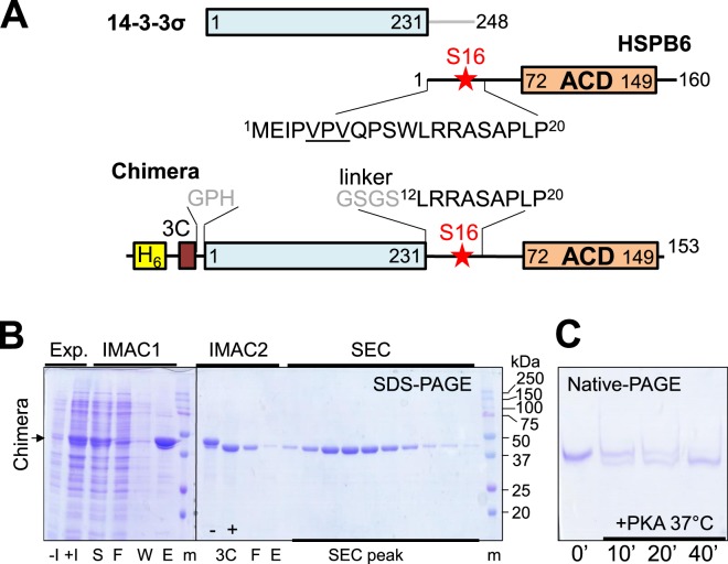
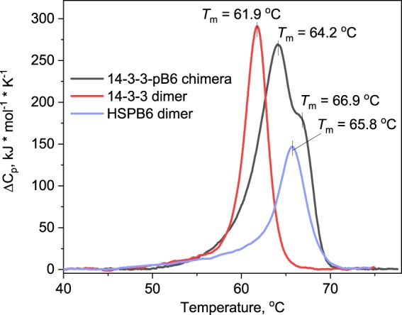
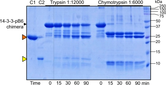
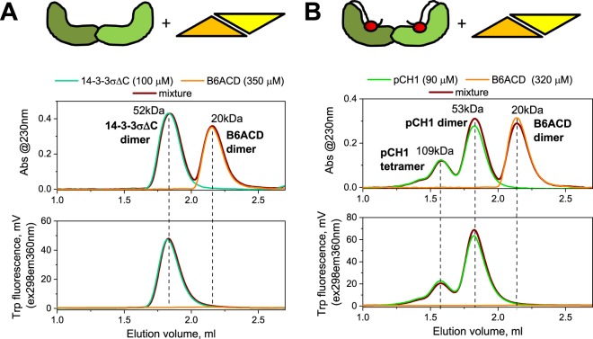
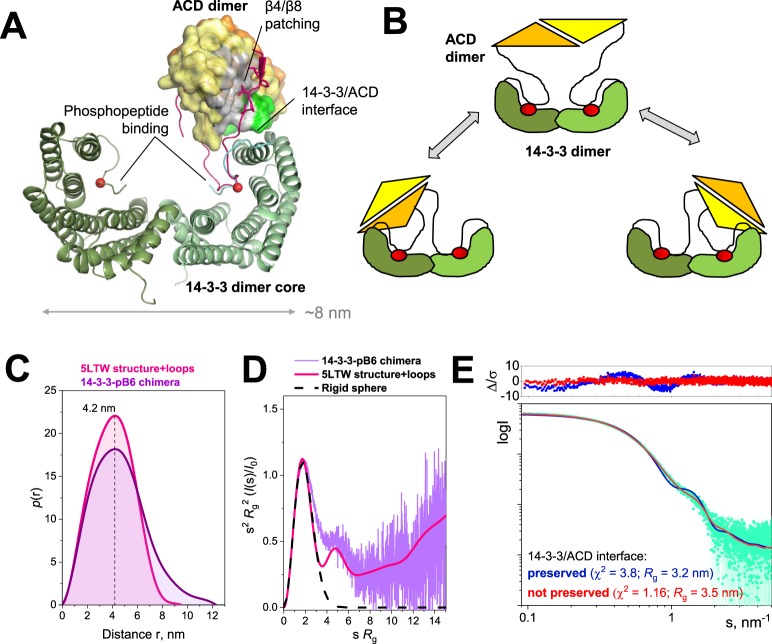
Regulatory 14-3-3 proteins interact with a plethora of phosphorylated partner proteins, however 14-3-3 complexes feature intrinsically disordered regions and often a transient type of interactions making structural studies difficult. Here we engineer and examine a chimera of human 14-3-3 tethered to a nearly complete partner HSPB6 which is phosphorylated by protein kinase A (PKA). HSPB6 includes a long disordered N-terminal domain (NTD), a phosphorylation motif around Ser16, and a core α-crystallin domain (ACD) responsible for dimerisation. The chosen design enables an unstrained binding of pSer16 in each 1433 subunit and secures the correct 2:2 stoichiometry. Differential scanning calorimetry, limited proteolysis and small-angle X-ray scattering (SAXS) support the proper folding of both the 14-3-3 and ACD dimers within the chimera, and indicate that the chimera retains the overall architecture of the native complex of 14-3-3 and phosphorylated HSPB6 that has recently been resolved using crystallography. At the same time, the SAXS data highlight the weakness of the secondary interface between the ACD dimer and the C-terminal lobe of 14-3-3 observed in the crystal structure. Applied to other 14-3-3 complexes, the chimeric approach may help probe the stability and specificity of secondary interfaces for targeting them with small molecules in the future.


Conflict of interest statement

The authors declare no competing interests.

Figures

Figure 1
Figure 1
Design and purification of the 14-3-3-pB6 chimera. (A) Schematics showing the primary structure of both individual proteins and the chimera. The N-terminal hexahistidine-tag cleavable by 3C protease is shown. (B) Expression (Exp.) and chromatographic (IMAC1, IMAC2, SEC) purification of the unphosphorylated chimera as analyzed by SDS-PAGE. Qualitatively similar results were obtained after co-expression with PKA, but with a lower yield (not shown). Non-induced (−I), induced (+I), soluble (S), flowthrough (F), wash (W), eluted (E) fractions, and fractions collected during SEC (SEC peak) are indicated below the gel. Protein markers (m) with the corresponding masses (in kDa) are also indicated. Position of the chimera band is shown by an arrow to the left. Note the shift after the 3C proteolysis (3C: “−”, “+”). (C) Kinetics of in vitro phosphorylation of the purified chimera by PKA analyzed by native-PAGE. Note the downward shift corresponding to phosphate group incorporation.
Figure 2
Figure 2
Oligomeric state of the 14-3-3-pB6 chimera. SEC profiles of either unphosphorylated (A) or PKA-phosphorylated (B) chimera loaded on a Superdex 200 Increase 5/150 column at different concentrations (indicated in mg/ml) and run at a 0.45 ml/min flow rate. MW values of the peaks were determined from column calibration. Dashed lines show the lowest concentration profiles scaled to the main peak of the highest concentration profiles for clarity. (C) Absolute MW values for the main two peaks of the phosphorylated chimera obtained by SEC-MALLS on a Superdex 200 Increase 10/300 column operated at a 0.5 ml/min flow rate. (D) Formation of chimera tetramers due to the domain swapping between two different chimera dimers. NTD IDRs – intrinsically disordered regions of the HSPB6 N-terminal domain.
Figure 3
Figure 3
Analysis of the presence of folded domains within the 14-3-3-pB6 chimera by DSC. The samples containing either the 14-3-3 dimer, the HSPB6 dimer, or the 14-3-3-pB6 chimera were subjected to DSC at a constant heating rate of 1 °C/min. Thermal transition temperatures for the peaks (Tm) are indicated as the positions of their maximum.
Figure 4
Figure 4
Kinetics of the 14-3-3-pB6 chimera cleavage by trypsin or chymotrypsin at indicated protease:substrate ratios. Time points are indicated below the gel in min at 37 °C. Positions of the intact chimera and protein markers bands (in kDa) are shown by arrows. C1 and C2 represent the 14-3-3σ∆C (26.1 kDa, orange triangle) and HSPB6∆N56 (11.0 kDa, yellow triangle) control lanes.
Figure 5
Figure 5
Lack of interaction between the isolated ACD and either 14-3-3σ∆C (A) or the 14-3-3-phosphopeptide chimera pCH1. (B) These two scenarios, shown schematically on top of the panels, were examined by size-exclusion chromatography with simultaneous detection by 230 nm absorbance and Trp-specific fluorescence (excitation 298 nm, emission 360 nm). Concentrations of 14-3-3 and ACD are indicated in µM. Dashed lines show the positions of the main peaks on different profiles for clarity.
Figure 6
Figure 6
Structural analysis of the 14-3-3-pB6 chimera using SAXS. (A) Crystal structure of the 14-3-3σ∆C–pHSPB6∆C complex (PDB 5LTW). The 14-3-3 dimer is shown as cartoon. The HSPB6 ACD dimer is shown as a molecular surface, with residues involved in the interface with 14-3-3 highlighted in light green. The partially ordered NTD of one HSPB6 chain is shown in magenta. In addition, the N-terminal arms of the peptidic pCH1 chimera (when superimposed on the full complex) are drawn as cyan and green lines. Phosphoserines are represented by red spheres. (B) Schematics showing that in the absence of the β4/β8 patching, the 14-3-3/ACD interface may be preserved or not preserved. (C,D) Comparison of the p(r) functions (C) and the dimensionless Kratky plots (D) for the chimera (experimental SAXS data processed by GNOM) and the 5LTW structure supplemented with the missing loops (calculated from the model by CRYSOL and GNOM). (E) The fits of the best among each of the two types of models depending on whether the 14-3-3/ACD is preserved or not preserved to the SAXS data and the associated residuals (∆/σ) shown on top.

References

    1. Aitken A. 14-3-3 proteins: a historic overview. Semin. Canc. Biol. 2006;16:162–172. doi: 10.1016/j.semcancer.2006.03.005. - DOI - PubMed
    1. Aitken A. Functional specificity in 14-3-3 isoform interactions through dimer formation and phosphorylation. Chromosome location of mammalian isoforms and variants. Plant Mol Biol. 2002;50:993–1010. doi: 10.1023/A:1021261931561. - DOI - PubMed
    1. Yang X, et al. Structural basis for protein-protein interactions in the 14-3-3 protein family. Proc Natl Acad Sci USA. 2006;103:17237–17242. doi: 10.1073/pnas.0605779103. - DOI - PMC - PubMed
    1. Sluchanko NN, Gusev NB. Oligomeric structure of 14-3-3 protein: what do we know about monomers? FEBS Lett. 2012;586:4249–4256. doi: 10.1016/j.febslet.2012.10.048. - DOI - PubMed
    1. Boston PF, Jackson P, Thompson RJ. Human 14-3-3 protein: radioimmunoassay, tissue distribution, and cerebrospinal fluid levels in patients with neurological disorders. J Neurochem. 1982;38:1475–1482. doi: 10.1111/j.1471-4159.1982.tb07928.x. - DOI - PubMed

Publication types

LinkOut - more resources