. 2019 Oct 18;9(1):14992.
doi: 10.1038/s41598-019-51506-0.

Impact of Early Life Antibiotic Exposure and Neonatal Hyperoxia on the Murine Microbiome and Lung Injury

Affiliations

Impact of Early Life Antibiotic Exposure and Neonatal Hyperoxia on the Murine Microbiome and Lung Injury

Melissa H Althouse et al. Sci Rep. .

Abstract

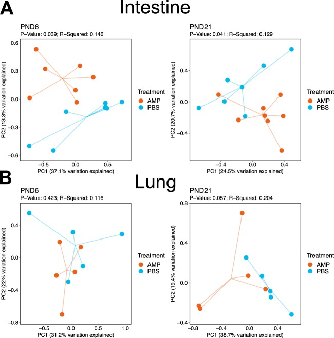
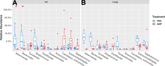
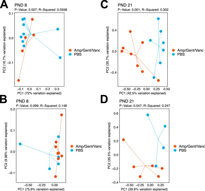
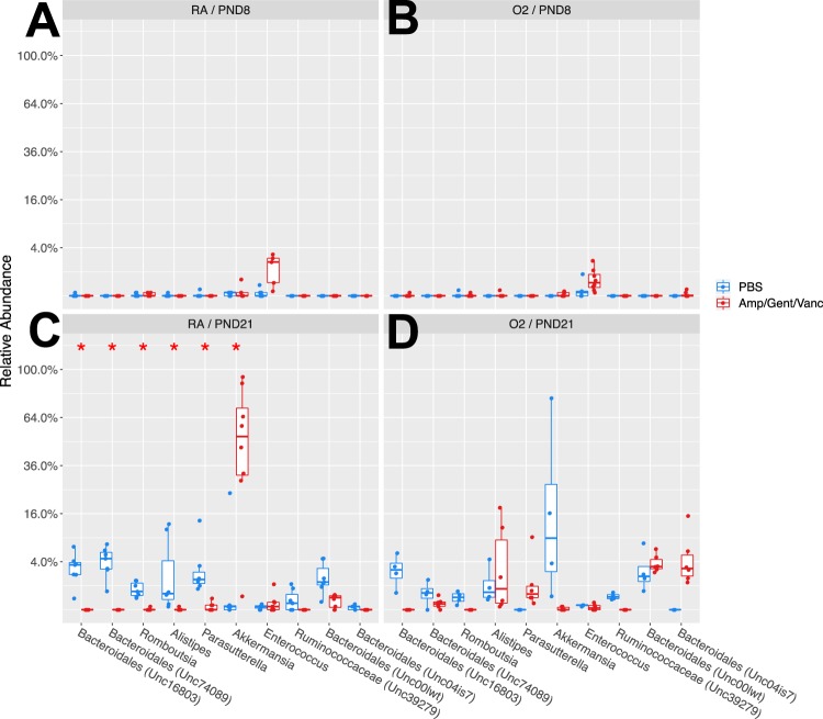
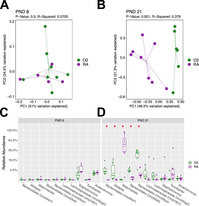
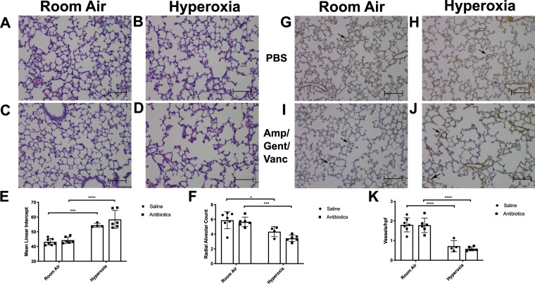
Cross talk between the intestinal microbiome and the lung and its role in lung health remains unknown. Perinatal exposure to antibiotics disrupts the neonatal microbiome and may have an impact on the preterm lung. We hypothesized that perinatal antibiotic exposure leads to long-term intestinal dysbiosis and increased alveolar simplification in a murine hyperoxia model. Pregnant C57BL/6 wild type dams and neonatal mice were treated with antibiotics before and/or immediately after delivery. Control mice received phosphate-buffered saline (PBS). Neonatal mice were exposed to 95% oxygen for 4 days or room air. Microbiome analysis was performed using 16S rRNA gene sequencing. Pulmonary alveolarization and vascularization were analyzed at postnatal day (PND) 21. Perinatal antibiotic exposure modified intestinal beta diversity but not alpha diversity in neonatal mice. Neonatal hyperoxia exposure altered intestinal beta diversity and relative abundance of commensal bacteria in antibiotic treated mice. Hyperoxia disrupted pulmonary alveolarization and vascularization at PND 21; however, there were no differences in the degree of lung injury in antibiotic treated mice compared to vehicle treated controls. Our study suggests that exposure to both hyperoxia and antibiotics early in life may cause long-term alterations in the intestinal microbiome, but intestinal dysbiosis may not significantly influence neonatal hyperoxic lung injury.


Conflict of interest statement

The authors declare no competing interests.

Figures

Figure 1
Figure 1
Early ampicillin exposure in neonatal mice alters intestinal microbial diversity. (A) Principal coordinate analysis plots represent intestinal beta diversity between ampicillin and PBS treated mice. Intestinal beta diversity was significantly different between ampicillin treated mice and controls at PND 6 (p = 0.039) and PND 21 (p = 0.041). (B) Lung beta diversity was not altered at either PND 6 (p = 0.42) or PND 21 (p = 0.057). Beta diversity was analyzed using both weighted and unweighted (Shown) UniFrac analysis. n = 5–8/group. AMP = ampicillin. PBS = phosphate buffered saline. PND = postnatal day.
Figure 2
Figure 2
Ampicillin alters microbial composition in the intestine but not the lung at PND 21. (A) Taxa box plot A represents relative abundance at the genus level in the intestine at PND 21. Bacteroides and Akkermansia are decreased in Ampicillin treated mice compared to PBS controls. (B) Taxa box plot B represents relative abundance at the genus level in the lung at PND 21. There were no significant differences in microbial composition between ampicillin treated mice and controls in the lung. AMP = ampicillin. PBS = phosphate buffered saline. Int = intestine. n = 5–8/group. Significant differences between the ampicillin and PBS treated groups is represented by *P < 0.05.
Figure 3
Figure 3
Perinatal antibiotic exposure with a three antibiotic regimen modifies intestinal beta diversity. Principal coordinate analysis plots represent intestinal beta diversity between three antibiotic (ampicillin, vancomycin, gentamicin) and PBS (control) treated mice stratified by oxygen exposure and timepoint. RA (A) and hyperoxia (B) groups exposed to antibiotics did not differ in beta diversity from controls at PND 8 (p = 0.51 and p = 0.099, respectively). At PND 21, intestinal beta diversity differed in both room air (p = 0.001; C) and hyperoxia (p = 0.047; D) exposed mice treated with antibiotics compared to PBS treated controls. n = 4–9/group. Amp/Gent/Vanc = Ampicillin, gentamicin, vancomycin antibiotic cocktail. PBS = phosphate buffered saline. PND = postnatal day.
Figure 4
Figure 4
Perinatal antibiotics modify relative abundance of intestinal commensal bacteria at PND 21 in neonatal mice. At PND 8, there were no differences in relative abundance of genera between antibiotic exposed mice and PBS controls in either room air (A) or hyperoxia (B). At PND 21 in the room air group (C) there is a decrease in Bacteroidales, Romboutsia, and Parasutterella and an increase in Akkermansia seen in the antibiotic exposed mice when compared to controls (*p < 0.05). (D) There were no significant differences seen in relative abundance of intestinal commensal organisms in the hyperoxia group at PND 21 between antibiotic exposed and controls. n = 4–9/group. Amp/Gent/Vanc = Ampicillin, gentamicin, vancomycin antibiotic cocktail. PBS = phosphate buffered saline. PND = postnatal day. RA = Room Air, O2 = Hyperoxia.
Figure 5
Figure 5
Hyperoxia modifies intestinal beta diversity and relative abundance of commensal bacteria in antibiotic exposed mice. (A) At PND 8, beta diversity was unchanged between room air and hyperoxia exposed mice (p = 0.5). (B) At PND 21, mice exposed to hyperoxia and antibiotics develop a distinctly different microbiota than those exposed to room air and antibiotics and beta diversity was statistically significant (p = 0.001). (C) No differences in relative abundance of different taxa were seen on PND 8. (D) At PND 21, there were significant increases in the genera Bacteroidales and Alistipes and a decrease in Akkermansia in the hyperoxia-exposed mice compared to room air. Analyzed using weighted UniFrac analysis. N = 5–8/group. PND = postnatal day. RA = room air. O2 = Hyperoxia.
Figure 6
Figure 6
Hyperoxia reduces pulmonary alveolarization and vascularization but the addition of antibiotics does not worsen lung injury phenotype. (AD) Representative lung sections with H&E at 20x magnification from neonatal mice exposed to RA or hyperoxia in addition to PBS or antibiotics. Mice exposed to hyperoxia (B,D) have decreased alveolarization. Mean linear intercept (E) was increased and radial alveolar count (F) was decreased in hyperoxia exposed mice. (GJ) Representative lung sections shown at 20x magnification subjected to immunohistochemistry for Von Willebrand factor. (K) Angiogenesis was assessed via microvessel count per high-powered field. Mice exposed to hyperoxia displayed a decreased vessel count/ high powered field when compared to controls. Scale bars represent 100 microns. (n = 4–7/group; Mean ± SEM). Data analyzed by 2-way ANOVA. Significant differences shown by ****P < 0.0001 and ***P < 0.001 and *P < 0.05. hpf = high powered field. PBS = phosphate buffered saline. Amp/Gent/Vanc = ampicillin, gentamicin, vancomycin antibiotic cocktail.

References

    1. Jobe AH. The new bronchopulmonary dysplasia. Curr. Opin. Pediatr. 2011;23:167–172. doi: 10.1097/MOP.0b013e3283423e6b. - DOI - PMC - PubMed
    1. Davidson L, Berkelhamer S. Bronchopulmonary Dysplasia: Chronic Lung Disease of Infancy and Long-Term Pulmonary Outcomes. J. Clin. Med. 2017;6:4. doi: 10.3390/jcm6010004. - DOI - PMC - PubMed
    1. Kalikkot Thekkeveedu R, Guaman MC, Shivanna B. Bronchopulmonary dysplasia: A review of pathogenesis and pathophysiology. Respir. Med. 2017;132:170–177. doi: 10.1016/j.rmed.2017.10.014. - DOI - PMC - PubMed
    1. Collado MC, et al. Factors influencing gastrointestinal tract and microbiota immune interaction in preterm infants. Pediatr. Res. 2015;77:726–731. doi: 10.1038/pr.2015.54. - DOI - PubMed
    1. Berrington JE, Stewart CJ, Embleton ND, Cummings SP. Gut microbiota in preterm infants: Assessment and relevance to health and disease. Arch. Dis. Child. Fetal Neonatal Ed. 2013;98:F286–90. doi: 10.1136/archdischild-2012-302134. - DOI - PubMed

Publication types