. 2019 Oct 18;9(1):15008.
doi: 10.1038/s41598-019-51549-3.

Evaluation of Small Molecule Drug Uptake in Patient-Derived Prostate Cancer Explants by Mass Spectrometry

Affiliations

Evaluation of Small Molecule Drug Uptake in Patient-Derived Prostate Cancer Explants by Mass Spectrometry

Shadrack M Mutuku et al. Sci Rep. .

Abstract

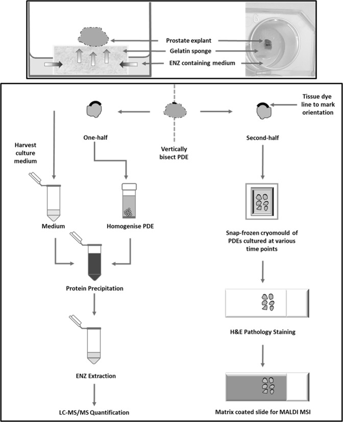
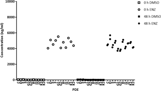
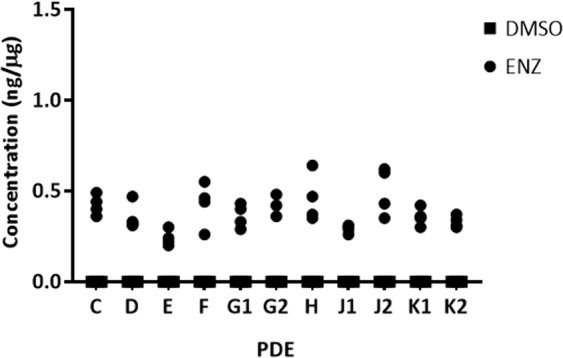
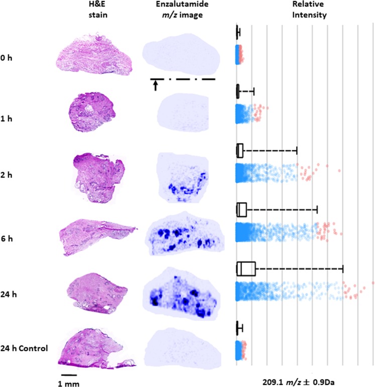
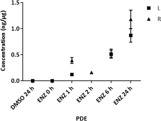
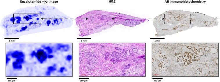
Patient-derived explant (PDE) culture of solid tumors is increasingly being applied to preclinical evaluation of novel therapeutics and for biomarker discovery. In this technique, treatments are added to culture medium and penetrate the tissue via a gelatin sponge scaffold. However, the penetration profile and final concentrations of small molecule drugs achieved have not been determined to date. Here, we determined the extent of absorption of the clinical androgen receptor antagonist, enzalutamide, into prostate PDEs, using liquid chromatography-tandem mass spectrometry (LC-MS/MS) and matrix-assisted laser/desorption ionisation (MALDI) mass spectrometry imaging (MSI). In a cohort of 11 PDE tissues from eight individual patients, LC-MS/MS quantification of PDE homogenates confirmed enzalutamide (10 µM) uptake by all PDEs, which reached maximal average tissue concentration of 0.24-0.50 ng/µg protein after 48 h culture. Time dependent uptake of enzalutamide (50 µM) in PDEs was visualized using MALDI MSI over 24-48 h, with complete penetration throughout tissues evident by 6 h of culture. Drug signal intensity was not homogeneous throughout the tissues but had areas of markedly high signal that corresponded to drug target (androgen receptor)-rich epithelial regions of tissue. In conclusion, application of MS-based drug quantification and visualization in PDEs, and potentially other 3-dimensional model systems, can provide a more robust basis for experimental study design and interpretation of pharmacodynamic data.


Conflict of interest statement

The authors declare no competing interests.

Figures

Figure 1
Figure 1
Schematic of drug uptake in ex vivo culture of human prostate tumors. Top panel. Schematic and photograph of a PDE on top of a gelatine sponge in a well with 500 µL medium. Bottom panel. Workflow chart for evaluation of ENZ uptake in human prostate PDEs by mass spectrometry.
Figure 2
Figure 2
LC-MS/MS quantification of ENZ in conditioned explant media. PDE’s were cultured in medium containing 10 µM ENZ or DMSO control over a 48h period. LC-MS/MS quantification of the predominant ENZ fragment ion, m/z 209.09 is shown. Drug concentration in medium expressed as ng/ml.
Figure 3
Figure 3
LC-MS/MS quantification of ENZ in PDE homogenates. PDEs were cultured in medium containing 10 µM ENZ or DMSO control over a 48h period. LC-MS/MS quantification of the ENZ MRM transition 465.1 → 209.09 is shown. Measured drug concentration in homogenate is normalized to total protein expressed as ng/µg of total protein. Intra-patient variability of 17.7% and intra-patient variability of 20.1%. n = 4 (two explant pieces per well containing medium with 10 µM ENZ or DMSO vehicle).
Figure 4
Figure 4
Time profile of ENZ penetration in ex vivo prostate tissue culture. Left panel. Six PDE’s were cultured in medium containing 50 µM ENZ over a 24 h period or DMSO control (24 h). MALDI MSI MS/MS images of the predominant ENZ fragment ion, m/z 209.09, are shown with normalization to TIC next to a serial section stained by H&E. The arrow at the bottom of the image at 0 h indicates the contact of the PDE with the gelatin sponge surface. H&E sections are 30 µm adjacent to the MALDI image tissue section. Right panel. Relative intensity of m/z 209.09 at 0 h, 1 h, 2 h, 6, h and 24 h/24 h control of respective MS/MS images (A) show a time-dependent increase in drug concentration in the tissues during culture. Red dots represent the spectra of enzalutamide outside of the upper quartiles.
Figure 5
Figure 5
LC-MS/MS quantification of ENZ in PDE homogenates over a time course. Six PDEs from either the left (L) and right (R) prostate cores were cultured in 50 µM ENZ and harvested at 0 h, 1 h, 2 h, 6 h, 24 h or 24 h (DMSO control) and bisected. One-half was homogenized for LC-MS/MS analysis. Drug concentration is normalized to total protein expressed as ng/µg. *Note: PDE for L at 2 h lost during sample homogenization. Error bars indicate the 15% CV threshold for accuracy and precision of assay.
Figure 6
Figure 6
ENZ signal co-localization in prostate tissue epithelium. Comparison of the MS/MS image (ENZ fragment ion, m/z 209.09, left) to an H&E scan (middle) and immunohistochemical detection of the androgen receptor (AR). Lower panels: higher magnification images.

References

    1. Merriel SWD, Funston G, Hamilton W. Prostate Cancer in Primary Care. Adv Ther. 2018;35:1285–1294. doi: 10.1007/s12325-018-0766-1. - DOI - PMC - PubMed
    1. Barfeld SJ, Itkonen HM, Urbanucci A, Mills IG. Androgen-regulated metabolism and biosynthesis in prostate cancer. Endocr Relat Cancer. 2014;21:T57–66. doi: 10.1530/ERC-13-0515. - DOI - PubMed
    1. Montgomery B, et al. Neoadjuvant Enzalutamide Prior to Prostatectomy. Clin Cancer Res. 2017;23:2169–2176. doi: 10.1158/1078-0432.CCR-16-1357. - DOI - PMC - PubMed
    1. Hoda MR, Kramer MW, Merseburger AS, Cronauer MV. Androgen deprivation therapy with Leuprolide acetate for treatment of advanced prostate cancer. Expert Opin Pharmacother. 2017;18:105–113. doi: 10.1080/14656566.2016.1258058. - DOI - PubMed
    1. Schalken J, Fitzpatrick JM. Enzalutamide: targeting the androgen signalling pathway in metastatic castration-resistant prostate cancer. BJU Int. 2016;117:215–225. doi: 10.1111/bju.13123. - DOI - PMC - PubMed

Publication types