. 2020 Jun;46(4):375-390.
doi: 10.1111/nan.12584. Epub 2019 Nov 6.

Oxidative DNA damage is concurrently repaired by base excision repair (BER) and apyrimidinic endonuclease 1 (APE1)-initiated nonhomologous end joining (NHEJ) in cortical neurons

Affiliations

Oxidative DNA damage is concurrently repaired by base excision repair (BER) and apyrimidinic endonuclease 1 (APE1)-initiated nonhomologous end joining (NHEJ) in cortical neurons

J-L Yang et al. Neuropathol Appl Neurobiol. 2020 Jun.

Abstract

Aims: Accumulating studies have suggested that base excision repair (BER) is the major repair pathway of oxidative DNA damage in neurons, and neurons are deficient in other DNA repair pathways, including nucleotide excision repair and homologous recombination repair. However, some studies have demonstrated that neurons could efficiently repair glutamate- and menadione-induced double-strand breaks (DSBs), suggesting that the DSB repair mechanisms might be implicated in neuronal health. In this study, we hypothesized that BER and nonhomologous end joining (NHEJ) work together to repair oxidative DNA damage in neurons.

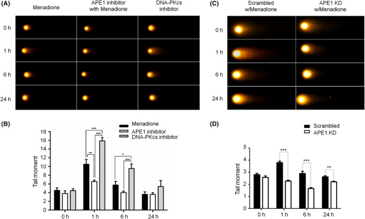
Methods: Immunohistochemistry and confocal microscopy were employed to examine the colocalization of apyrimidinic endonuclease 1 (APE1), histone variant 2AX (γH2AX) and phosphorylated p53-binding protein (53BP1). APE1 inhibitor and shRNA were respectively applied to suppress APE1 activity and protein expression to determine the correlation of APE1 and DSB formation. The neutral comet assay was used to determine and quantitate the formation of DSB.

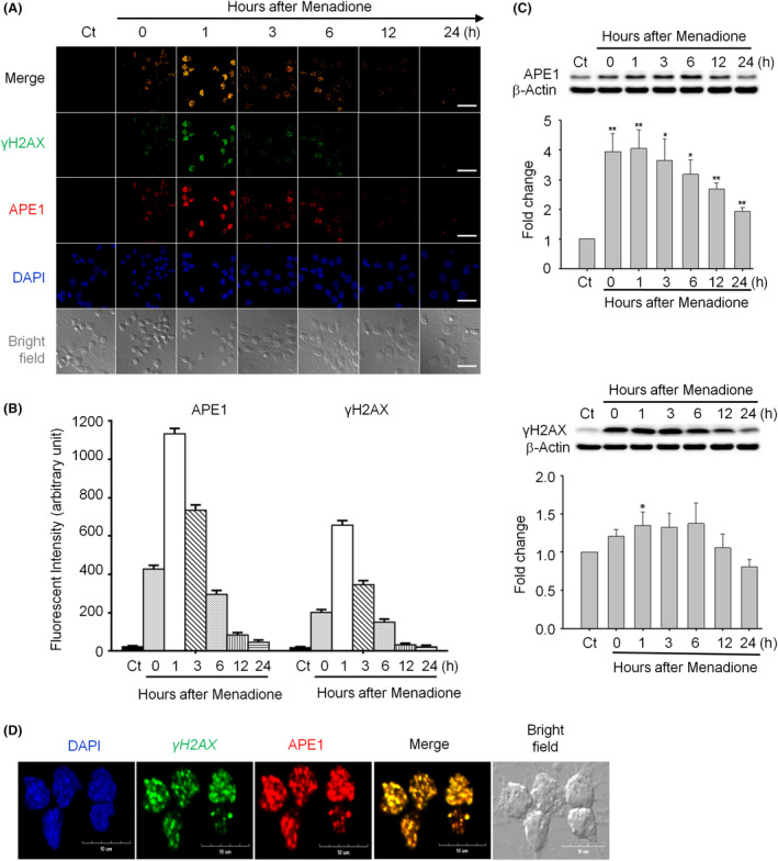
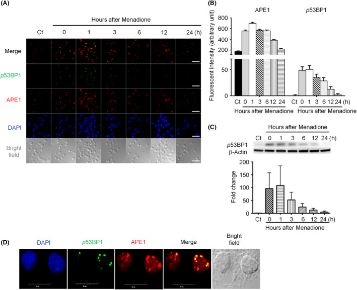
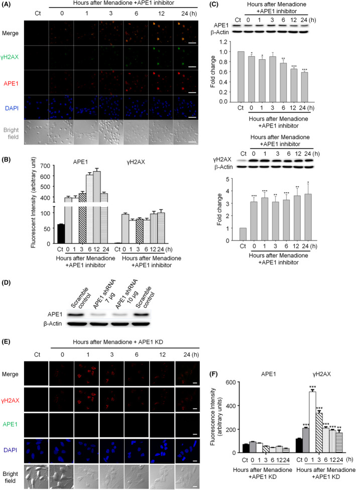
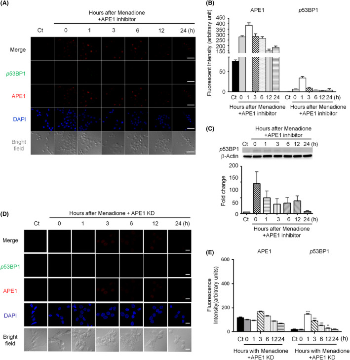
Results: Both γH2AX and 53BP1 were upregulated and colocalized with APE1 in the nuclei of rat cortical neurons subjected to menadione-induced oxidative insults. Phospho53BP1 foci were efficiently abolished, but γH2AX foci persisted following the suppression of APE1 activity. Comet assays demonstrated that the inhibition of APE1 decreased the DSB formation.

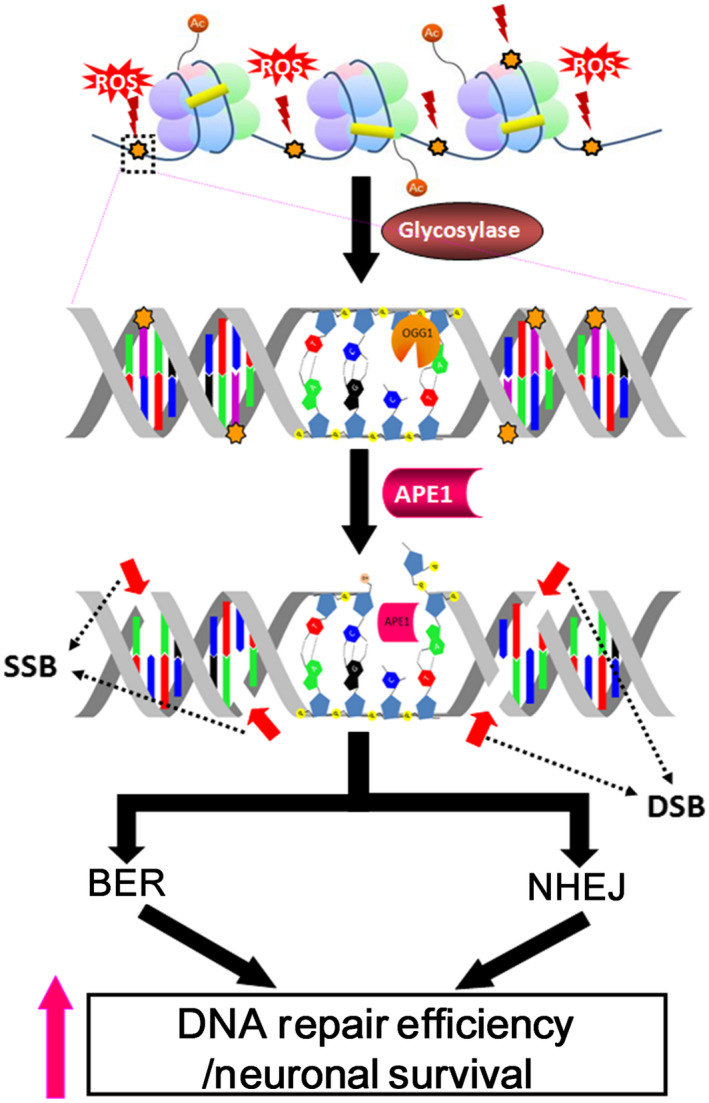
Conclusions: Our results indicate that APE1 can engage the NHEJ mechanism in the repair of oxidative DNA damage in neurons. These findings provide insights into the mechanisms underlying the efficient repair of oxidative DNA damage in neurons despite the high oxidative burden.

Keywords: apurinic/apyrimidinic endonuclease 1 (APE1); base excision repair (BER); neuron; nonhomologous end joining (NHEJ); oxidative DNA damage.


Conflict of interest statement

The authors declare no conflict of interest.

Figures

Figure 1
Figure 1
Apurinic/apyrimidinic endonuclease 1 (APE1) and phosphorylated histone 2AX (γH2AX) foci are colocalized and co‐induced in neurons subjected to an oxidative insult. (A) The levels of APE1 (red) and γH2AX (green) were rapidly elevated in the nuclei (DAPI, blue) of rat cortical neurons after transient exposure to 40 μM menadione and returned to the background levels at 12‐h post‐treatment (scale bar: 50 nm). (B) Quantitation of the data in (A). (C) Western blotting showed that both APE1 and γH2AX levels peaked at 1 h, started decreasing at 6 h and returned to the basal/control levels at 24 h after menadione treatment. (D) The enlarged image (1‐h postmenadione treatment) demonstrates colocalization of APE1 and γH2AX foci (scale bar: 10 nm). All statistics were based on comparison with the control group. Data are shown as means ± SE (n = 3). *P < 0.05; **P < 0.01.
Figure 2
Figure 2
P53‐binding protein 1 (53BP1) colocalizes with apurinic/apyrimidinic endonuclease 1 (APE1) after menadione‐induced oxidative DNA damage in rat cortical neurons. (A) APE1 (red) and 53BP1 (green) accumulated in the nuclei (blue) immediately after transient exposure to 40 μM menadione, and colocalized 53BP1 and APE1 foci were observed. 53BP1 foci were smaller and less intense than APE1 foci (scale bar: 50 nm). (B) Both APE1 and 53BP1 focus intensities peaked at 1 h, started decreasing at 6 h, and returned to the basal/control levels at 24 h after the oxidative insult. (C) Immunoblotting demonstrated that the phosphorylated 53BP1 level immediately increased, peaked at 1 h after oxidative stress, and then gradually decreased. (D) The enlarged image of 1‐h postmenadione treatment shows colocalization of APE1 and 53BP1 foci (scale bar: 10 nm). Data are shown as means ± SE (n = 3).
Figure 3
Figure 3
Suppression of apurinic/apyrimidinic endonuclease 1 (APE1) activity or knockdown of APE1 leads to persistent nuclear histone 2AX (γH2AX) foci in neurons subjected to an oxidative insult. (A) Both APE1 and γH2AX foci immediately accumulated and persisted in the nuclei for up to 24 h in APE1 inhibitor‐treated neurons after a transient menadione insult (scale bar: 50 nm). (B) Quantitation of the data in (A). (C) Western blotting demonstrated that the suppression of APE1 endonuclease activity resulted in high‐phosphorylated γH2AX and APE1 levels in the nuclei for at least 24 h after the oxidative insult. However, the total protein level of APE1 significantly decreased. (D) Western blotting showed that APE1 shRNA vector (7 μg)‐produced lentiviruses were sufficient to silence APE1 expression in primary cortical neurons. (E) In the APE1‐knockdown neurons, γH2AX foci promptly accumulated, peaked at 1 h and persisted in the nuclei for up to 24 h after a transient menadione insult (scale bar: 10 nm). (F) Quantitation of the data in (E). All statistics were based on comparison with the control group. Data are shown as means ± SE (n = 3). *P < 0.05; **P < 0.01; ***P < 0.001.
Figure 4
Figure 4
Suppression of apurinic/apyrimidinic endonuclease 1 (APE1) activity or knockdown of APE1 reduces phosphorylated p53‐binding protein 1 (53BP1) foci in the nuclei of rat cortical neurons subjected to oxidative stress. (A) Phosphorylated 53BP1 foci (green) were dramatically repressed when APE1 activity was inhibited following menadione treatment. APE1 (red) accumulation appeared immediately and persisted in the nuclei (blue) for at least 24 h (scale bar: 50 nm). (B) Quantitation of the data in (A). (C) Immunoblotting images showed that the levels of phosphorylated 53BP1 foci (green) significantly decreased in APE1‐knockdown neurons (scale bar: 10 nm). (D) Quantitation of the data in (C). Data are shown as means ± SE (n = 3).
Figure 5
Figure 5
Inhibition of apurinic/apyrimidinic endonuclease 1 (APE1) activity or knockdown of APE1 suppresses double‐stranded break (DSB) formation in oxidatively damaged neurons. (A) Comet assay demonstrated that DSB formation was increased at 1 and 6 h and then decreased to the basal level at 24‐h posttreatment in cortical neurons after a menadione insult. Comet tails were reduced in APE1 inhibitor‐treated cortical neurons compared with both control (menadione only) and DNA‐dependent protein kinase catalytic subunit (DNA‐PKcs) inhibitor‐treated neurons. Neurons treated with the DNA‐PKcs inhibitor showed the largest comet tails at 1 and 6 h, which were reduced to the control levels at 24 h. (B) Quantitation of the data in (A) Comet images showed that DSB formation was significantly less in APE1 knockdown neurons than in scrambled control neurons at 1, 6 and 24 h after an oxidative insult. (D) Quantitation of the data in (C). Data are shown as means ± SE (n = 3). *P < 0.05; **P < 0.01; ***P < 0.001.
Figure 6
Figure 6
Suppression of apurinic/apyrimidinic endonuclease 1 (APE1) or DNA‐dependent protein kinase catalytic subunit (DNA‐PKcs) activity significantly increases neuronal death after oxidative insults. (A) Suppression of APE1 endonuclease activity induced a similar rate of neuronal death to that induced by a menadione oxidative insult at 24‐h post‐treatment and an even higher rate than that induced by menadione at 48 h. Neuronal viability dramatically decreased from 6 h after menadione and APE1 inhibitor treatments and continued to decrease for 48 h. (B) DNA‐PKcs inhibitor was not toxic to neurons under a normal condition; however, when neurons were treated with both menadione and DNA‐PKcs inhibitor, a higher rate of neuronal death was observed than that observed in cells treated with menadione alone. Data are shown as means ± SE (n = 4).
Figure 7
Figure 7
Proposed model of apurinic/apyrimidinic endonuclease 1 (APE1) involvement in both base excision repair (BER) and nonhomologous end joining (NHEJ) in response to oxidative DNA damage. After glycosylase removes the oxidised base, APE1, a key BER enzyme, incises the DNA backbone 5′ of the abasic site. The APE1‐produced nick results in spontaneous single‐strand breaks (SSBs) in the same strand as well as nearby nicks in the opposite strand, producing double‐strand break (DSB)‐like damage. Confocal microscopy and comet assay results suggest that APE1 plays a central role in the generation of DSBs that trigger the NHEJ repair pathway in the repair process of oxidative DNA damage in neurons. The concurrent activation of BER and NHEJ increases oxidative damage repair efficiency and promotes neuronal survival.

References

    1. Bohr VA, Ottersen OP, Tonjum T. Genome instability and DNA repair in brain, ageing and neurological disease. Neuroscience 2007; 145: 1183–6 - PubMed
    1. Mandavilli BS, Rao KS. Accumulation of DNA damage in aging neurons occurs through a mechanism other than apoptosis. J. Neurochem 1996; 67: 1559–65 - PubMed
    1. Martin LJ. DNA damage and repair: relevance to mechanisms of neurodegeneration. J. Neuropathol Exp Neurol 2008; 67: 377–87 - PMC - PubMed
    1. Martin LJ, Liu Z, Pipino J, Chestnut B, Landek MA. Molecular regulation of DNA damage‐induced apoptosis in neurons of cerebral cortex. Cereb Cortex 2009; 19: 1273–93 - PMC - PubMed
    1. Hegde ML, Mantha AK, Hazra TK, Bhakat KK, Mitra S, Szczesny B. Oxidative genome damage and its repair: implications in aging and neurodegenerative diseases. Mech Ageing Dev 2012; 133: 157–68 - PMC - PubMed

Publication types

Substances