Review
. 2020 Feb 13;35(1):51-60.
doi: 10.1093/mutage/gez026.

Altered DNA ligase activity in human disease

Affiliations
Review

Altered DNA ligase activity in human disease

Alan E Tomkinson et al. Mutagenesis. .

Abstract

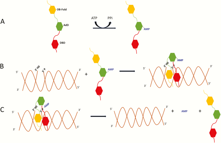
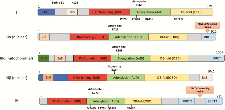
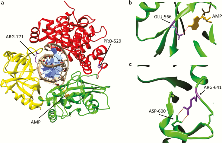
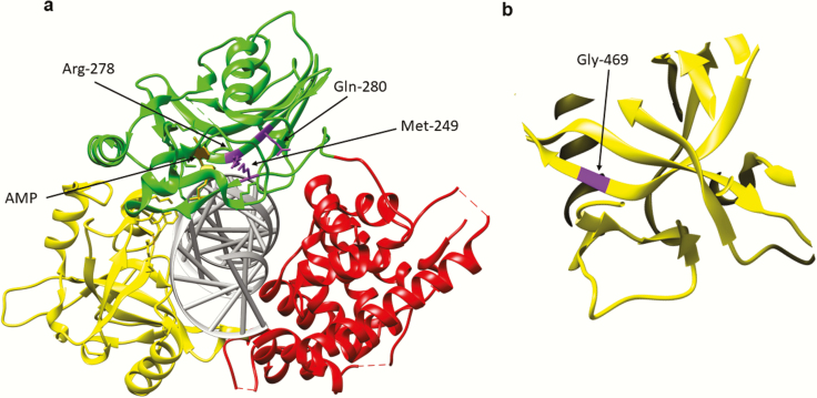
The joining of interruptions in the phosphodiester backbone of DNA is critical to maintain genome stability. These breaks, which are generated as part of normal DNA transactions, such as DNA replication, V(D)J recombination and meiotic recombination as well as directly by DNA damage or due to DNA damage removal, are ultimately sealed by one of three human DNA ligases. DNA ligases I, III and IV each function in the nucleus whereas DNA ligase III is the sole enzyme in mitochondria. While the identification of specific protein partners and the phenotypes caused either by genetic or chemical inactivation have provided insights into the cellular functions of the DNA ligases and evidence for significant functional overlap in nuclear DNA replication and repair, different results have been obtained with mouse and human cells, indicating species-specific differences in the relative contributions of the DNA ligases. Inherited mutations in the human LIG1 and LIG4 genes that result in the generation of polypeptides with partial activity have been identified as the causative factors in rare DNA ligase deficiency syndromes that share a common clinical symptom, immunodeficiency. In the case of DNA ligase IV, the immunodeficiency is due to a defect in V(D)J recombination whereas the cause of the immunodeficiency due to DNA ligase I deficiency is not known. Overexpression of each of the DNA ligases has been observed in cancers. For DNA ligase I, this reflects increased proliferation. Elevated levels of DNA ligase III indicate an increased dependence on an alternative non-homologous end-joining pathway for the repair of DNA double-strand breaks whereas elevated level of DNA ligase IV confer radioresistance due to increased repair of DNA double-strand breaks by the major non-homologous end-joining pathway. Efforts to determine the potential of DNA ligase inhibitors as cancer therapeutics are on-going in preclinical cancer models.


Figures

Fig. 1.
Fig. 1.
Three-step ligation reaction. (A) DNA ligases in an extended conformation interact with ATP to generate a covalent AMP-ligase intermediate with the AMP moiety linked to specific lysine residues in the Adenylation Domain (AdD, green). (B) When the DNA ligase recognises a ligatable nick, the catalytic region composed of the DNA Binding Domain (DBD, red) and Oligonucleotide/Oligosaccharide Binding-fold Domain (OBD, yellow) in addition to the AdD changes conformation, encircling the DNA nick with each of the three domains contacting the DNA. Within this structure, the covalently bound AMP is transferred to the 5′ termini of the nicked DNA. (C) Lastly, using the OH group at the 3′ termini as a nucleophile, non-adenylated DNA ligase catalyses the phosphodiester bond formation releasing the bound AMP.
Fig. 2.
Fig. 2.
Domain organisation of the DNA ligases encoded by the human LIG1, LIG3 and LIG4 genes. The Adenylation Domain (AdD, green) and Oligonucleotide/Oligosaccharide Binding-fold Domain (OBD, yellow) domains comprise the catalytic core that contains the key active site lysine residue. The less conserved DNA Binding Domain (DBD, red) is N-terminal to this core. The non-catalytic N-terminal region of DNA ligase I contains PCNA interaction peptide, PIP (blue) and nuclear localisation signal (beige). The three isoforms of DNA ligase III have an N-terminal Zinc finger domain (light orange: ZnF). Mitochondrial DNA ligase IIIα has a mitochondrial localisation signal, MLS (dark green) at the N-terminus. DNA ligase IIIα and DNA ligase IV contain one and two C-terminal BRCT domains (blue), respectively. The DNA ligase IIIα BRCT domain is required for interaction with XRCC1 and whereas XRCC4 interacts with the region between the DNA ligase IV BRCT domains. Amino acid substitutions identified in DNA ligase deficiency syndromes are indicated below the DNA ligase polypeptides.
Fig. 3.
Fig. 3.
Amino acid substitutions identified in individuals with DNA ligase I deficiency syndrome. (a) Ribbon diagram showing the Adenylation domain (AdD, green), OB-fold domain (OBD, yellow) and DNA binding domain (DBD, red) of DNA ligase I encircling a nicked DNA duplex (grey). The AMP group (gold) linked to the 5-phosphate terminus of the DNA nick held within the AdD is indicated. Substitution of Arg771 (purple) in the OBD alters interaction with DNA, resulting in reduced enzymatic activity. with Trp and Pro529 depicted in purple. Substitution of Pro529 (purple) in the DBD with Leu has no effect on enzymatic activity. (b) Glu566 residue (purple) forms a hydrogen bond with N6 of the AMP moiety (gold). Replacement of Glu566 Lys inactivates enzyme activity. (c) Arg641 (purple) forms a salt bridge with Asp600 within the AdD domain. Replacement of R641 with Leu disrupts the salt bridge, resulting in reduced enzymatic activity that appears to be due to defective DNA binding.
Fig. 4.
Fig. 4.
Amino acid substitutions identified in individuals with DNA ligase IV deficiency syndrome. (a) Ribbon diagram showing the Adenylation domain (AdD, green), OB-fold domain (OBD, yellow) and DNA binding domain (DBD, red) of DNA ligase IV encircling a nicked DNA duplex (grey). The AMP group (gold) linked to the 5-phosphate terminus of the DNA nick held within the AdD is indicated. Amino acid substitutions identified in the AdD; R278H, Q280R, M249V (purple) are indicated. (b) Replacement of Gly469 (purple) with Glu likely destabilises the OBD by disrupting hydrophobic interactions between β sheets within the OBD.

References

    1. Ho C. K., Wang L. K., Lima C. D. and Shuman S (2004) Structure and mechanism of RNA ligase. Structure, 12, 327–339. - PubMed
    1. Shuman S. and Schwer B (1995) RNA capping enzyme and DNA ligase: a superfamily of covalent nucleotidyl transferases. Mol. Microbiol., 17, 405–410. - PubMed
    1. Ellenberger T. and Tomkinson A. E (2008) Eukaryotic DNA ligases: structural and functional insights. Annu. Rev. Biochem., 77, 313–338. - PMC - PubMed
    1. Chen S. H., and Yu X (2018) Human DNA ligase IV is able to use NAD+ as an alternative adenylation donor for DNA ends ligation. Nucleic Acids Res 47, 1321–1334 - PMC - PubMed
    1. Ahel I., Rass U., El-Khamisy S. F., Katyal S., Clements P. M., McKinnon P. J., Caldecott K. W. and West S. C (2006) The neurodegenerative disease protein aprataxin resolves abortive DNA ligation intermediates. Nature, 443, 713–716. - PubMed

Publication types