Review
. 2020 Jun;160(2):106-115.
doi: 10.1111/imm.13136. Epub 2019 Nov 11.

Immune responses in the human female reproductive tract

Affiliations
Review

Immune responses in the human female reproductive tract

Leticia Monin et al. Immunology. 2020 Jun.

Abstract

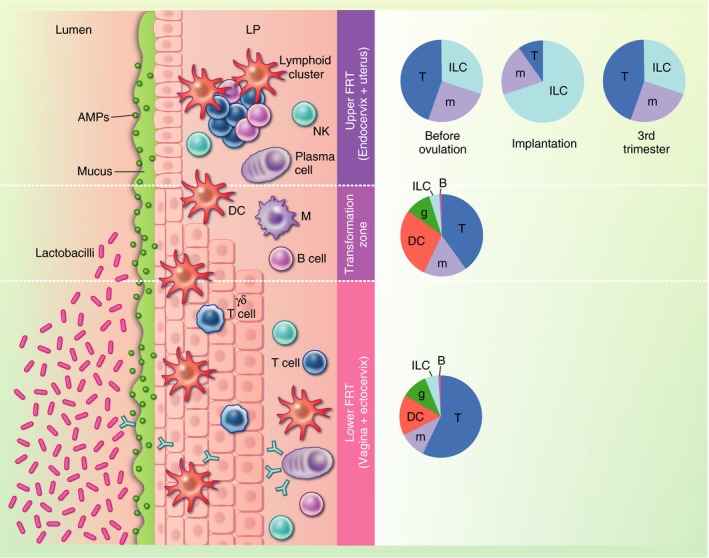
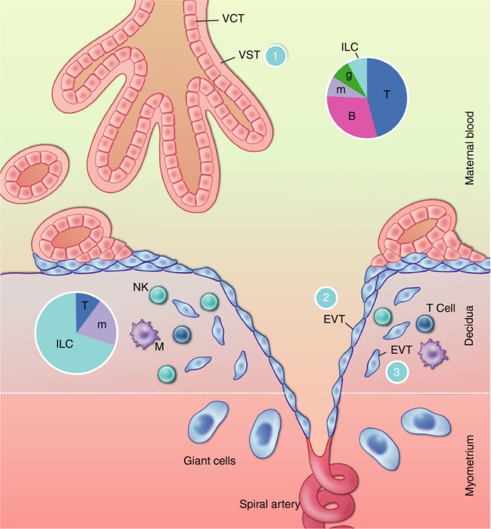
Mucosal surfaces are key interfaces between the host and its environment, but also constitute ports of entry for numerous pathogens. The gut and lung mucosae act as points of nutrient and gas exchange, respectively, but the physiological purpose of the female reproductive tract (FRT) is to allow implantation and development of the fetus. Our understanding of immune responses in the FRT has traditionally lagged behind our grasp of the situation at other mucosal sites, but recently reproductive immunologists have begun to make rapid progress in this challenging area. Here, we review current knowledge of immune responses in the human FRT and their heterogeneity within and between compartments. In the commensal-rich vagina, the immune system must allow the growth of beneficial microbes, whereas the key challenge in the uterus is allowing the growth of the semi-allogeneic fetus. In both compartments, these objectives must be balanced with the need to eliminate pathogens. Our developing understanding of immune responses in the FRT will help us develop interventions to prevent the spread of sexually transmitted diseases and to improve outcomes of pregnancy for mothers and babies.

Keywords: ILC; T-cells; macrophages; mucosa; uterus.


Conflict of interest statement

The authors declare no competing interests.

Figures

Figure 1
Figure 1
Immune and non‐immune barriers in the female reproductive tract (FRT). The FRT can be broadly divided into an upper section, comprising the uterus and the endocervix, and a lower section, which includes the vagina and ectocervix. The upper reproductive tract is lined by a single layer of columnar epithelium, while the lower reproductive tract is lined by stratified squamous epithelium. The zone where the two types of epithelia meet is called the transformation zone. Several non‐immune barriers form a first line of defence against pathogen invasion: the presence of tight junctions constitutes a physical barrier, mucus and antimicrobial peptides form a chemical barrier, and the Lactobacillus‐rich vaginal milieu creates a biological barrier. This multi‐layered defence strategy is further reinforced by a variety of immune cells that reside within the epithelium and the lamina propria (LP), patrolling for invading microorganisms. Pie charts indicate the composition of immune cells along the FRT. In the upper FRT, the composition differs by stage of the menstrual cycle or pregnancy where ‘Implantation’ represents the composition in the secretory phase of the menstrual cycle and early pregnancy. AMPs, antimicrobial peptides; DC, dendritic cell; FRT, female reproductive tract; g, granulocyte; ILC, innate lymphoid cell; LP, lamina propria; m, monocyte/macrophage; NK, natural killer.
Figure 2
Figure 2
Interfaces between the placenta and the maternal immune system. The placental villi are bathed in the maternal blood. The outer layer of the villi is covered by VST with an underlying layer of mononuclear VCT. Villous trophoblast (VT) is protected from recognition by T‐cells, which are frequent in the maternal blood, by their complete lack of MHC expression (interface 1). They are further protected by immune cell recognition by their syncytial nature and thick glycocalyx. The inner layer of VCT grows out of the villi to anchor the placenta to the maternal decidua. VCT differentiates to extravillous trophoblast (EVT), some of which migrates down the spiral arteries, replacing the endothelial cells as far as the inner third of the myometrium. This process aids in the transformation of the spiral arteries, allowing blood to flow to the placenta at low pressure. These cells are in contact with the maternal blood (interface 2). Some EVT cells are also present in the decidua, where they interface with the unique immune cells present in this microenvironment (interface 3). EVT is largely protected from T‐cell recognition because they do not express the major TCR ligands HLA‐A and ‐B, but they do express the natural killer (NK) cell ligands HLA‐C, ‐E and G. At interface 2, this may protect EVT from recognition by blood NK cells. At interface 3, the expression of HLA‐C is likely to allow recognition by decidual NK cells, which are not cytotoxic but rather seem to have a role in tissue remodelling. Pie charts indicate the composition of maternal immune cells in the blood and decidua. VCT, villous cytotrophoblast; VST, villous syncytiotrophoblast; EVT, extravillous trophoblast; T, T‐cells; B, B‐cells; m, monocytes/macrophages; g, granulocytes; ILC, innate lymphoid cells.

References

    1. Fisk NM, Atun R. Systematic analysis of research underfunding in maternal and perinatal health. BJOG 2009; 116:347–56. - PubMed
    1. Tsevat DG, Wiesenfeld HC, Parks C, Peipert JF. Sexually transmitted diseases and infertility. Am J Obstet Gynecol 2017; 216:1–9. - PMC - PubMed
    1. Goldenberg RL, Hauth JC, Andrews WW. Intrauterine infection and preterm delivery. N Engl J Med 2000; 342:1500–7. - PubMed
    1. Lai SK, Wang Y‐Y, Wirtz D, Hanes J. Micro‐ and macrorheology of mucus. Adv Drug Deliv Rev 2009; 61:86–100. - PMC - PubMed
    1. Iwasaki A. Antiviral immune responses in the genital tract: clues for vaccines. Nat Rev Immunol 2010; 10:699–711. - PMC - PubMed

Publication types

MeSH terms