. 2019 Nov 7;105(5):1030-1039.
doi: 10.1016/j.ajhg.2019.09.022. Epub 2019 Oct 17.

De Novo Mutations in FOXJ1 Result in a Motile Ciliopathy with Hydrocephalus and Randomization of Left/Right Body Asymmetry

Affiliations

De Novo Mutations in FOXJ1 Result in a Motile Ciliopathy with Hydrocephalus and Randomization of Left/Right Body Asymmetry

Julia Wallmeier et al. Am J Hum Genet. .

Abstract

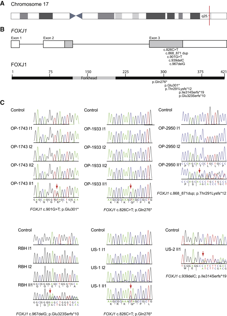
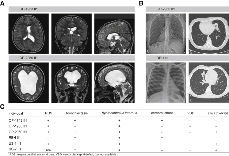
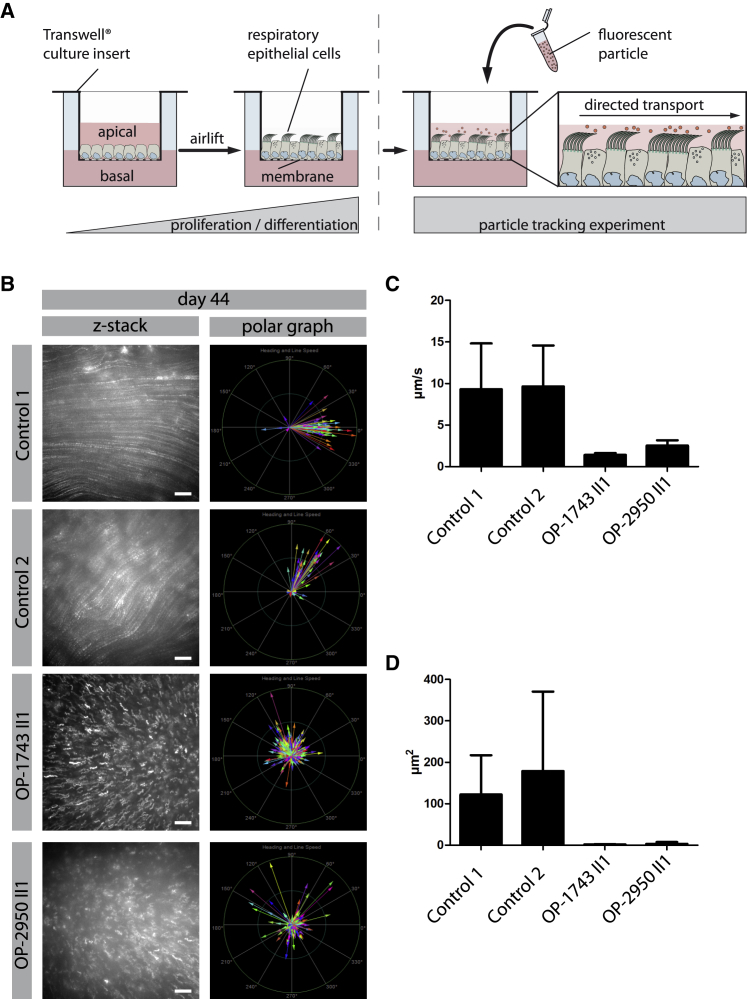
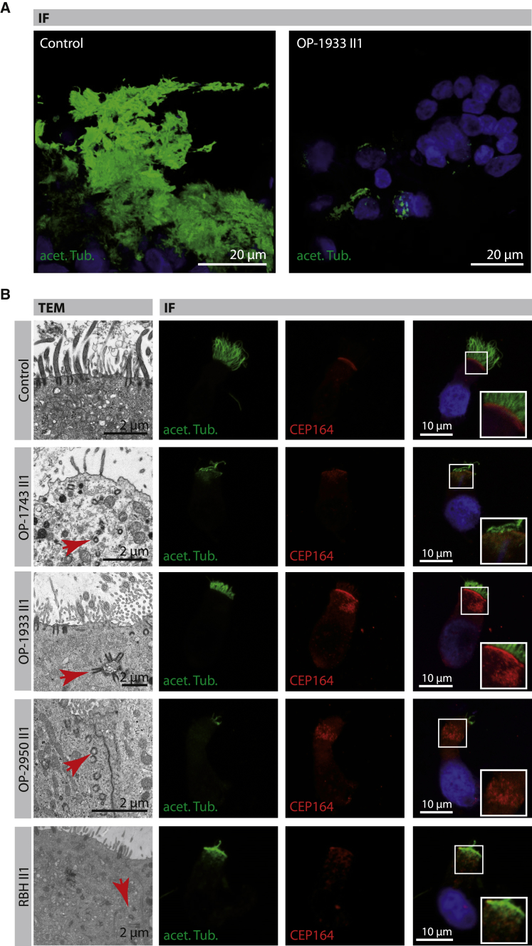
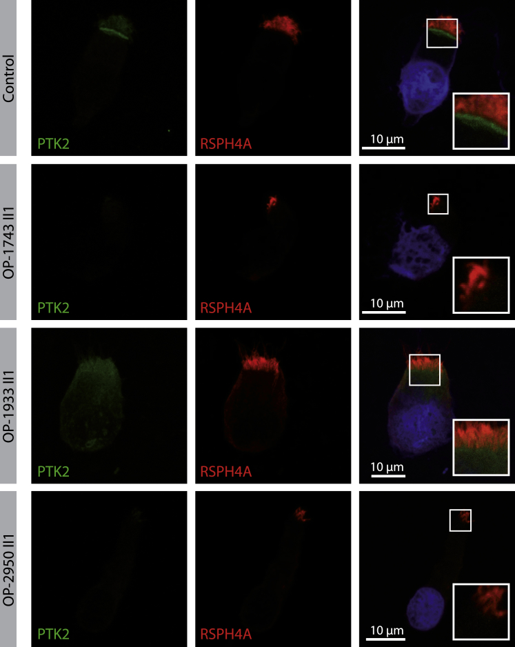
Hydrocephalus is one of the most prevalent form of developmental central nervous system (CNS) malformations. Cerebrospinal fluid (CSF) flow depends on both heartbeat and body movement. Furthermore, it has been shown that CSF flow within and across brain ventricles depends on cilia motility of the ependymal cells lining the brain ventricles, which play a crucial role to maintain patency of the narrow sites of CSF passage during brain formation in mice. Using whole-exome and whole-genome sequencing, we identified an autosomal-dominant cause of a distinct motile ciliopathy related to defective ciliogenesis of the ependymal cilia in six individuals. Heterozygous de novo mutations in FOXJ1, which encodes a well-known member of the forkhead transcription factors important for ciliogenesis of motile cilia, cause a motile ciliopathy that is characterized by hydrocephalus internus, chronic destructive airway disease, and randomization of left/right body asymmetry. Mutant respiratory epithelial cells are unable to generate a fluid flow and exhibit a reduced number of cilia per cell, as documented by high-speed video microscopy (HVMA), transmission electron microscopy (TEM), and immunofluorescence analysis (IF). TEM and IF demonstrate mislocalized basal bodies. In line with this finding, the focal adhesion protein PTK2 displays aberrant localization in the cytoplasm of the mutant respiratory epithelial cells.

Keywords: FOXJ1; cilia; ciliogenesis; ependyma; hydrocephalus; lung disease.


Conflict of interest statement

The authors declare no competing interests.

Figures

Figure 1
Figure 1
Heterozygous De Novo Pathogenic Variants in FOXJ1 in Affected with Hydrocephalus and Chronic Destructive Airway Disease (A) Schematic overview of chromosome 17. FOXJ1 (CCDS32739, GenBank: NM_001454.4) is located on chromosome 17q25.1 (red mark). (B) FOXJ1 consists of two coding exons and one alternative first exon encoding a 2,641 bp transcript and 421 amino acid protein. (C) Electropherograms of Sanger sequencing results for family OP-1933, OP-1743, OP-2950, RBH, and US-1. Consistent with de novo mutations, none of the variants were identified in either the parents or non-affected siblings. In US-2 no parental DNA was available.
Figure 2
Figure 2
Affected with Pathogenic FOXJ1 Variants Display Obstructive Hydrocephalus, Randomization of Left/Right Body Asymmetry, and a Chronic Destructive Airway Disease (A) Cranial magnetic resonance imaging of OP-1933 II1 was performed after shunt insertion (right lateral ventricle) to relieve raised intracranial pressure. The left lateral ventricle and the third ventricle are dilated. The lateral view documents stenosis of the aqueduct of Sylvius and a small fourth ventricle. OP-2950 II1 shows massively dilated brain ventricles. Lateral view indicates a patent aqueduct and a dilated fourth ventricle due to closure of the foramen of Magendii and the lateral apertures. (B) Chest X-ray of OP-2950 II1 shows situs inversus totalis. The computed tomography scan of OP-2950 II1 and RBH II1 exhibit atelectasis and bronchiectasis of the middle lobe. (C) Summary of clinical findings in the affected individuals.
Figure 3
Figure 3
Air Liquid Interface (ALI) Cultures of FOXJ1-Mutant Respiratory Epithelial Cells Are Unable to Generate a Directed Fluid Flow (A) Schematic depicts the experimental set-up of particle tracking analyses performed on ALI-cultured respiratory epithelial cells. Respiratory epithelial cells from FOXJ1-mutants (OP-1743 II1, OP-2950 II1) as well as healthy control subjects were cultured under ALI conditions. After complete differentiation (30 days, 37 days, and 44 days after airlift), 0.5 μm fluorescent particles were added to the apical compartments of the cells. (B–D) Tracking videos are represented as z stack projections, while the transport direction of each particle is summarized in polar graphs (B). Under healthy conditions the fluorescent particles were transported in a linear direction along the cell layer, whereas the particle transport in FOXJ1 mutant cells (OP-1743 II1, OP-2950 II1) was non-oriented, highly reduced in speed (μm/s) (C) and in mean squared displacement (μm2) (D). For statistical evaluation, 15 videos per person were analyzed. Thereby, 253 particles were tracked per video on average. Scale bars represent 20 μm.
Figure 4
Figure 4
FOXJ1 Mutant Respiratory Epithelial Cells Show a Reduced Number of Cilia and Mislocalized Basal Bodies by Transmission Electron Microscopy (TEM) and Immunofluorescence Microscopy Analysis (IF) (A) Respiratory epithelial cells cultured as spheroids from a healthy control subject and OP-1933 II1. Cilia are stained with antibodies targeting acetylated α-tubulin (acet. Tub.; green) after complete differentiation. Cells of OP-1933 II1 demonstrate a variable reduction of cilia in comparison to the control. Nuclei are stained with Hoechst33342 (blue). (B) Transmission electron microscopy (TEM) photographs of multiciliated cells (MCC; first row) from a healthy control subject show basal bodies attached to the apical membrane and nucleating multiple motile cilia. Respiratory epithelial cells from mutant individuals with pathogenic FOXJ1 variants (OP-1743 II1, OP-1933 II1, OP-2950 II1, RBH II1) exhibit mislocalized basal bodies (representative examples shown by red arrows) within the cytoplasm, consistent with a basal body docking defect. Respiratory epithelial cells are stained with antibodies targeting acetylated α-tubulin (acet. Tub.; green) and antibodies targeting mother centrioles (CEP164, red). In control cells, basal bodies (red) are aligned at the apical cell region, whereas in FOXJ1 mutant cells they are mainly mislocalized within the cytoplasm, consistent with TEM findings. Right row shows higher magnification images of regions of CEP164-positive basal bodies. Nuclei were stained with Hoechst33342 (blue).
Figure 5
Figure 5
PTK2, a Member of the Subapical Protein Network, Shows Abnormal Localization in FOXJ1 Mutant Cells by Immunofluorescence Microscopy Analysis (IF) Respiratory epithelial cells from control and FOXJ1 mutant individuals (OP-1743 II1, OP-1933 II1, OP-2950 II1) are stained with antibodies targeting PTK2 (green). PTK2, which forms complexes named ciliary adhesions that are associated with basal bodies and striated rootlets, shows reduced localization in FOXJ1 mutant cells compared to the control. Regions around the subapical cell membrane showing PTK2 at higher magnification (right row). Nuclei are stained with Hoechst33342 (blue).
Figure 6
Figure 6
FOXJ1 Is an Essential Component in Signaling Pathways for the Generation of Motile Cilia Schematics illustrating the function of FOXJ1 in the generation of motile cilia in the NOTCH1- and NOTO-dependent pathway in (A) multiciliated cells and (B) the ciliated cells of the embryonic node, respectively. Pathogenic variants in MCIDAS and CCNO (marked in green) are known to cause a ciliogenesis defect in multiple motile cilia causing a mucociliary clearance disorder referred to as reduced generation of multiple motile cilia, (RGMC).

References

    1. Kahle K.T., Kulkarni A.V., Limbrick D.D., Jr., Warf B.C. Hydrocephalus in children. Lancet. 2016;387:788–799. - PubMed
    1. Fliegauf M., Benzing T., Omran H. When cilia go bad: cilia defects and ciliopathies. Nat. Rev. Mol. Cell Biol. 2007;8:880–893. - PubMed
    1. Altmüller J., Motameny S., Becker C., Thiele H., Chatterjee S., Wollnik B., Nürnberg P. A systematic comparison of two new releases of exome sequencing products: the aim of use determines the choice of product. Biol. Chem. 2016;397:791–801. - PubMed
    1. Wheway G., Mitchison H.M., Genomics England Research Consortium Opportunities and Challenges for Molecular Understanding of Ciliopathies-The 100,000 Genomes Project. Front. Genet. 2019;10:127. - PMC - PubMed
    1. Lek M., Karczewski K.J., Minikel E.V., Samocha K.E., Banks E., Fennell T., O’Donnell-Luria A.H., Ware J.S., Hill A.J., Cummings B.B., Exome Aggregation Consortium Analysis of protein-coding genetic variation in 60,706 humans. Nature. 2016;536:285–291. - PMC - PubMed

Publication types

Substances