

A Comprehensive Resource for Induced Pluripotent Stem Cells from Patients with Primary Tauopathies
- PMID: 31631020
- PMCID: PMC6895712
- DOI: 10.1016/j.stemcr.2019.09.006
A Comprehensive Resource for Induced Pluripotent Stem Cells from Patients with Primary Tauopathies
Abstract
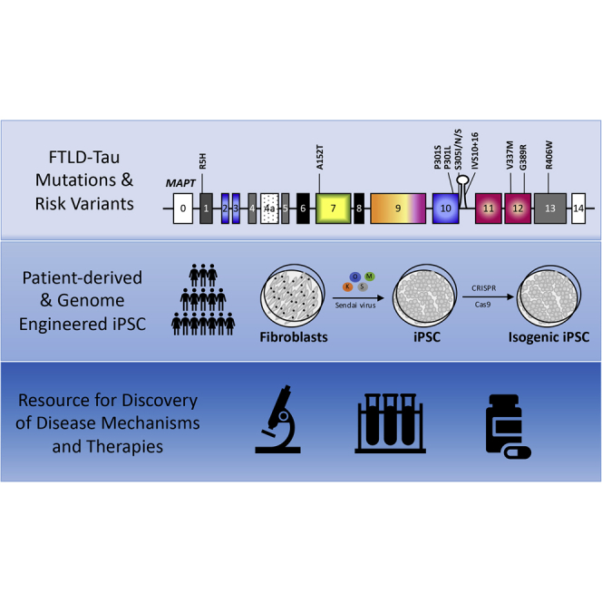
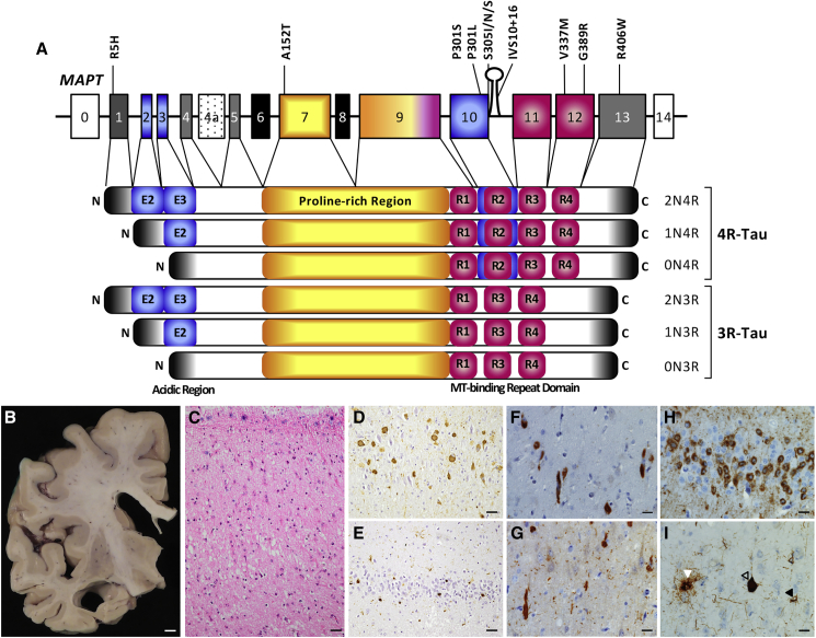
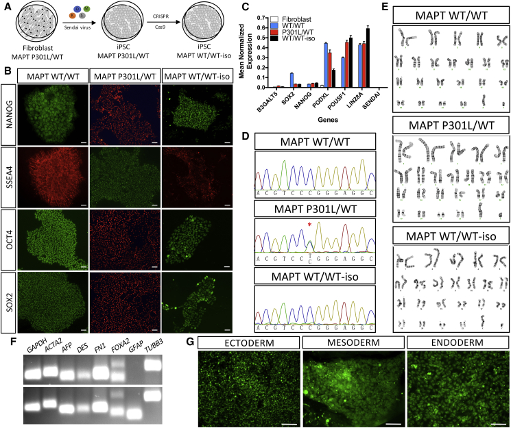
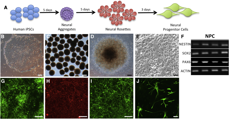
Primary tauopathies are characterized neuropathologically by inclusions containing abnormal forms of the microtubule-associated protein tau (MAPT) and clinically by diverse neuropsychiatric, cognitive, and motor impairments. Autosomal dominant mutations in the MAPT gene cause heterogeneous forms of frontotemporal lobar degeneration with tauopathy (FTLD-Tau). Common and rare variants in the MAPT gene increase the risk for sporadic FTLD-Tau, including progressive supranuclear palsy (PSP) and corticobasal degeneration (CBD). We generated a collection of fibroblasts from 140 MAPT mutation/risk variant carriers, PSP, CBD, and cognitively normal controls; 31 induced pluripotent stem cell (iPSC) lines from MAPT mutation carriers, non-carrier family members, and autopsy-confirmed PSP patients; 33 genome engineered iPSCs that were corrected or mutagenized; and forebrain neural progenitor cells (NPCs). Here, we present a resource of fibroblasts, iPSCs, and NPCs with comprehensive clinical histories that can be accessed by the scientific community for disease modeling and development of novel therapeutics for tauopathies.
Keywords: CRISPR/Cas9; MAPT; corticobasal degeneration; fibroblasts; frontotemporal dementia; induced pluripotent stem cells; neural progenitor cells; progressive supranuclear palsy; tau; tauopathy.
Copyright © 2019 The Authors. Published by Elsevier Inc. All rights reserved.
Figures
References
-
- Arai T., Ikeda K., Akiyama H., Nonaka T., Hasegawa M., Ishiguro K., Iritani S., Tsuchiya K., Iseki E., Yagishita S. Identification of amino-terminally cleaved tau fragments that distinguish progressive supranuclear palsy from corticobasal degeneration. Ann. Neurol. 2004;55:72–79. - PubMed
-
- Arai T., Ikeda K., Akiyama H., Tsuchiya K., Yagishita S., Takamatsu J. Intracellular processing of aggregated tau differs between corticobasal degeneration and progressive supranuclear palsy. Neuroreport. 2001;12:935–938. - PubMed
-
- Behnam M., Ghorbani F., Shin J.H., Kim D.S., Jang H., Nouri N., Sedghi M., Salehi M., Ansari B., Basiri K. Homozygous MAPT R406W mutation causing FTDP phenotype: a unique instance of a unique mutation. Gene. 2015;570:150–152. - PubMed
Publication types
MeSH terms
Substances
Grants and funding
- P30 AG066444/AG/NIA NIH HHS/United States
- T32 AG023481/AG/NIA NIH HHS/United States
- R01 AG058233/AG/NIA NIH HHS/United States
- U24 AG021886/AG/NIA NIH HHS/United States
- K12 HD001459/HD/NICHD NIH HHS/United States
- R01 AG054008/AG/NIA NIH HHS/United States
- P30 AG062422/AG/NIA NIH HHS/United States
- P50 AG023501/AG/NIA NIH HHS/United States
- P01 AG019724/AG/NIA NIH HHS/United States
- U54 NS092089/NS/NINDS NIH HHS/United States
- R01 AG051390/AG/NIA NIH HHS/United States
- K08 AG052648/AG/NIA NIH HHS/United States
- R01 NS095252/NS/NINDS NIH HHS/United States
- P01 AG003991/AG/NIA NIH HHS/United States
- P50 AG005681/AG/NIA NIH HHS/United States
- U01 AG045390/AG/NIA NIH HHS/United States
- R35 NS097277/NS/NINDS NIH HHS/United States
LinkOut - more resources
Full Text Sources
Other Literature Sources
Miscellaneous
