. 2019 Oct 7:13:1973-1980.
doi: 10.2147/OPTH.S185184. eCollection 2019.

Update On Cenegermin Eye Drops In The Treatment Of Neurotrophic Keratitis

Affiliations

Update On Cenegermin Eye Drops In The Treatment Of Neurotrophic Keratitis

Hosam Sheha et al. Clin Ophthalmol. .

Abstract

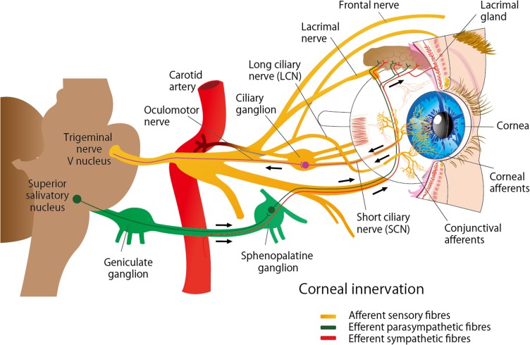
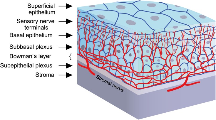
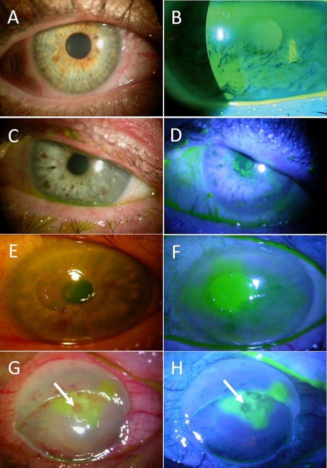
Neurotrophic keratitis is an underdiagnosed degenerative condition induced by impairment to the corneal nerves which may lead to persistent epithelial defects and corneal blindness. Current medical and surgical treatments are only supportive and poorly tackle the underlying problem of corneal anesthesia; hence, fail to provide a permanent cure. Cenegermin is a newly introduced recombinant human nerve growth factor (rhNGF) that may address this issue. Preliminary clinical trials have demonstrated the safety and efficacy of topical cenegermin in patients with moderate to severe neurotrophic keratitis; however, the clinical experience with this drug is still limited. This review summarizes the pathogenesis and management of neurotrophic keratitis as well as the mechanism of action, uses, and limitations of cenegermin eye drops in the treatment of neurotrophic keratitis.

Keywords: cenegermin; corneal nerves; nerve growth factors; neurotrophic keratitis; persistent epithelial defect.


Conflict of interest statement

The authors report no conflicts of interest in this work.

Figures

Figure 1
Figure 1
Nerve supply of the cornea. The cornea is innervated by the ophthalmic branch of the trigeminal nerve (V1) and by sympathetic and parasympathetic autonomic nerves.
Figure 2
Figure 2
Corneal nerves distribution: Nerve fibers penetrate the corneal periphery at the limbus and approach toward the central cornea at the level of the anterior stroma while penetrating the Bowman’s membrane to create the sub-basal nerve plexus, at the level of the basal epithelial cells and basement membrane of the epithelium. Terminal branches from the sub-basal plexus pass anteriorly into the epithelial cell layers, terminating within or in between epithelial cells.
Figure 3
Figure 3
Stages of neurotrophic keratitis: Impaired cornea sensitivity and lack of trophic support trigger nonspecific epithelial irregularity and tear film changes (A, B). Stage 1- mild (C, D) is characterized by corneal punctate keratopathy due to focal epithelial damage and loss of tight junctions. It is associated with mild stromal edema with or without corneal neovascularization. Stage 2 – moderate (E, F) is distinguished by the presence of central persistent epithelial defect, surrounded by edematous, cloudy, and poorly adherent epithelium. Stage 3- severe (G, H) is characterized by stromal ulceration and thinning that may progress to melting and perforation (arrows).

References

    1. Muller LJ, Marfurt CF, Kruse F, Tervo TM. Corneal nerves: structure, contents and function. Exp Eye Res. 2003;76(5):521–542. doi:10.1016/s0014-4835(03)00050-2 - DOI - PubMed
    1. Al-Aqaba MA, Fares U, Suleman H, Lowe J, Dua HS. Architecture and distribution of human corneal nerves. Br J Ophthalmol. 2010;94(6):784–789. doi:10.1136/bjo.2009.173799 - DOI - PubMed
    1. Stepp MA, Tadvalkar G, Hakh R, Pal-Ghosh S. Corneal epithelial cells function as surrogate Schwann cells for their sensory nerves. Glia. 2017;65(6):851–863. doi:10.1002/glia.23102 - DOI - PMC - PubMed
    1. Versura P, Giannaccare G, Pellegrini M, Sebastiani S, Campos EC. Neurotrophic keratitis: current challenges and future prospects. Eye Brain. 2018;10:37–45. doi:10.2147/EB.S117261 - DOI - PMC - PubMed
    1. Al-Aqaba MA, Anis FS, Mohammed I, Dua HS. Nerve terminals at the human corneoscleral limbus. Br J Ophthalmol. 2018;102(4):556–561. doi:10.1136/bjophthalmol-2017-311146 - DOI - PubMed