. 2019 Oct 9:13:3529-3538.
doi: 10.2147/DDDT.S210220. eCollection 2019.

Aucubin Protects Chondrocytes Against IL-1β-Induced Apoptosis In Vitro And Inhibits Osteoarthritis In Mice Model

Affiliations

Aucubin Protects Chondrocytes Against IL-1β-Induced Apoptosis In Vitro And Inhibits Osteoarthritis In Mice Model

Bo-Wei Wang et al. Drug Des Devel Ther. .

Abstract

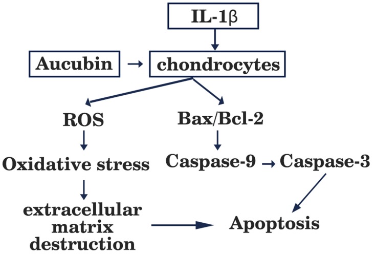
Objective: Chondrocyte apoptosis has also been strongly correlated with the severity of cartilage damage and matrix depletion in an osteoarthritis (OA) joint. Therefore, pharmacological inhibitors of apoptosis may provide a novel treatment option for patients with OA. Aucubin, a natural compound isolated from Eucommia ulmoides, has been proved to possess antioxidative and anti-apoptotic properties. However, anti-osteoarthritis effect of aucubin in animal model and anti-apoptotic response of aucubin in OA chondrocytes remain unclear. This study aimed to determine whether aucubin could slow progression of OA in a mouse model and inhibit the IL-1β-induced chondrocyte apoptosis.

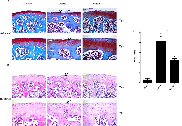
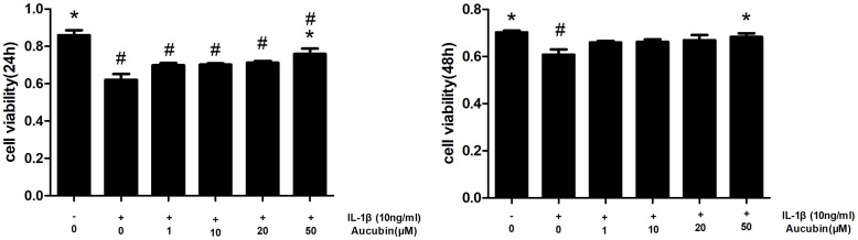
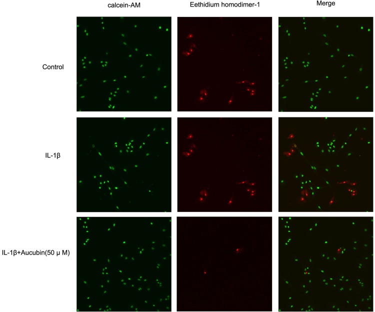
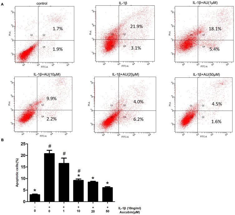
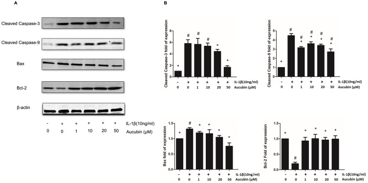
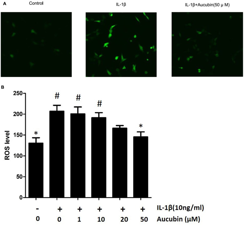
Methods: OA severity and articular cartilage degradation were evaluated by Safranin-O staining, Hematoxylin-eosin (H&E) staining, and Osteoarthritis Research Society International (OARSI) standards. Chondrocyte viability was observed by Cell Counting Kit-8 (CCK8) and live/dead cells assay; the apoptotic rate of chondrocytes was evaluated by flow cytometry (FCM) with Annexin V-FITC/PI kit. Mediators of apoptosis were tested by Western blot of Bax, caspase-3, caspase-9, and Bcl-2 expression. The intracellular levels of Reactive oxygen species (ROS) were assessed by the probe of 2,7-Dichlorofluorescin diacetate (DCFH-DA).

Results: The articular cartilage in the limb with destabilization of the medial meniscus (DMM) exhibited early OA-like manifestations characterized by proteoglycan loss, cartilage fibrillation, and erosion, with lower OARSI score. Oral administration of aucubin remarkably attenuated the loss of proteoglycan and the articular cartilage erosion and decreased the OARSI scores underwent DMM surgery. Aucubin treatment significantly reverses IL-1β-induced cytotoxicity and attenuated the IL-1β-induced chondrocyte apoptosis. In addition, aucubin can significantly inhibit mediators of apoptosis in rat primary chondrocytes. Furthermore, aucubin remarkably attenuated the IL-1β-induced intracellular ROS production.

Conclusion: Our findings suggest that aucubin has a protective effect on articular cartilage and slowing progression of OA in a mouse model. This protective effect may result from inhibiting chondrocyte apoptosis and excessive ROS production.

Keywords: IL-1β; ROS; apoptosis; aucubin; osteoarthritis.


Conflict of interest statement

The authors report no conflicts of interest in this work.

Figures

Figure 1
Figure 1
Oral administration of aucubin preserved articular cartilage. (A) Safranin O staining. Solid arrows show proteoglycan loss and cartilage destruction at 8-week post operation. Scale bar, 50 μM (top), 25 μM (bottom). (B) H&E staining where hyaline cartilage (HC) thickness is declined and marked by arrows. Scale bars, 50 μM (top), 25μM (bottom). (C) OARSI scores of articular cartilage at 8-week post operation. Sham: sham-surgery; Vehicle: DMM-surgery treated with vehicle; Aucubin: DMM-surgery treated with aucubin. n=6 per group. Significant differences analysis in all groups compared with the sham group (*P<0.05), compared with the vehicle group (#P<0.05).
Figure 2
Figure 2
Aucubin attenuates cytotoxicity of IL-1β on chondrocyte viability. Chondrocytes were pretreated with various concentrations of aucubin (1, 10, 20, and 50 μM) for 2 hrs prior to IL-1β (10 ng/mL) for 24 hrs and 48 hrs time of point, and analyzed with the CCK8. Data are represented as mean ± S E M for three independent experiments. *Statistically significant difference (P < 0.05) versus the IL-1β group. #Statistically significant results (P < 0.05) versus control.
Figure 3
Figure 3
Aucubin suppresses IL-1β impaired chondrocytes viability. After treatment with the culture medium, IL-1β alone, pre-treatment with indicated concentration of aucubin (50 μM) followed by IL-1β for 24 hr, respectively, chondrocytes viability was assessed and visualized by live/dead assay with fluorescent dyes to distinguish live and dead cells.
Figure 4
Figure 4
Aucubin inhibited IL-1β-induced chondrocyte apoptosis. (A) Apoptotic chondrocytes were quantified by flow cytometry analysis with fluorescence staining. (B) The data indicated the percentages of apoptotic cells, which were statistically analysis and detected by flow cytometry. Data are expressed as mean ± S E M for three independent experiments. *Statistically significant difference (P < 0.05) versus the IL-1β group. #Statistically significant results (P < 0.05) versus control.
Figure 5
Figure 5
Apoptosis-related protein changes in aucubin treated or IL-1β-induced chondrocytes. (A) The expression levels of Bax, cleaved caspase-3, cleaved caspase-9, and Bcl-2 were detected by Western blot. (B) Quantitative result of Western blotting. All bands were measured in triplicate and normalized to GAPDH. Data are presented as mean ± S E M for three independent experiments. *Statistically significant difference (P < 0.05) versus the IL-1β group. #Statistically significant results (P < 0.05) versus control.
Figure 6
Figure 6
Aucubin attenuates ROS generation in IL-1β-induced chondrocytes. The ROS level represented by fluorescence in chondrocytes visualized under confocal microscopy (A) and cellular ROS levels were measured by spectrophotometer (B) Data are expressed as mean ± SEM for three independent experiments. *Statistically significant difference (P<0.05) versus the IL-1β group. #Statistically significant results (P <0.05) versus control.
Figure 7
Figure 7
Schematic diagram showing apoptotic and ROS signaling pathway induced by IL-1β in articular chondrocytes.

References

    1. Centers for Disease C, Prevention. Prevalence of doctor-diagnosed arthritis and arthritis-attributable activity limitation–United States, 2010–2012. MMWR. 2013;62(44):869–873. - PMC - PubMed
    1. Mistry D, Oue Y, Chambers MG, Kayser MV, Mason RM. Chondrocyte death during murine osteoarthritis. Osteoarthritis Cartilage. 2004;12(2):131–141. - PubMed
    1. Thomas CM, Fuller CJ, Whittles CE, Sharif M. Chondrocyte death by apoptosis is associated with cartilage matrix degradation. Osteoarthritis Cartilage. 2007;15(1):27–34. doi:10.1016/j.joca.2006.06.012 - DOI - PubMed
    1. Sharif M, Whitehouse A, Sharman P, Perry M, Adams M. Increased apoptosis in human osteoarthritic cartilage corresponds to reduced cell density and expression of caspase-3. Arthritis Rheum. 2004;50(2):507–515. doi:10.1002/art.20020 - DOI - PubMed
    1. Musumeci G, Loreto C, Carnazza ML, Strehin I, Elisseeff J. OA cartilage derived chondrocytes encapsulated in poly(ethylene glycol) diacrylate (PEGDA) for the evaluation of cartilage restoration and apoptosis in an in vitro model. Histol Histopathol. 2011;26(10):1265–1278. doi:10.14670/HH-26.1265 - DOI - PubMed

MeSH terms