. 2019 Oct 9:14:8073-8094.
doi: 10.2147/IJN.S220740. eCollection 2019.

CD-340 functionalized doxorubicin-loaded nanoparticle induces apoptosis and reduces tumor volume along with drug-related cardiotoxicity in mice

Affiliations

CD-340 functionalized doxorubicin-loaded nanoparticle induces apoptosis and reduces tumor volume along with drug-related cardiotoxicity in mice

Laboni Mondal et al. Int J Nanomedicine. .

Erratum in

Abstract

Background and objective: Targeted drug delivery of nanoparticles decorated with site-specific recognition ligands is of considerable interest to minimize cytotoxicity of chemotherapeutics in the normal cells. The study was designed to develop CD-340 antibody-conjugated polylactic-co-glycolic acid (PLGA) nanoparticles loaded with a highly water-soluble potent anticancer drug, doxorubicin (DOX), to specifically deliver entrapped DOX to breast cancer cells.

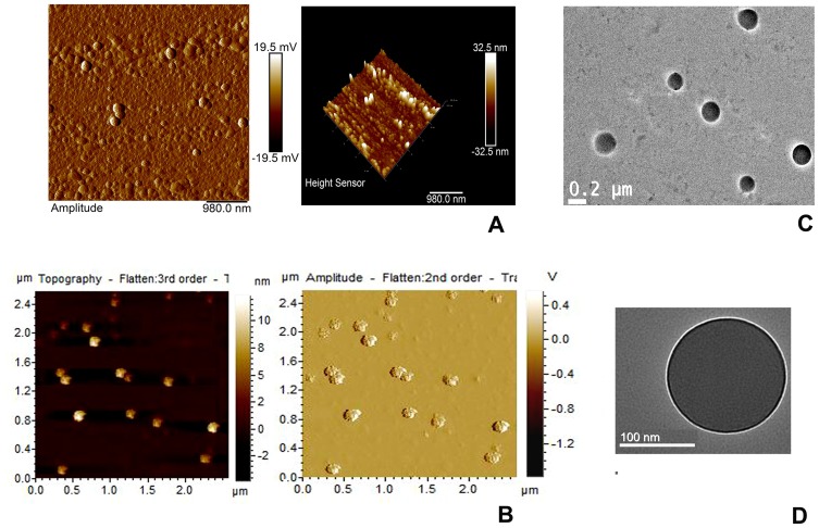
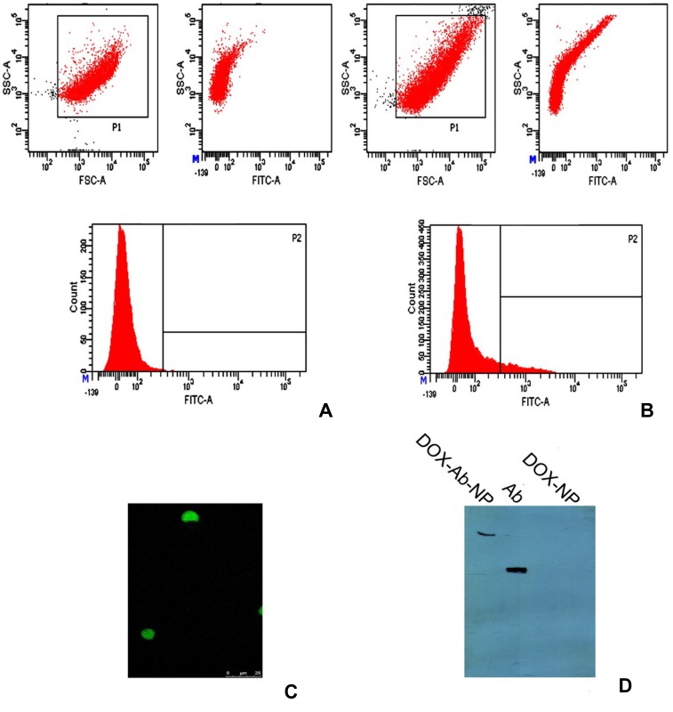
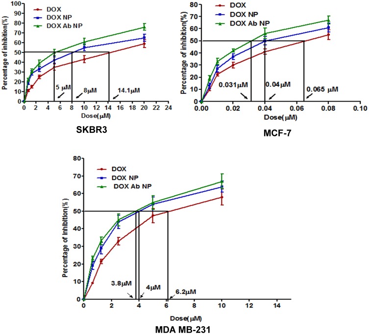
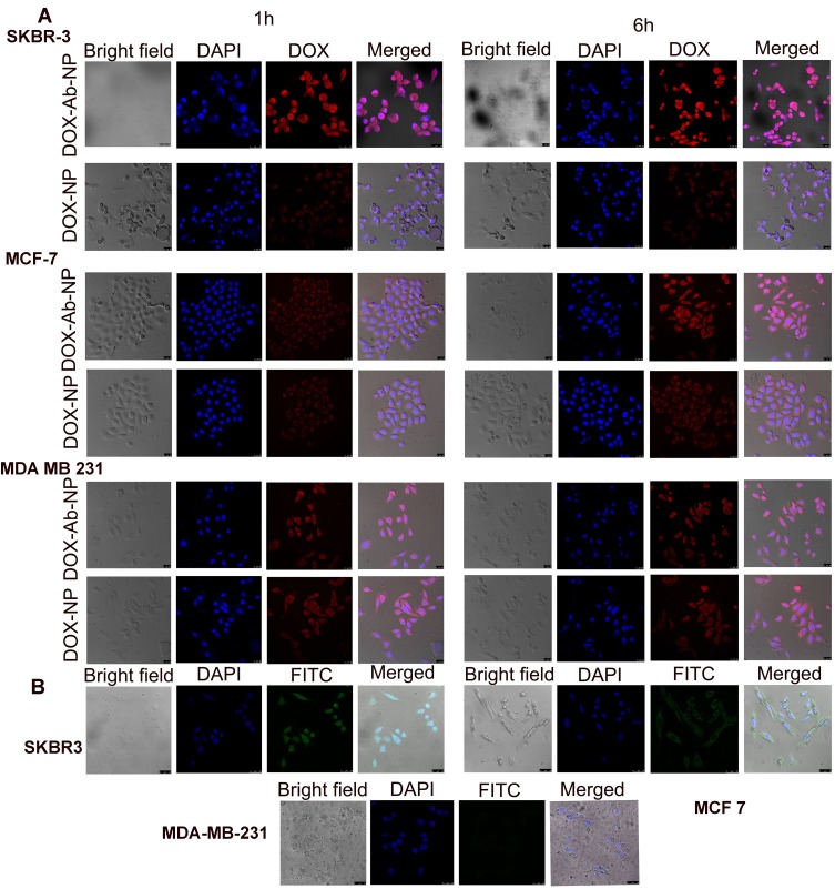
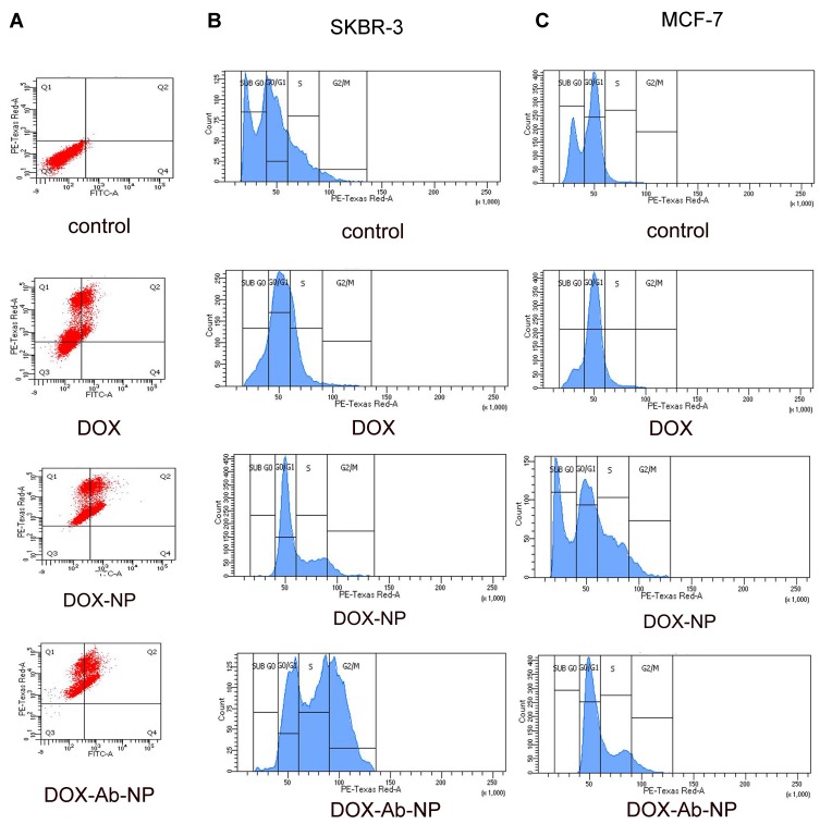
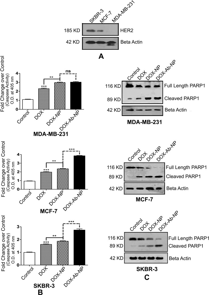
Methods: The study showed how to incorporate water-soluble drug in a hydrophobic PLGA (85:15) based matrix which otherwise shows poor drug loading due to leaching effect. The optimized formulation was covalently conjugated to anti-human epidermal growth factor receptor-2 (HER2) antibody (CD-340). Surface conjugation of the ligand was assessed by flow cytometry, confocal microscopy, and gel electrophoresis. Selectivity and cytotoxicity of the experimental nanoparticles were tested on human breast cancer cells SKBR-3, MCF-7, and MDA-MB-231. Both CD-340-conjugated and unconjugated nanoparticles were undergone in vitro and in vivo characterization.

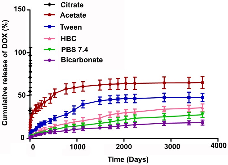
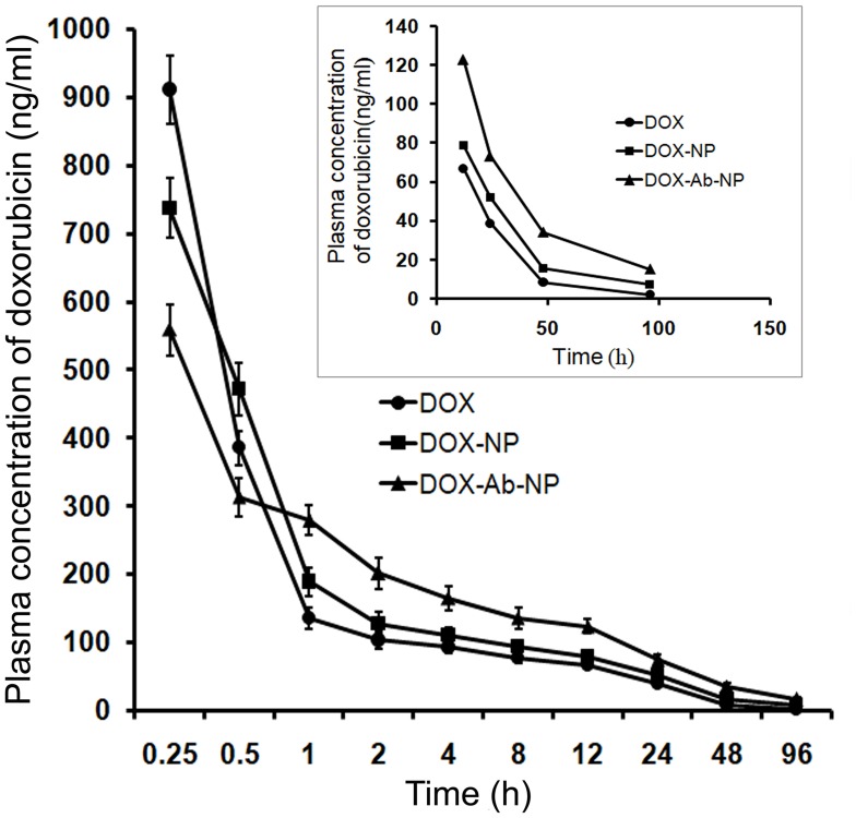
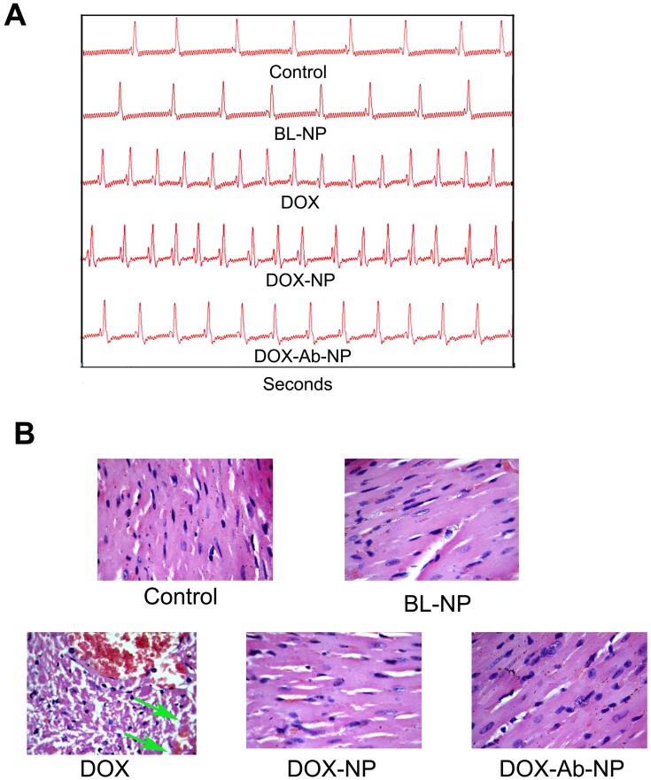
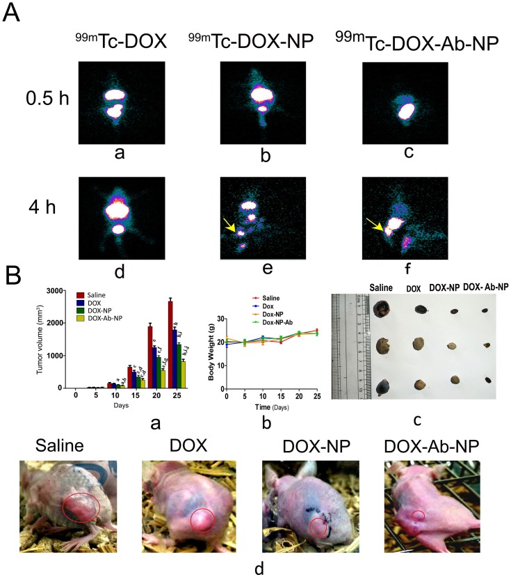
Result: Higher level of incorporation of DOX (8.5% W/W), which otherwise shows poor drug loading due to leaching effect of the highly water-soluble drug, was seen in this method. In HER2-overexpressing tumor xenograft model, radiolabeled antibody-conjugated nanoparticles showed preferentially more of the formulation accumulation in the tumor area when compared to the treatments with the unconjugated one or with the other control groups of mice. The ligand conjugated nanoparticles showed considerable potential in reduction of tumor growth and cardiac toxicity of DOX in mice, a prominent side-effect of the drug.

Conclusion: In conclusion, CD-340-conjugated PLGA nanoparticles containing DOX preferentially delivered encapsulated drug to the breast cancer cells and in breast tumor and reduced the breast tumor cells by apoptosis. Site-specific delivery of the formulation to neoplastic cells did not affect normal cells and showed a drastic reduction of DOX-related cardiotoxicity.

Keywords: breast cancer; ligand; nanoparticles; targeting; tumor.


Conflict of interest statement

The authors of this article have no conflict of interest to declare with regard to this work.

Figures

Figure 1
Figure 1
AFM and TEM images of experimental nanoparticles. Notes: AFM (A) DOX-NP, (B) DOX-Ab-NP and TEM (C) DOX-NP and (D) DOX-Ab-NP. Abbreviations: PLGA, polylactic-co-glycolic acid; PVA, poly (vinyl alcohol); AFM, Atomic force microscopy; DOX, doxorubicin; Ab, antibody; NP, nanoparticle; TEM, transmission electron microscopy.
Figure 2
Figure 2
In-vitro drug release profile of DOX from DOX-NP in different buffers (citrate buffer (pH 3), acetate buffer (pH 5), phosphate buffer (pH 7.4), and bicarbonate buffer (pH 10)) and different media (PBS release media added with hydroxy propyl β-cyclodextrin (HBC)/TWEEN, tween 80). Data show mean±SD (n=3). Abbreviations: PVA, poly (vinyl alcohol); TPGS, tocopherol polyethylene glycol succinate; DOX, doxorubicin; Ab, antibody; NP, nanoparticle.
Figure 3
Figure 3
Different studies for confirmation of antibody conjugation on the surface of polylactic-co-glycolic acid (PLGA) nanoparticles. Notes: (A) Flow cytometry data for unconjugated nanoparticles, (B) for antibody-conjugated nanoparticles, (C) laser-scanning confocal microscopy, showing FITC labeled CD-340 antibody-conjugated scattered nanoparticles and (D) SDS-PAGE gel electrophoresis, showing CD-340 antibody-conjugated NP,  antibody unconjugated NP and free-antibody. Abbreviations: DOX, doxorubicin; Ab, antibody; NP, nanoparticle.
Figure 4
Figure 4
In-vitro cytotoxicity data of DOX, DOX-NP, and DOX-Ab-NP determined in SKBR-3 cells, MCF-7 cells, and MDA-MB-231 cells. Data show mean ±SD (n=3). Abbreviations: DOX, doxorubicin; Ab, antibody; NP, nanoparticle.
Figure 5
Figure 5
Cellular uptake of antibody conjugated and unconjugated nanoparticles in various cell lines. Notes: (A) Cellular uptake of DOX-NP and DOX-Ab-NP in SKBR-3, MCF-7, and MDA-MB-231 cells observed by confocal microscopy at 1 and 6 hrs, respectively (B) Cellular uptake of antibody-conjugated blank nanoparticles in SKBR-3, MCF-7, and MDA-MB-231 cells observed by confocal microscopy at 6 hrs. Abbreviations: DOX, doxorubicin; Ab, antibody; NP, nanoparticle.
Figure 6
Figure 6
Apoptosis and cell cycle analysis data of breast cancer cells treated with experimental formulations and free-drug. Notes: (A) Apoptosis study by flow cytometry of SKBR-3 cells with the treatment of free DOX, DOX –NP, and DOX-Ab-NP after 24 hrs. Cell cycle analysis by flow cytometry for (B) SKBR-3 cells and (C) MCF-7 cells treated with DOX, DOX-NP, and DOX-Ab-NP for 24 hrs. Abbreviations: DOX, doxorubicin; Ab, antibody; NP, nanoparticle.
Figure 7
Figure 7
Comparative HER2 levels, caspase activity, and PARP cleavages in SKBR-3 cells, MCF-7 cells, and MDA-MB-231 cells. Notes: (A) Determination of the relative level of HER2 in SKBR-3 cells, MCF-7 cells, and MDA-MB-231 cells. (B) Activation of Caspase in MDA-MB-231 cells, MCF-7 cells, and SKBR-3 cells. (C) Detection of PARP cleavage in MDA-MB-231 cells, MCF-7 cells, and SKBR-3 cells. The values of p show statistical level of significance, **p<0.05, ***p<0.01, comparison between the groups are shown by over-head lines. Abbreviations: DOX, doxorubicin; Ab, antibody; NP, nanoparticle.
Figure 8
Figure 8
Plasma drug concentration upon IV bolus injection (at a dose of 10 mg/kg body weight) of DOX, DOX-NP, and DOX-Ab-NP. The same curve (time point 12–96 hrs) has been shown in the magnified version in the inset. Data show mean± SD (n=3). Abbreviations: DOX, doxorubicin; Ab, antibody; NP, nanoparticle.
Figure 9
Figure 9
Comparative cardiotoxicity profiles by the treatment of DOX, BL-NP, DOX-NP, and DOX-Ab-NP. Notes: (A) Echocardiography images. (B) Histopathology of cardiac tissue (Vascularization due to free-drug treatment was shown by the green arrow in the respective image). Abbreviations: DOX, doxorubicin; Ab, antibody; NP, nanoparticle.
Figure 10
Figure 10
Biodistribution of drug/formulation data and, tumor and tumor-related data in experimental animals. Notes: (A) Biodistribution and accumulation data of 99mTc labeled drug/formulations in experimental mice (a) time-dependent biodistribution and accumulation of 99mTc-DOX at 0.5 hrs (d) and at 4 hrs, (b) 99mTc -DOX-NP at 0.5 hrs (e) and 4 hrs, and (c) 99mTc -DOX-Ab-NP at 0.5 hrs and (f) 4 hrs, in xenograft mice-bearing breast tumor. (B) Changes in tumor volume, animal body weight, and excised tumors from the experimental mice along with the tumor growth-related representative visual image data. (a) Relative changes in tumor volume with time. Data showed mean tumor volume (mean ± SD, n=3) in different experimental and control mice. The value p<0.05 shows the level of statistical significance. Prefix of p (i.e., a–j), indicates as mentioned below: “a” indicates statistical level of significance when data were compared against normal saline treated (control) mice on day 10; “b” indicates statistical level of significance when data were compared against DOX-treated mice on day 10; “c” indicates statistical level of significance when data were compared against normal saline treated (control) mice on day 15; “d” indicates statistical level of significance when data were compared against DOX-treated mice on day 15; “e” indicates statistical level of significance when data were compared against normal saline treated (control) mice on day 20; “f” indicates statistical level of significance when data were compared against DOX-treated mice on day 20; “g” indicates statistical level of significance when data were compared against DOX-NP-treated mice on day 20; “h” indicates statistical level of significance when data were compared against normal saline treated (control) mice on day 25; “i” indicates statistical level of significance when data were compared against DOX-treated mice on day 25; “j” indicates statistical level of significance when data were compared against DOX-NP-treated mice on day 25. (b) Change of body weight of nude mice-bearing tumor. Data represent mean ± SD (n=3); data showed that there was no statistically significant variation between the groups of mice. (c) Tumors excised from the experimental mice after treatment with saline/DOX/DOX-NP/DOX-Ab-NP. (d) Images on day 25 of nude mice bearing SKBR-3 tumor after treatment with saline/DOX/DOX-NP/DOX-Ab-NP. Tumor areas have been marked with red circles in the figure. Abbreviations: 99mTc-DOX: radiolabeled free-doxorubicin, 99mTc-DOX -NP: radiolabeled doxorubicin loaded nanoparticles, 99mTc-DOX-Ab-NP: radiolabeled doxorubicin loaded antibody conjugated nanoparticles; DOX, doxorubicin; Ab, antibody; NP, nanoparticle.

Similar articles

Cited by

References

    1. Siegel RL, Miller KD, Jemal A. Cancer statistics, 2016. CA Cancer J Clin. 2016;66:7–30. doi:10.3322/caac.21332 - DOI - PubMed
    1. Mirakabad FST, Nejati-Koshki K, Akbarzadeh A, et al. PLGA-based nanoparticles as cancer drug delivery systems. Asian Pac J Cancer Prev. 2014;15(2):517–535. doi:10.7314/apjcp.2014.15.2.517 - DOI - PubMed
    1. Mukherjee B, Das S, Chakraborty S, et al. Potentials of polymeric nanoparticle as drug carrier for cancer therapy: with a special reference to pharmacokinetic parameters. Curr Drug Metab. 2015;15(6):565–580. doi:10.2174/1389200215666140605150703 - DOI - PubMed
    1. Alatrash G, Molldrem JJ. Tumor-associated antigens In: Socié G, Blazar BR editors. Immune Biology of Allogeneic Hematopoietic Stem Cell Transplantation.Models in Discovery and Translation. Academic Press; ISBN: 978-0-12-416004-0; 2013. 143–164.
    1. Mukherjee B. Nanosize drug delivery system. Curr Pharm Biotechnol. 2013;14(15):1221. - PubMed

MeSH terms