. 2019 Oct 3:10:1059.
doi: 10.3389/fneur.2019.01059. eCollection 2019.

The Association Between Distinct Frontal Brain Volumes and Behavioral Symptoms in Mild Cognitive Impairment, Alzheimer's Disease, and Frontotemporal Dementia

Affiliations

The Association Between Distinct Frontal Brain Volumes and Behavioral Symptoms in Mild Cognitive Impairment, Alzheimer's Disease, and Frontotemporal Dementia

Antti Cajanus et al. Front Neurol. .

Abstract

Our aim was to investigate the association between behavioral symptoms of agitation, disinhibition, irritability, elation, and aberrant motor behavior to frontal brain volumes in a cohort with various neurodegenerative diseases. A total of 121 patients with mild cognitive impairment (MCI, n = 58), Alzheimer's disease (AD, n = 45) and behavioral variant frontotemporal dementia (bvFTD, n = 18) were evaluated with a Neuropsychiatric Inventory (NPI). A T1-weighted MRI scan was acquired for each participant and quantified with a multi-atlas segmentation method. The volumetric MRI measures of the frontal lobes were associated with neuropsychiatric symptom scores with a linear model. In the regression model, we included CDR score and TMT B time as covariates to account for cognitive and executive functions. The brain volumes were corrected for age, gender and head size. The total behavioral symptom score of the five symptoms of interest was negatively associated with the volume of the subcallosal area (β = -0.32, p = 0.002). High disinhibition scores were associated with reduced volume in the gyrus rectus (β = -0.30, p = 0.002), medial frontal cortex (β = -0.30, p = 0.002), superior frontal gyrus (β = -0.28, p = 0.003), inferior frontal gyrus (β = -0.28, p = 0.005) and subcallosal area (β = -0.28, p = 0.005). Elation scores were associated with reduced volumes of the medial orbital gyrus (β = -0.30, p = 0.002) and inferior frontal gyrus (β = -0.28, p = 0.004). Aberrant motor behavior was associated with atrophy of frontal pole (β = -0.29, p = 0.005) and the subcallosal area (β = -0.39, p < 0.001). No significant associations with frontal brain volumes were found for agitation and irritability. We conclude that the subcallosal area may be common neuroanatomical area for behavioral symptoms in neurodegenerative diseases, and it appears to be independent of disease etiology.

Keywords: Alzheimer's disease; behavioral symptoms; frontotemporal dementia; magnetic resonance imaging; mild cognitive impairment.


Figures

Figure 1
Figure 1
Hierarchical clustering of the NPI symptoms shown as a dendrogram. The clustering was done with between-groups linkage using Pearson's correlation as the distance between the NPI items.
Figure 2
Figure 2
Group comparison of brain structure volumes of different diagnostic groups. Top row illustrates the comparison between bvFTD and MCI groups. The bvFTD group shows frontal and temporal atrophy and also reduced volume of deep gray matter structures. The middle row shows AD group's temporal and parietal atrophy compared to the MCI group. The lowest row shows the bvFTD group having reduced orbitofrontal, medial frontal and thalamic volumes compared to AD. The AD group showed no volume reduction compared to the bvFTD group. Comparison was done with two-tailed t-test and only significant areas of p ≤ 0.001 are highlighted.
Figure 3
Figure 3
Association of the volume of the subcallosal area and total behavioral scores. A regression analysis with CDR score and TMT-B time as a confounders revealed a significant association between the volume of the subcallosal area and the total behavioral score (β = −0.32, p = 0.002).
Figure 4
Figure 4
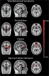
Correlation of the total and individual behavioral symptoms with brain volumes. In the first row, total behavioral symptom scores show a correlation with the subcallosal area and left superior frontal gyrus. All three symptom scores show a correlation with ventromedial prefrontal cortex. The left superior frontal gyrus showed a correlation with elation and disinhibition, while aberrant motor behavior was associated with right superior frontal gyrus. In addition to frontal volumes, disinhibition scores were associated with the bilateral temporal pole. The subcallosal area is shown in cyan circle in the top row. Irritability and agitation showed no correlation with any focal brain volume. Negative correlations are shown in red. No positive correlations were detected.

References

    1. Srikanth S, Nagaraja AV, Ratnavalli E. Neuropsychiatric symptoms in dementia-frequency, relationship to dementia severity and comparison in Alzheimer's disease, vascular dementia and frontotemporal dementia. J Neurol Sci. (2005) 236:43–8. 10.1016/j.jns.2005.04.014 - DOI - PubMed
    1. Mega MS, Cummings JL, Fiorello T, Gornbein J. The spectrum of behavioral changes in Alzheimer's disease. Neurology. (1996) 46:130–5. - PubMed
    1. do Ferreira MC, Abreu MJ, Machado C, Santos B, Machado Á, Costa AS. Neuropsychiatric profile in early versus late onset Alzheimer's Disease. Am J Alzheimer's Dis Other Dementiasr. (2018) 33:93–9. 10.1177/1533317517744061 - DOI - PMC - PubMed
    1. Travis Seidl JN, Massman PJ, Seidl JNT, Massman PJ. Cognitive and functional correlates of NPI-Q Scores and Symptom Clusters in Mildly Demented Alzheimer Patients. Alzheimer Dis Assoc Disord. (2016) 30:145–51. 10.1097/WAD.0000000000000104 - DOI - PubMed
    1. Borroni B, Cosseddu M, Pilotto A, Premi E, Archetti S, Gasparotti R, et al. . Early stage of behavioral variant frontotemporal dementia: clinical and neuroimaging correlates. Neurobiol Aging. (2015) 36:3108–15. 10.1016/J.NEUROBIOLAGING.2015.07.019 - DOI - PubMed

LinkOut - more resources