. 2019 Sep 26:11:1758835919875555.
doi: 10.1177/1758835919875555. eCollection 2019.

Novel multi-drugs incorporating hybrid-structured nanofibers enhance alkylating agent activity in malignant gliomas

Affiliations

Novel multi-drugs incorporating hybrid-structured nanofibers enhance alkylating agent activity in malignant gliomas

Shih-Jung Liu et al. Ther Adv Med Oncol. .

Abstract

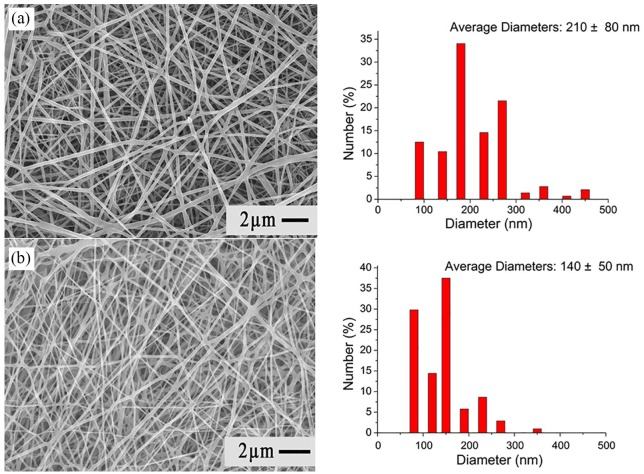
Background: Malignant gliomas (MGs) are highly chemotherapy-resistant. Temozolomide (TMZ) and carmustine (BiCNU) are alkylating agents clinically used for treating MGs. However, their effectiveness is restrained by overexpression of the DNA repair protein O6-methylguanine-DNA methyltransferase (MGMT) in tumors. O6-benzylguanine (O6-BG) is a nonreversible inhibitor of MGMT, it promotes the cytotoxicity of alkylating chemotherapy. The authors have developed a hybrid-structured nanofibrous membrane (HSNM) that sequentially delivers high concentrations of O6-BG, BiCNU, and TMZ in an attempt to provide an alternative to the current therapeutic options for MGs.

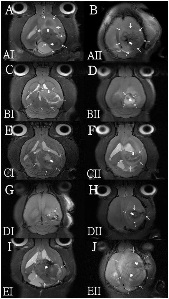
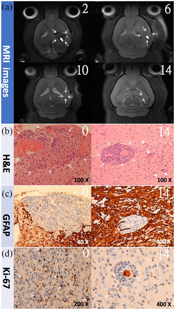
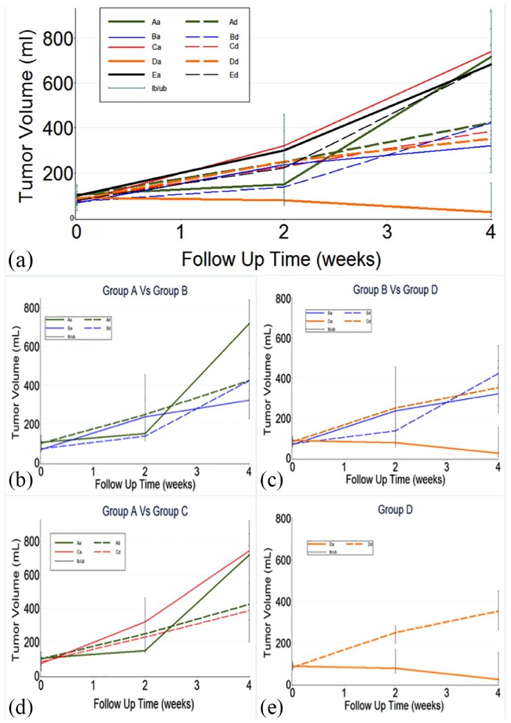
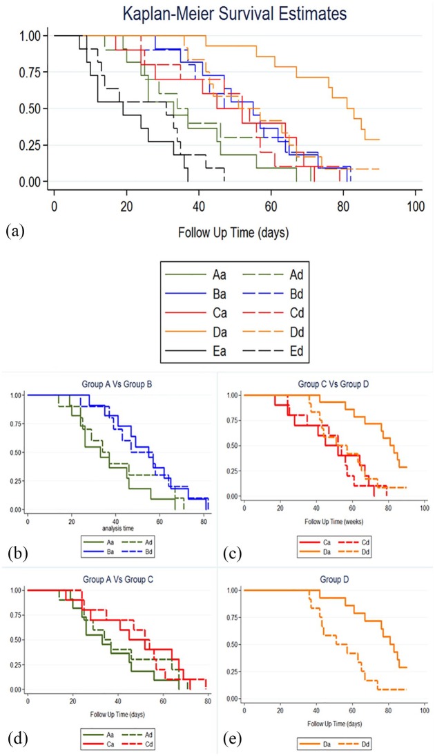
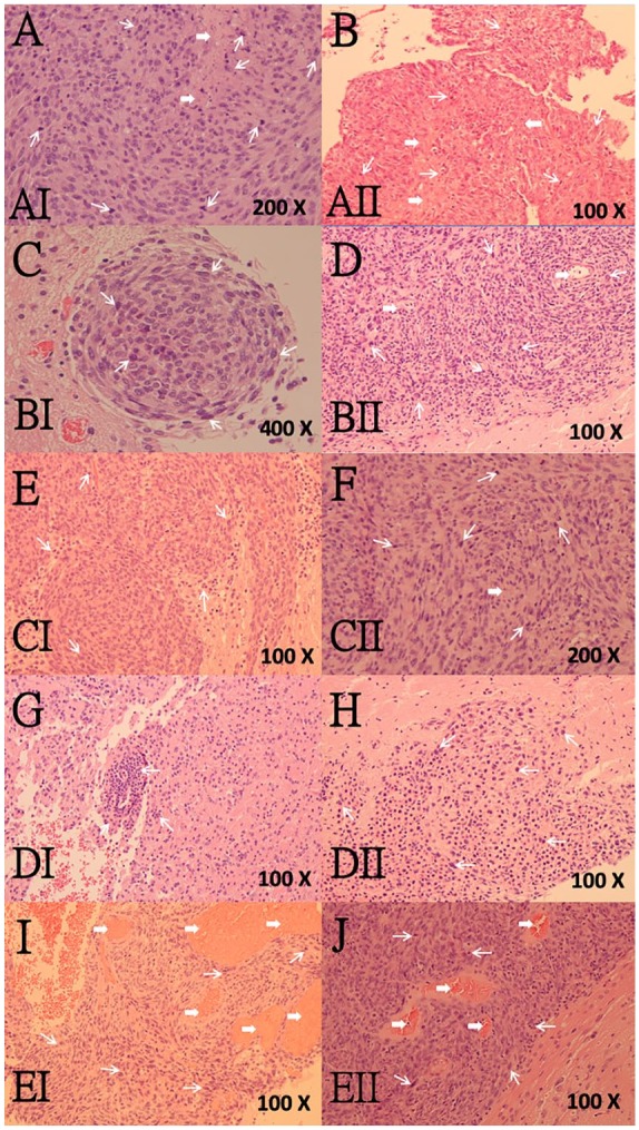
Methods: The HSNMs were implanted onto the cerebral surface of pathogen-free rats following surgical craniectomy, while the in vivo release behaviors of O6-BG, TMZ, and BiCNU from the HSNMs were explored. Subsequently, the HSNMs were surgically implanted onto the brain surface of two types of tumor-bearing rats. The survival rate, tumor volume, malignancy of tumor, and apoptotic cell death were evaluated and compared with other treatment regimens.

Results: The biodegradable HSNMs sequentially and sustainably delivered high concentrations of O6-BG, BiCNU, and TMZ for more than 14 weeks. The tumor-bearing rats treated with HSNMs demonstrated therapeutic advantages in terms of retarded and restricted tumor growth, prolonged survival time, and attenuated malignancy.

Conclusion: The results demonstrated that O6-BG potentiates the effects of interstitially transported BiCNU and TMZ. Therefore, O6-BG may be required for alkylating agents to offer maximum therapeutic benefits for the treatment of MGMT-expressing tumors. In addition, the HSNM-supported chemoprotective gene therapy enhanced chemotherapy tolerance and efficacy. It can, therefore, potentially provide an improved therapeutic alternative for MGs.

Keywords: O6-benzylguanine (O6-BG); O6-methylguanine-DNA methyltransferase (MGMT); chemoresistance; malignant glioma; nanofibrous membrane.


Conflict of interest statement

Conflict of interest statement: The authors declare no conflicts of interest in preparing this article.

Figures

Figure 1.
Figure 1.
The surgical procedure, tumor cell, and in vivo drug study. (a) 1. Craniectomy (approximately 10 mm × 10 mm), 2. one-twentieth Gliadel wafer, and 3. HSNM (10 mm × 10 mm) surgically implanted onto the brain surface of rats. (b) F98 cells expressed approximately seven times higher levels of MGMT than 9L cells. (c) In vivo release curve for O6-BG, BiCNU, and TMZ liberation from the HSNMs.
Figure 2.
Figure 2.
SEM image and fiber size distribution: (a) blend nanofibers; (b) sheath-core nanofibers.
Figure 3.
Figure 3.
FTIR spectra of pure PLGA nanofibrous membranes and HSNMs. The spectra assay confirmed that the pharmaceuticals were successfully embedded in the PLGA membranes.
Figure 4.
Figure 4.
Brain MRI 4 weeks after treatment in each subgroup. The letters on the lower-left corner of each image indicate the subgroup. Central necrosis and tumor are indicated with thick and thin arrows, respectively. The tumor fulminant expansion caused severe mass effect and midline shift in subgroup EI I and EII J, whereas rapid growth in subgroups AI A and AII B resulted in ventricle compression and midline shift. In subgroups BI C, BII D, CI E, and CII F, tumor growth caused mild-to-moderate mass effect and midline shift. TV decreased without midline shift in subgroup DI G and there was a slight increase in subgroup DII H.
Figure 5.
Figure 5.
The study results of subgroup DI. The number on the upper-right corner indicates weeks after HSNM implantation. (a) Serial MRI scans. The tumor decreasing in size with time and no tumor regrowth was noted. (b) H&E staining image. The tumor area is restricted and localized with a low number of satellite tumor cells outside the tumor mass (thick arrows). (c) GFAP immunocytochemical staining. GFAP-positive glial cells (thin arrows) are distinct and surround the shrinking tumor. (d) The Ki-67 labeling index. Approximately 12.21% before and 4.93% 14 weeks after HSNM implantation.
Figure 6.
Figure 6.
The repeated-measures mixed model was used to evaluate differences in the mean 4-week TVs between each subgroup. (a) TVs increased at various rates, except in group DI. The TV decreased after HSNM implantation. (b) TVs were significantly different between subgroups BI and AI (p = 0.016), but not between subgroups BII and AII (p = 0.810). (c) and (d) TVs were significantly different between subgroups DI and CI as well as DI and BI (p < 0.05), but not between subgroups DII and CII as well as DII and BII (p > 0.05). (e) TVs were significantly different between subgroups of DI and DII (p < 0.001).
Figure 7.
Figure 7.
Overall survival of tumor-bearing rats in each subgroup. (a) Of the five groups, group D had the longest survival time, followed by groups B, C, A, and E. (b) The survival time was significantly different between subgroups BII and AII (p = 0.039). (c) The survival time was significantly different between subgroups CI and AI as well as CII and AII. (d) The survival time was significantly different between subgroups DI and CI (p = 0.001), but not between subgroups DII and CII (p = 0.420). (e) The survival time was significantly shorter in subgroup DII than in subgroup DI (p < 0.05).
Figure 8.
Figure 8.
H&E staining. The letters in the lower-left corner of each image indicate the subgroup. Karyorrhectic tumor cells and central (coagulation) necrosis are indicated with thin and thick arrows, respectively. Diffuse karyorrhectic tumor cells with central necrosis were noted in subgroups EI I, EII J, AI A, AII B, and BII D. Restricted tumor area and little central necrosis were observed in subgroups BI C, CI E, and CII F. The tumor cells tended to localize in small areas in subgroups DI G and DII H.
Figure 9.
Figure 9.
GFAP immunocytochemical staining. The letters in the lower-left corner of each image indicate the subgroup. GFAP-positive glial cells are indicated with arrows. No GFAP expression was noted in subgroups EI I and EII j. A small number of scattered GFAP-positive glial cells were detected in subgroups AI A, AII B, and CI E. A number of scattered GFAP-positive glial cells were detected in subgroups BI C, BII D, and CII F. Coarse, dendritic GFAP-positive glial cells were detected in subgroups DI G and DII H.
Figure 10.
Figure 10.
The Ki-67 labeling index by MIB-1 immunostaining in each subgroup. The letters in the lower-left corner of each image indicate the subgroup, followed by its Ki-67 labeling index (percentage). The arrows indicate Ki-67-positive cells. An extremely high Ki-67 labeling index was detected in subgroups EI I and EII J. The Ki-67 labeling index in subgroups AI A, AII B, BI C, and BII D was 49.43%, 35.42%, 42.42%, and 39.43%, respectively. The Ki-67 labeling index was low in subgroup CI E (approximately 20.73%) and was 29.19% in subgroup CII F. The Ki-67 labeling index decreased to less than 10% in subgroups DI G and DII H.
Figure 11.
Figure 11.
Evaluation of apoptosis of glioma cells by TUNEL assay in each subgroup. The letter at the lower-left corner of each image denotes the subgroup, while the black arrows indicate TUNEL-positive apoptotic cells. A limited number of apoptotic nuclei were found in subgroups AI A, AII B, BI C, and BII D, but subgroups CI E and CII F displayed some apoptotic nuclei in the intratumor area. A significant increase in the number of TUNEL-positive apoptotic nuclei was observed in subgroups DI G and DII H. However, no TUNEL-positive apoptotic nucleus were detected in the tumor cells in subgroups EI I and EII J. (Magnification, 100×).

References

    1. Wen PY, Kesari S. Malignant gliomas in adults. N Engl J Med 2008; 359: 492–507. - PubMed
    1. Wong ET, Hess KR, Gleason MJ, et al. Outcomes and prognostic factors in recurrent glioma patients enrolled onto phase II clinical trials. J Clin Oncol 1999; 17: 2572–2578. - PubMed
    1. Stummer W, Reulen HJ, Meinel T, et al. Extent of resection and survival in glioblastoma multiforme: identification of and adjustment for bias. Neurosurgery 2008; 62: 564–576; discussion -76. - PubMed
    1. Bobola MS, Tseng SH, Blank A, et al. Role of O6-methylguanine-DNA methyltransferase in resistance of human brain tumor cell lines to the clinically relevant methylating agents temozolomide and streptozotocin. Clin Cancer Res 1996; 2: 735–741. - PubMed
    1. Quinn JA, Jiang SX, Reardon DA, et al. Phase II trial of temozolomide plus O6-benzylguanine in adults with recurrent, temozolomide-resistant malignant glioma. J Clin Oncol 2009; 27: 1262–1267. - PMC - PubMed