. 2019 Sep 15;11(9):5560-5572.
eCollection 2019.

Effects of Tianqijiangtang capsule on survival, self-renewal and differentiation of hippocampal neural stem cells of embryonic rats cultured in high glucose medium

Affiliations

Effects of Tianqijiangtang capsule on survival, self-renewal and differentiation of hippocampal neural stem cells of embryonic rats cultured in high glucose medium

Chun-Yong Han et al. Am J Transl Res. .

Abstract

Objective: This study aims to investigate the effects of Tianqijiangtang capsule on the survival, self-renewal and differentiation of hippocampal neural stem cells (NSCs) of embryonic rats cultured in high glucose medium.

Materials and methods: A cell model of diabetic encephalopathy was established. Cell viability was assessed to screen the optimal concentration of glucose for the cell model of diabetic encephalopathy. Then, the effects of Tianqijiangtang capsule on the proliferation and differentiation of NSCs, and the expression of vascular endothelial growth factor (VEGF) and brain-derived neurotrophic factor (BDNF) in the culture medium and cells were detected.

Results: High glucose significantly reduced the ability of survival, proliferation and differentiation of NSCs, which was statistically significant, when compared to the control group (P < 0.05 or 0.01). Tianqijiangtang capsule significantly enhanced the survival, proliferation and differentiation of NSCs cultured in high glucose medium, which was statistically significant, when compared with the high glucose group (P < 0.05 or 0.01). The high glucose culture resulted in a significant decrease in VEGF and BDNF levels in culture medium and cells of NSCs. Tianqijiangtang capsule significantly increased the level of VEGF nuclear BDNF in cells and the culture medium, which was significantly higher, when compared to that in the high glucose group (P < 0.05 or 0.01).

Conclusion: Tianqijiangtang capsule enhances the level of neurotrophic factor synthesized and secreted by hippocampal NSCs cultured with high glucose through the autocrine and paracrine pathway, promotes the NSC survival, replication and differentiation of new neurons and astrocytes, and reduces the degeneration and necrosis of nerve cells.

Keywords: Tianqijiangtang capsule; brain-derived neurotrophic factor; diabetic encephalopathy; neural stem cells; vascular endothelial growth factor.


Conflict of interest statement

None.

Figures

Figure 1
Figure 1
The culture of NSCs of embryonic rat hippocampus and Nestin positive cells of NSCs of embryonic rat hippocampus (600×). A: Primary culture for one day for NSCs obtained from embryonic rat hippocampus, 100×; B: Primary culture for three days for NSCs obtained from embryonic rat hippocampus forming neurospheres, 200×; C: Passage culture for one day for NSCs obtained from embryonic rat hippocampus, which formed neurospheres, 100×; D: Passage culture for three days for NSCs obtained from embryonic rat hippocampus forming neurospheres, 200×; E: Adherent culture for three days of passage for NSCs obtained from embryonic rat hippocampus, the edge of neurospheres appear with the processes, 100×; F: Adherent culture for five days of passage for NSCs obtained from embryonic rat hippocampus, NSCs becoming differentiated, 100×; G: Monoclonal culture for three days for NSCs obtained from embryonic rat hippocampus, single cell proliferation into neurospheres, 200×; H: Nestin positive cells of NSCs obtained from embryonic rat hippocampus (red); I: DAPI nucleus of NSCs obtained from embryonic rat hippocampus (blue); J: The merge of nestin and DAPI for NSCs obtained from embryonic rat hippocampus.
Figure 2
Figure 2
Nestin positive cells for NSCs obtained from embryonic rat hippocampus (A, red), Brdu positive cells for NSCs obtained from embryonic rat hippocampus (B, green), DAPI nucleus for NSCs obtained from embryonic rat hippocampus (C, blue), and the merge (D) of nestin, BrdU and DAPI for NSCs obtained from embryonic rat hippocampus (600×). Nestin positive cells for NSCs obtained from embryonic rat hippocampus (E, red), DAPI nucleus for NSCs obtained from embryonic rat hippocampus (F, blue), Tuj-1 positive cells for NSCs obtained from embryonic rat hippocampus (G, green), and the merge (H) of nestin, Tuj-1 and DAPI for NSCs obtained from embryonic rat hippocampus (600×). Nestin positive cells for NSCs obtained from embryonic rat hippocampus (I, red), vimentin positive cells for NSCs obtained from embryonic rat hippocampus (J, green), DAPI nucleus for NSCs obtained from embryonic rat hippocampus (K, blue), and the merge (L) of nestin, vimentin and DAPI for NSCs obtained from embryonic rat hippocampus (600×).
Figure 3
Figure 3
A. The screening of the optimal glucose concentration for the model of NSCs of embryonic rat hippocampus. The optimal concentration was 8,500 μg/ml. B. The screening of the optimal concentration of Tianqijiangtang capsule in promoting the proliferation of NSCs. The best concentration for the Tianqijiangtang capsule was 50 μg/ml. C. The screening of the best time point of 50 μg/ml of Tianqijiangtang capsule in promoting the proliferation of NSCs. The best time point was nine hours.
Figure 4
Figure 4
In control group, BrdU positive cells for NSCs obtained from embryonic rat hippocampus (A, red), Nestin positive cells for NSCs obtained from embryonic rat hippocampus (B, green), DAPI nucleus for NSCs obtained from embryonic rat hippocampus (C, blue), and the merge (D) of BrdU, nestin and DAPI for NSCs obtained from embryonic rat hippocampus (600×). In the high glucose group, Brdu positive cells for NSCs obtained from embryonic rat hippocampus (A, red), Nestin positive cells for NSCs obtained from embryonic rat hippocampus (B, green), DAPI nucleus for NSCs obtained from embryonic rat hippocampus (C, blue), and the merge (D) of BrdU, nestin and DAPI for NSCs obtained from embryonic rat hippocampus (600×). In the Tianqijiangtang capsule group, BrdU positive cells for NSCs obtained from embryonic rat hippocampus (A, red), Nestin positive cells for NSCs obtained from embryonic rat hippocampus (B, green), DAPI nucleus for NSCs obtained from embryonic rat hippocampus (C, blue), and the merge (D) of BrdU, nestin and DAPI for NSCs obtained from embryonic rat hippocampus (600×). Statistical results of the area and optical density, and the number of nestin, BrdU positive cells in the control, high glucose and Tianqijiangtang capsule group. Compared with the control group, *P < 0.05, **P < 0.01; compared with the model group: #P < 0.05, ##P < 0.01.
Figure 5
Figure 5
In the control group, Brdu positive cells for NSCs obtained from embryonic rat hippocampus (A, red), Tuj-1 positive cells for NSCs obtained from embryonic rat hippocampus (B, green), DAPI nucleus for NSCs obtained from embryonic rat hippocampus (C, blue), and the merge (D) of BrdU, Tuj-1 and DAPI for NSCs obtained from embryonic rat hippocampus (600×). In the high glucose group, Brdu positive cells for NSCs obtained from embryonic rat hippocampus (A, red), Tuj-1 positive cells for NSCs obtained from embryonic rat hippocampus (B, green), DAPI nucleus for NSCs obtained from embryonic rat hippocampus (C, blue), and the merge (D) of BrdU, Tuj-1 and DAPI for NSCs obtained from embryonic rat hippocampus (600×). In the Tianqijiangtang capsule group, BrdU positive cells for NSCs obtained from embryonic rat hippocampus (A, red), Tuj-1 positive cells for NSCs obtained from embryonic rat hippocampus (B, green), DAPI nucleus for NSCs obtained from embryonic rat hippocampus (C, blue), and the merge (D) of BrdU, Tuj-1 and DAPI for NSCs obtained from embryonic rat hippocampus (600×). The results of the statistical analyses of the area and optical density, and number of Tuj-1 and BrdU positive cells in the control, high glucose and Tianqijiangtang capsule group. Compared with the control group, *P < 0.05, **P < 0.01; compared with the model group, #P < 0.05, ##P < 0.01.
Figure 6
Figure 6
In the control group, BrdU positive cells for NSCs obtained from embryonic rat hippocampus (red), vimentin positive cells for NSCs obtained from embryonic rat hippocampus (green), DAPI nucleus for NSCs obtained from embryonic rat hippocampus (blue), and the merge of BrdU, vimentin and DAPI for NSCs obtained from embryonic rat hippocampus (600×). In the high glucose group, BrdU positive cells for NSCs obtained from embryonic rat hippocampus (red), vimentin positive cells for NSCs obtained from embryonic rat hippocampus (green), DAPI nucleus for NSCs obtained from embryonic rat hippocampus (blue), and the merge of BrdU, vimentin and DAPI for NSCs obtained from embryonic rat hippocampus (600×). In the Tianqijiangtang capsule group, Brdu positive cells for NSCs obtained from embryonic rat hippocampus (red), vimentin positive cells for NSCs obtained from embryonic rat hippocampus (green), DAPI nucleus for NSCs obtained from embryonic rat hippocampus (blue), and the merge of BrdU, vimentin and DAPI for NSCs obtained from embryonic rat hippocampus (600×). Statistical results of the area and optical density, and number of vimentin and BrdU positive cells in the control, high glucose and Tianqijiangtang capsule group. Compared with the control group: *P < 0.05, **P < 0.01; Compared with the model group, #P < 0.05, ##P < 0.01.
Figure 7
Figure 7
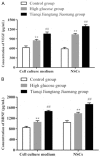
A. The results of the statistical analyses of the content of VEGF and BDNF in the culture medium. Compared with the control group: *P < 0.05, **P < 0.01; Compared with the model group: #P < 0.05, ##P < 0.01. B. The results of the statistical analyses of the content of VEGF and BDNF in cells. Compared with the control group, *P < 0.05, **P < 0.01; Compared with the model group: #P < 0.05, ##P < 0.01.

References

    1. Cumbie BC, Hermayer KL. Current concepts in targeted therapies for the pathophysiology of diabetic microvascular complications. Vasc Health Risk Manag. 2007;3:823–832. - PMC - PubMed
    1. Khan ZA, Farhangkhoee H, Chakrabarti S. Towards newer molecular targets for chronic diabetic complications. Curr Vasc Pharmacol. 2006;4:45–57. - PubMed
    1. Fekete A, Rosta K, Wagner L, Prokai A, Degrell P, Ruzicska E, Vegh E, Toth M, Ronai K, Rusai K, Somogyi A, Tulassay T, Szabo AJ, Ver A. Na+, K+-ATPase is modulated by angiotensin II in diabetic rat kidney--another reason for diabetic nephropathy? J Physiol. 2008;586:5337–5348. - PMC - PubMed
    1. Mijnhout GS, Scheltens P, Diamant M, Biessels GJ, Wessels AM, Simsek S, Snoek FJ, Heine RJ. Diabetic encephalopathy: a concept in need of a definition. Diabetologia. 2006;49:1447–1448. - PubMed
    1. Miles WR, Root HF. Psychologic tests applied in diabetic patients. Arch Intern Med. 1922;30:767–770.

LinkOut - more resources